

Case Reports
doi: 10.3201/eid2403.170772.
Invasive Infections Caused by Nannizziopsis spp. Molds in Immunocompromised Patients
Affiliations
- PMID: 29460742
- PMCID: PMC5823334
- DOI: 10.3201/eid2403.170772
Item in Clipboard
Case Reports
Invasive Infections Caused by Nannizziopsis spp. Molds in Immunocompromised Patients
Emerg Infect Dis.
2018 Mar.
Abstract
We report 2 new cases of invasive infections caused by Nannizziopsis spp. molds in France. Both patients had cerebral abscesses and were immunocompromised. Both patients had recently spent time in Africa.
Keywords: Nannizziopsis; central nervous system fungal infection; emerging disease; fungi; immunocompromised patients; invasive fungal infection; molds; opportunistic fungal pathogen.
Figures
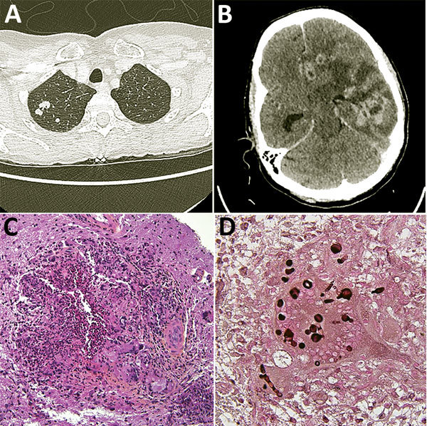
Diagnostic testing of a 52-year-old woman from France living in Mali who had Nannizziopsis spp. fungal infection. A) Thoracic-abdominal-pelvic scan shows pseudo-nodular lesions in the apex of the right lung, of which one is excavated. B) Cerebral computed tomography scan shows contrast enhancement on several hemispheric nodules on the left and in frontal, parietal, and temporal regions, responsible for large surrounding edema and compression of the left lateral ventricle. The median line is deviated to the right with a subfalcorial herniation. C) Hematoxylin-eosin-saffron stain of brain biopsy containing mononuclear inflammatory infiltrates; giant cell granulomas; histiocytes, sometimes with an epithelioid appearance; and neutrophils (original magnification ×200). D) Grocott stain showing thick bulbous mycelial filaments in the cytoplasm of certain giant cells/histiocytes (original magnification ×600). Round shapes correspond to cross-sections of bulbous territories.
References
-
- Stillwell WT, Rubin BD, Axelrod JL. Chrysosporium, a new causative agent in osteomyelitis. A case report. Clin Orthop Relat Res. 1984; (184):190–2. - PubMed
-
- Sigler L, Hambleton S, Paré JA. Molecular characterization of reptile pathogens currently known as members of the chrysosporium anamorph of Nannizziopsis vriesii complex and relationship with some human-associated isolates. J Clin Microbiol. 2013;51:3338–57. 10.1128/JCM.01465-13 - DOI - PMC - PubMed
Publication types
MeSH terms
Substances
LinkOut - more resources
Full Text Sources
Other Literature Sources
Molecular Biology Databases
Miscellaneous
