Review
. 2018 Apr;15(4):222-234.
doi: 10.1038/nrurol.2018.9. Epub 2018 Feb 20.

Clinical implications of PTEN loss in prostate cancer

Affiliations
Review

Clinical implications of PTEN loss in prostate cancer

Tamara Jamaspishvili et al. Nat Rev Urol. 2018 Apr.

Abstract

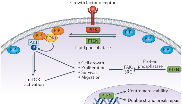
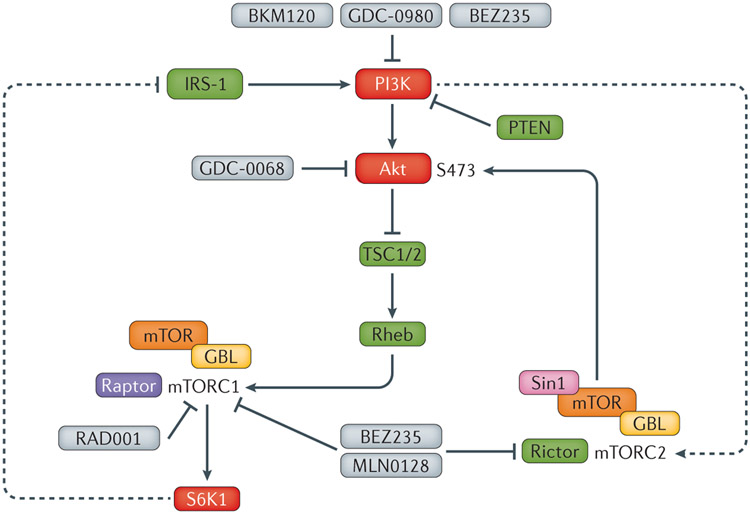
Genomic aberrations of the PTEN tumour suppressor gene are among the most common in prostate cancer. Inactivation of PTEN by deletion or mutation is identified in ∼20% of primary prostate tumour samples at radical prostatectomy and in as many as 50% of castration-resistant tumours. Loss of phosphatase and tensin homologue (PTEN) function leads to activation of the PI3K-AKT (phosphoinositide 3-kinase-RAC-alpha serine/threonine-protein kinase) pathway and is strongly associated with adverse oncological outcomes, making PTEN a potentially useful genomic marker to distinguish indolent from aggressive disease in patients with clinically localized tumours. At the other end of the disease spectrum, therapeutic compounds targeting nodes in the PI3K-AKT-mTOR (mechanistic target of rapamycin) signalling pathway are being tested in clinical trials for patients with metastatic castration-resistant prostate cancer. Knowledge of PTEN status might be helpful to identify patients who are more likely to benefit from these therapies. To enable the use of PTEN status as a prognostic and predictive biomarker, analytically validated assays have been developed for reliable and reproducible detection of PTEN loss in tumour tissue and in blood liquid biopsies. The use of clinical-grade assays in tumour tissue has shown a robust correlation between loss of PTEN and its protein as well as a strong association between PTEN loss and adverse pathological features and oncological outcomes. In advanced disease, assessing PTEN status in liquid biopsies shows promise in predicting response to targeted therapy. Finally, studies have shown that PTEN might have additional functions that are independent of the PI3K-AKT pathway, including those affecting tumour growth through modulation of the immune response and tumour microenvironment.


Conflict of interest statement

Competing interests statement

T.L.L. has received research support from Ventana Medical Systems. D.M.B. has received financial support from Myriad Genetics and Metamark Genetics.

Figures

Figure 1 ∣
Figure 1 ∣. The diverse cellular roles of PTEN.
PTEN acts as lipid phosphatase, converting phosphatidylinositol 3,4,5-trisphosphate [PI(3,4,5)P3 or PIP3] into phosphatidylinositol 4,5-bisphosphate [PI(4,5)P2 or PIP2]. In this capacity, PTEN antagonizes the function of Class I PI3K activity, which converts PIP2 to PIP3. This lipid phosphatase activity of PTEN suppresses the activation of the downstream oncogenic AKT and mTOR signalling cascades. However, PTEN also has several other noncanonical functions, including weak protein phosphatase activity with known kinase substrates such as FAK and SRC. Finally, PTEN probably functions in the nucleus in a PI3K-independent manner to promote chromosome stability and DNA repair .
Figure 2 ∣
Figure 2 ∣. Prostate cancer samples with variable PTEN protein expression by IHC and corresponding PTEN FISH.
a ∣ Prostate tumour with intact PTEN identified with IHC with two intact PTEN alleles detected using FISH. PTEN IHC demonstrates intact PTEN (top) and four-colour FISH image from an adjacent section (bottom) shows two intact PTEN alleles (red) with two intact copies of flanking genes, WAPAL (green) and FAS (aqua) as well as chromosome 10 centromeres (pink). b ∣ Prostate tumour showing PTEN expression using IHC with hemizygous PTEN deletion using FISH. PTEN IHC demonstrates intact PTEN protein (top), with four-colour FISH image (bottom) from an adjacent section showing a hemizygous PTEN deletion with loss of one PTEN gene (one red signal). As both centromeres (pink) and the WAPAL (green) and FAS (aqua) probes that flank either side of PTEN are retained, this hemizygous deletion is likely to be interstitial and restricted to the PTEN region.c ∣ Prostate tumour showing absence of PTEN expression by IHC with homozygous PTEN gene deletion detected in intraductal tumour by FISH. PTEN IHC image (top) shows loss of PTEN in tumour glands. Four-colour FISH image from an adjacent section (bottom) shows a homozygous deletion with loss of both PTEN genes (red). The retention of the centromeres (pink) and both WAPAL genes (green), but the presence of only one copy of FAS (aqua) indicates that one of the deletions involved both PTEN and FAS. d ∣ Prostate tumour showing PTEN protein loss by IHC with hemizygous PTEN gene deletion by FISH. PTEN IHC demonstrates complete PTEN loss (top), with four-colour FISH image (bottom) from an adjacent section showing a hemizygous PTEN deletion with loss of one PTEN gene (red) along with flanking genes WAPAL (green) and FAS (aqua). This tumour demonstrates complex hemizygous PTEN deletion, in which PTEN is deleted along with adjacent genes (WAPAL and FAS) located on both sides of PTEN.
Figure 3 ∣
Figure 3 ∣. Heterogeneous immunohistochemical expression of ERG and PTEN in prostate tumours.
a ∣ Heterogeneous PTEN loss is observed in some tumour glands (N), with intact staining in other tumour glands (P) whereas the same areas are uniformly positive for ERG expression indicating that clonal ERG genomic rearrangement is probably present with subsequent subclonal PTEN loss. b ∣ Heterogeneous PTEN loss is seen in some tumour glands (N), with intact staining in other tumour glands (P), whereas the same areas are uniformly negative for ERG expression (ERG genomic rearrangement is absent).
Figure 4 ∣
Figure 4 ∣. Algorithm for when to determine PTEN status on diagnostic biopsy material using IHC and FISH
a ∣ Prostate biopsy is indicated to confirm or exclude cancer in patients with elevated serum PSA and/or abnormal digital rectal exam (DRE). Patients with persistent PSA elevation might require repeat biopsy.. b) If cancer is detected and is low-to-intermediate risk (Grade group 1 or 2; Gleason score 3+3 or 3+4), PTEN status can be ascertained using immunohistochemical (IHC) staining. If PTEN protein expression is intact, the prognosis reflects the patient’s clinicopathological variables (e.g. Grade Group, PSA, age, DRE). If IHC demonstrates complete PTEN loss, biomarker status should be considered in the patient’s prognosis along with clinicopathological variables and FISH is not necessary. However, if PTEN loss is incomplete or ambiguous (e.g. negative PTEN staining in cancer glands along with negative PTEN expression in internal control, such as benign glands and/or stroma), FISH is recommended and bears similar connotations to PTEN loss determined by IHC. c ∣ If cancer is detected and is high risk (Grade group 3, 4 and 5; Gleason score 4+3 and 4+4, ≥4+5), PTEN status does not need to be determined, because prognosis will be strongly driven by clinicopathological variables.
Figure 5 ∣
Figure 5 ∣. Proposed management options using clinicopathological variables at biopsy and PTEN status.
Patients with Grade Group 1 (GG1) cancer and no loss of PTEN on biopsy (by IHC and/or FISH) should be considered for active surveillance. Patients with GG1 cancer with PTEN loss should be considered for definitive treatment using radiotherapy or prostatectomy. In some clinical contexts, patients with GG2 cancer with no loss of PTEN could be considered for active surveillance, particularly if they have low-volume disease and a low percentage of Gleason pattern 4 (indicated by dashed arrow). Patients with GG2 tumours with PTEN loss or tumours >GG2 should be considered for definitive treatment in most cases.
Figure 6 ∣
Figure 6 ∣. Selected drugs in clinical trials targeting the PI3K/AKT pathway that have been used in combination with androgen deprivation therapy.
PI3K inhibitors (GDC-0980, BKM120), combined PI3K/mTOR inhibitors (BEZ235), mTORC1 inhibitors (RAD001) and mTOR kinase inhibitors (CC-115) have been used in clinical trials in combination with novel and conventional androgen deprivation therapies. Whereas some trials have been largely negative (BKM-120) or poorly tolerated (BEZ235), others are promising and suggest that PTEN status might be a useful predictive biomarker for response in some contexts (eg, GDC-0068 or ipatasertib).

References

    1. Siegel RL, Miller KD & Jemal A Cancer Statistics, 2017. CA Cancer J Clin 67, 7–30, doi:10.3322/caac.21387 (2017). - DOI - PubMed
    1. Tosoian JJ et al. Intermediate and Longer-Term Outcomes From a Prospective Active-Surveillance Program for Favorable-Risk Prostate Cancer. J Clin Oncol 33, 3379–3385, doi:10.1200/JCO.2015.62.5764 (2015). - DOI - PMC - PubMed
    1. Cancer Genome Atlas Research, N. The Molecular Taxonomy of Primary Prostate Cancer. Cell 163, 1011–1025, doi:10.1016/j.cell.2015.10.025 (2015). - DOI - PMC - PubMed
    1. Taylor BS et al. Integrative genomic profiling of human prostate cancer. Cancer Cell 18, 11–22, doi:10.1016/j.ccr.2010.05.026 (2010). - DOI - PMC - PubMed
    1. Maehama T & Dixon JE The tumor suppressor, PTEN/MMAC1, dephosphorylates the lipid second messenger, phosphatidylinositol 3,4,5-trisphosphate. J Biol Chem 273, 13375–13378 (1998). - PubMed

Publication types