Review
. 2018 Feb 20;8(2):35.
doi: 10.3390/brainsci8020035.

Surgical Considerations of Intractable Mesial Temporal Lobe Epilepsy

Affiliations
Review

Surgical Considerations of Intractable Mesial Temporal Lobe Epilepsy

Warren W Boling. Brain Sci. .

Abstract

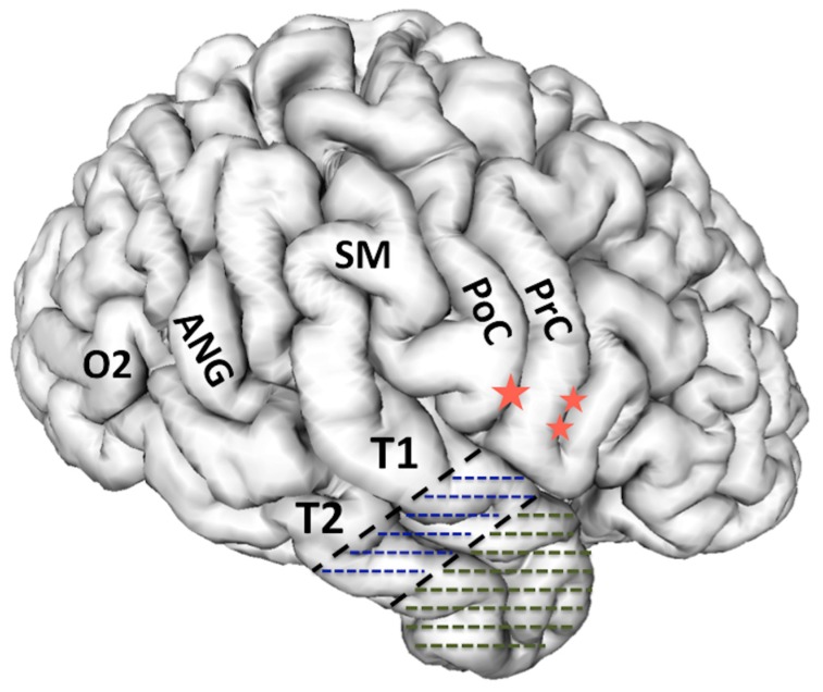
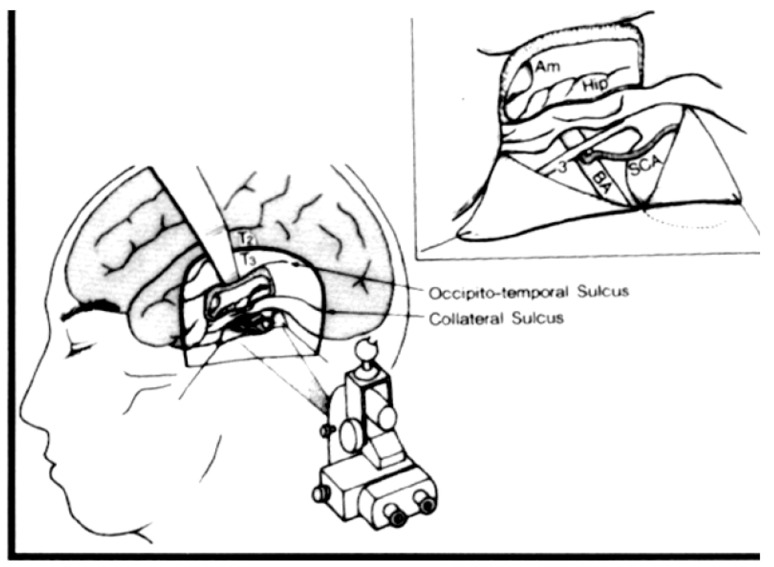
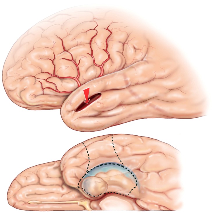
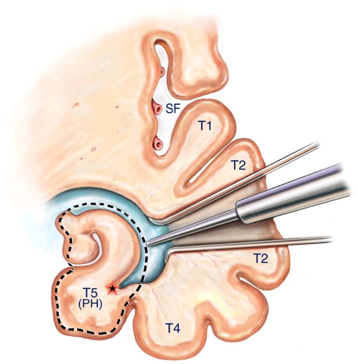
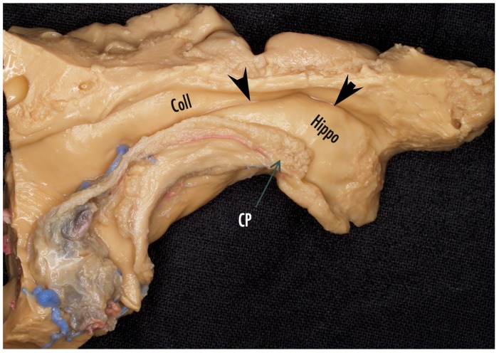
Surgery of temporal lobe epilepsy is the best opportunity for seizure freedom in medically intractable patients. The surgical approach has evolved to recognize the paramount importance of the mesial temporal structures in the majority of patients with temporal lobe epilepsy who have a seizure origin in the mesial temporal structures. For those individuals with medically intractable mesial temporal lobe epilepsy, a selective amygdalohippocampectomy surgery can be done that provides an excellent opportunity for seizure freedom and limits the resection to temporal lobe structures primarily involved in seizure genesis.

Keywords: epilepsy surgery; mesial temporal lobe epilepsy; selective amygdalohippocampectomy; temporal lobe epilepsy.


Conflict of interest statement

The author reports no conflicts of interest.

Figures

Figure 1
Figure 1
3-D reconstruction of the non-dominant right hemisphere. The dashed line illustrates the anatomical resection approach of cortico-amygdalohippocampectomy. In the non-dominant hemisphere, the resection is taken posteriorly to the level of the central sulcus along T1. In the dominant hemisphere, the T1 resection is no further posterior than the precentral sulcus in order to respect and preserve posterior language areas. Single star = central sulcus, double star = pre-central sulcus, T1 = superior temporal gyrus, T2 = middle temporal gyrus, PoC = post central gyrus, PrC = precentral gyrus, SM = supramarginal gyrus, ANG = angular gyrus, O2 = second occipital gyrus, which is the gyral continuum of T2 in the occipital lobe.
Figure 2
Figure 2
Reproduced with permission from Temporal Lobe Epilepsy: A Colloquium Sponsored by the National Institute of Neurological Diseases and Blindness, National Institutes of Health, Bethesda, Maryland, in Cooperation with the International League Against Epilepsy [45] Copyright©1959 American Medical Association. All rights reserved. In his presentation at the colloquium, Niemeyer described the convincing evidence from experimental research and clinical experience that the mesial temporal structures have a critical role in epileptogeneis of TLE. Niemeyer described a surgical technique he developed to selectively remove the mesial temporal structures via a trans-T2 trans-ventricular approach.
Figure 3
Figure 3
Reprinted and Modified by permission from Springer Nature: Adv Tech Stand Neurosurg, Yaşargil MG, Teddy PJ, Roth P. Selective amygdalohippocampectomy: operative anatomy and surgical technique. Copyright, 1985 [55]. (A) The Sylvian fissure is opened widely to expose the inferior delineation of the insula called the circular sulcus with the planned white matter incision marked in a dashed line between two middle cerebral opercular temporal arteries; (B) Surgeon’s view into the opened ventricle illustrating the choroid plexus (C), hippocampus (H), collateral eminence (star), middle cerebral vessels (M1 and M2), and amygdala (A). Numbers represent steps in the extra-pial removal of mesial temporal structures: coagulation and division of the hippocampal vessels (steps 1 and 2), disconnection of the hippocampal body from the hippocampal tail (step 3), opening the collateral eminence to empty and remove the parahippocampus (steps 4 and 5), and remove uncus and amygdala (step 6).
Figure 3
Figure 3
Reprinted and Modified by permission from Springer Nature: Adv Tech Stand Neurosurg, Yaşargil MG, Teddy PJ, Roth P. Selective amygdalohippocampectomy: operative anatomy and surgical technique. Copyright, 1985 [55]. (A) The Sylvian fissure is opened widely to expose the inferior delineation of the insula called the circular sulcus with the planned white matter incision marked in a dashed line between two middle cerebral opercular temporal arteries; (B) Surgeon’s view into the opened ventricle illustrating the choroid plexus (C), hippocampus (H), collateral eminence (star), middle cerebral vessels (M1 and M2), and amygdala (A). Numbers represent steps in the extra-pial removal of mesial temporal structures: coagulation and division of the hippocampal vessels (steps 1 and 2), disconnection of the hippocampal body from the hippocampal tail (step 3), opening the collateral eminence to empty and remove the parahippocampus (steps 4 and 5), and remove uncus and amygdala (step 6).
Figure 4
Figure 4
Reproduced with permission from: Figure 1. Tomokatsu Hori et al. Subtemporal Amygdalohippocampectomy for Treating Medically Intractable Temporal Lobe Epilepsy. Neurosurgery (1993) 33 (1): 50–57, [56]. Published by Oxford University Press on behalf of the Congress of Neurological Surgeons. Hori developed the subtemporal approach to SAH. The illustration shows a retractor elevating T3 (inferior temporal gyrus). This approach requires a gyrectomy of the fusiform gyrus to obtain access to the mesial structures. Hori, et al. also demonstrated in the inset figure that incising and reflecting the tentorium benefits accessing the mesial temporal structures for removal.
Figure 5
Figure 5
SAH corticectomy of about 2.5 cm is made along T2 just below the superior temporal sulcus. A white matter corridor is fashioned that follows the superior temporal sulcus down to the temporal horn, which is opened to visualize directly the mesial temporal structures.
Figure 6
Figure 6
Reprinted from Journal of Clinical Neuroscience 17 (9), Boling W, Minimal access keyhole surgery for mesial temporal lobe epilepsy, 1180–1184, Copyright 2010, with permission from Elsevier, [59]. Coronal view of the temporal lobe and nearby structures. A retractor is placed along the white matter corridor after opening the ventricle to fully expose its contents. The first step in the SAH is to enter the lateral ventricular sulcus (star) that lies between the bulges into the ventricle of the hippocampus and collateral eminence in order to empty the parahippocampus in a subpial fashion. Dotted line illustrates mesial temporal structures to be removed in this view, namely the hippocampal complex and parahippocampus. SF = Sylvian Fissure, T1= superior temporal gyrus, T2 = middle temporal gyrus, T4 = Fusiform gyrus, T5(PH) = parahippocampal gyrus.
Figure 7
Figure 7
Cadaver dissection of the mesial temporal lobe cut longitudinally along the temporal horn to illustrate the mesial temporal structures. Coll = collateral eminence, arrowheads point to the lateral ventricular sulcus, Hippo = head of the hippocampus, CP = choroid plexus.
Figure 8
Figure 8
View of a fixed and injected brain from the orbital frontal surface looking posteriorly. The normal relationships of structures lying adjacent to the mesial temporal lobe are visualized. The 3rd cranial nerve is normally abutting the pia of the anterior uncus. The posterior cerebral artery can be recognized along its course beneath the transparent pia of the uncus and the parahippocampus. The close association of the ICA and its bifurcation with the uncus and amygdala are illustrated. T4 = fusiform gyrus of the temporal lobe, rh = rhinal sulcus, Ent = entorhinal cortex (most anterior extent of the parahippocampus), Un = uncus, III = oculomotor cranial nerve, ICA = internal carotid artery, PCA = posterior cerebral artery, OPT = optic chiasm, BA = basilar artery, MCA = middle cerebral artery.
Figure 9
Figure 9
Image guidance view of the navigation pointer at the posterior extent of the hippocampal removal, which corresponds to the level of the midbrain tectum.
Figure 10
Figure 10
The keyhole minimal access approach benefits the patient with smaller skin incision and cranial opening. A slight curve in the scalp incision helps exposure and reduces retraction forces on the temporalis muscle compared with a straight linear incision.
Figure 11
Figure 11
Reprinted from Journal of Clinical Neuroscience 17 (9), Boling W, Minimal access keyhole surgery for mesial temporal lobe epilepsy, 1180–1184, Copyright 2010, with permission from Elsevier, [59]. The corticectomy and trans-T2 trans-ventricular approach to resection of the mesial temporal structures is identical to the approach via a standard scalp incision and cranial opening. The exposure accomplished with a keyhole access approach is more than adequate to perform the maneuvers required to complete the SAH.

References

    1. Téllez-Zenteno J.F., Hernández-Ronquillo L. A Review of the Epidemiology of Temporal Lobe Epilepsy. Epilepsy Res. Treat. 2012:630853. doi: 10.1155/2012/630853. - DOI - PMC - PubMed
    1. Semah F., Picot M.C., Adam C., Broglin D., Arzimanoglou A., Bazin B., Cavalcanti D., Baulac M. Is the underlying cause of epilepsy a major prognostic factor for recurrence? Neurology. 1998;51:1256–1262. doi: 10.1212/WNL.51.5.1256. - DOI - PubMed
    1. Hauser W.A., Annegers J.F., Kurland L.T. Prevalence of epilepsy in Rochester, Minnesota: 1940–1980. Epilepsia. 1991;32:429–445. doi: 10.1111/j.1528-1157.1991.tb04675.x. - DOI - PubMed
    1. Papez J.W. A proposed mechanism of emotion. Arch. Neurol. Psychiatry. 1937;38:725–743. doi: 10.1001/archneurpsyc.1937.02260220069003. - DOI
    1. Duvernoy H.M., Cattin F., Risold P.Y. The Human Hippocampus Functional Anatomy, Vascularization and Serial Sections with MRI. Springer; New York, NY, USA: 2013.

LinkOut - more resources