. 2018 Mar;10(3):363-371.
doi: 10.1038/nchem.2919. Epub 2018 Jan 8.

Direct observation of the influence of cardiolipin and antibiotics on lipid II binding to MurJ

Affiliations

Direct observation of the influence of cardiolipin and antibiotics on lipid II binding to MurJ

Jani Reddy Bolla et al. Nat Chem. 2018 Mar.

Abstract

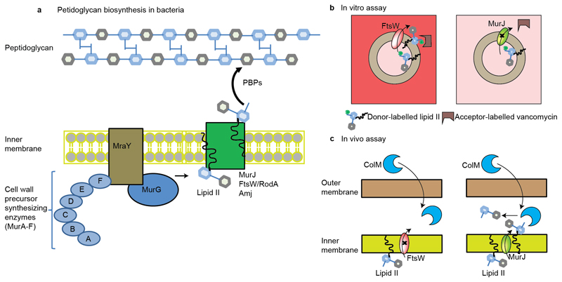
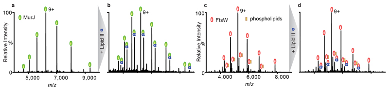
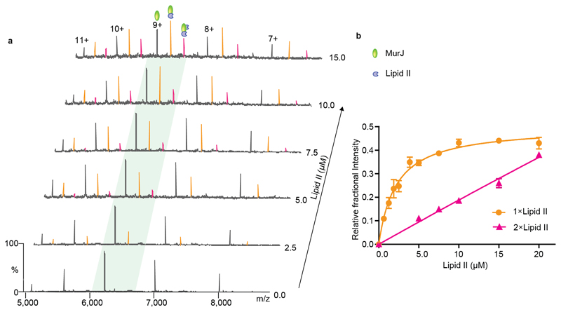
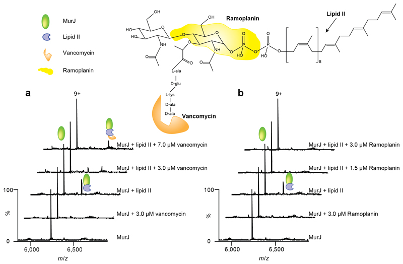
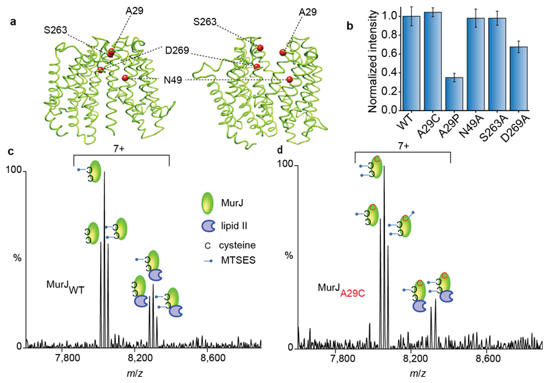
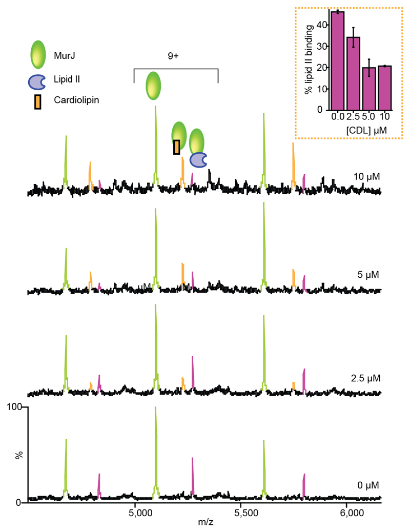
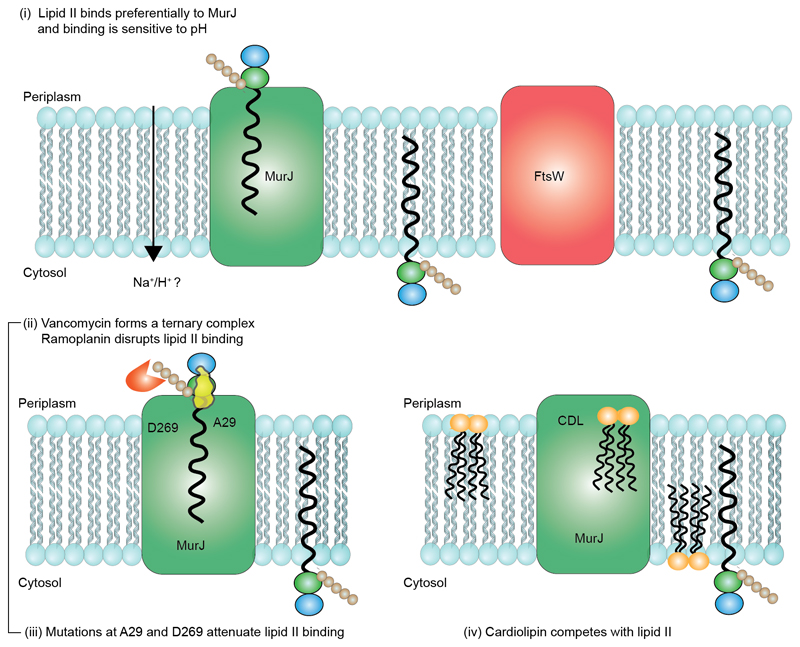
Translocation of lipid II across the cytoplasmic membrane is essential in peptidoglycan biogenesis. Although most steps are understood, identifying the lipid II flippase has yielded conflicting results, and the lipid II binding properties of two candidate flippases-MurJ and FtsW-remain largely unknown. Here we apply native mass spectrometry to both proteins and characterize lipid II binding. We observed lower levels of lipid II binding to FtsW compared to MurJ, consistent with MurJ having a higher affinity. Site-directed mutagenesis of MurJ suggests that mutations at A29 and D269 attenuate lipid II binding to MurJ, whereas chemical modification of A29 eliminates binding. The antibiotic ramoplanin dissociates lipid II from MurJ, whereas vancomycin binds to form a stable complex with MurJ:lipid II. Furthermore, we reveal cardiolipins associate with MurJ but not FtsW, and exogenous cardiolipins reduce lipid II binding to MurJ. These observations provide insights into determinants of lipid II binding to MurJ and suggest roles for endogenous lipids in regulating substrate binding.


Figures

Figure 1
Figure 1. Schematic representation of peptidoglycan biosynthesis and assays used to measure the translocation of lipid II.
a, Synthesis of the peptidoglycan begins with the formation of soluble precursors, produced from a series of cytoplasmic reactions, catalyzed by MurA-MurF. MraY-MurG complete the synthesis of the precursor lipid II. Lipid II is then translocated to the periplasm by different families of flippases (such as FtsW or RodA, MurJ, and Amj), where it is incorporated into the pre-existing cell wall by penicillin binding proteins and additional factors (–3). b, Assay used to measure the in vitro translocation of lipid II in liposomes (12). In this case, liposomes are loaded with donor-labeled lipid II (lipid II with green bulb) and when lipid II is flipped across the membrane (left panel with FtsW loaded), then an acceptor-labeled vancomycin derivative (U shaped box in brown) binds to it and a fluorescence signal is created. In this assay, fluorescence signal was created for lipisomes loaded with FtsW (left panel) but not when loaded with MurJ (right panel). c, Assay used to measure the in vivo translocation of lipid II in living E. coli (13). In this case, purified ColM toxin was added to actively growing E. coli cells. ColM cleaved lipid II product (disaccharide-pentapeptide) was then quantified in the periplasm. In this assay, flipping was observed only for MurJ (right panel) but not for FtsW (left panel).
Figure 2
Figure 2. Mass spectra of MurJ and FtsW before and after addtion of lipid II.
a, c, Mass spectra of MurJ and FtsW proteins (5 μM) in 0.05% (w/v) LDAO reveal a charge state series consistent with a single monomeric protein MurJ (green) and with some phospholipid binding (orange) to FtsW (red). Theoretical and observed masses for MurJ and FtsW are 56090 Da and 56089 Da, and 47172 Da and 47039 Da respectively. b,d Addition of lipid II (blue) at a final concentration of 5 μM to MurJ and 10 μM to FtsW, respectively, leads to formation of a complex with both proteins. A much greater degree of complex formation is observed for MurJ than FtsW at lower lipid II concentration. We find that lipid II binding does not vary significantly in LDAO concentrations ranging from 0.05 (2×CMC)–0.1% (4×CMC).
Figure 3
Figure 3. Determination of dissociation constants for lipid II binding to MurJ.
a Mass spectra recorded for solutions of MurJ with increasing concentrations of lipid II. At 2.5 μM lipid II a charge state series is observed (orange), corresponding to bound lipid II, which increases in intensity as the lipid II concentration is increased to 15 μM. A second lipid II binding peak (pink) emerges at concentrations above 5.0 μM. b A plot of the relative fractional intensity of the lipid binding peaks over the total peak intensity against the lipid II concentration (see Supplementary Methods) yields a curve for the first binding event and linear like fit for the second consistent with non-specific lipid II binding. Each data point and standard deviations are calculated from the average of the five observed charge states in three independent experiments. Error bars represent standard deviations (n=3).
Figure 4
Figure 4. Structure of lipid II showing binding sites for antibiotics and mass spectra recorded after addition of antibiotics to MurJ and to the Mur:lipid II complex.
Regions of the lipid II structure are coloured according to the vancomycin and ramoplanin binding sites (–38) (orange and yellow respectively). a Mass spectra of MurJ with vancomycin shows the 9+ charge state of MurJ and the absence of any appreciable adduct peaks corresponding to bound vancomycin. The mass spectra of MurJ and lipid II (at final concentrations of 5 μM and 3 μM respectively) confirm complex formation and addition of vancomycin (3 μM and 7 μM) leads to formation of the ternary complex. b Analogous spectra were recorded for ramoplanin. Addition of ramoplanin at low concentrations (1.5 μM and 3.0 μM) lead to disappearance of the MurJ:lipid II complex. All experiments were performed three times and spectra shown are representative.
Figure 5
Figure 5. Structures of MurJ with residues investigated through mutation, their effects on lipid II binding and spectra recorded for wild type and A29C after treating with MTSES in the presence of lipid II.
a Structures of MurJ in inward and outward facing forms were generated using I-TASSER and the structure PDB 5T77 and outward parameters of MurJTA (17). Mutated residues are shown as red spheres. b The effect of mutations on lipid II binding normalised to the wild type. N49A and S263A have no effect whereas A29P and D269A both have attenuated lipid II binding. Error bars represent standard deviations (n=3). c Mass spectrum recorded for MTSES treated wild-type MurJ. Both native cysteine residues are modified (second and third peak in the left set) and some unmodified protein remains leading to a triplet. All forms of the protein retain competency to bind lipid II (three peaks in the right hand set). d Mass spectrum for A29C MurJ variant with all three cysteine residues modified by MTSES (first, second and third peaks in the left set). In this case only two of the three modified sites are capable of binding to lipid II (first and second peaks in the right set) implying that the A29C mutant is unable to bind lipid II. All experiments were performed three times and spectra are representative.
Figure 6
Figure 6. Mass spectra reveal the effect of increasing cardiolipin concentration on lipid II binding to MurJ.
In the absence of cardiolipin the MurJ:lipid II (5 μM:3 μM) complex is formed (relative intensity ~45% of the MurJ peak). As the concentration of cardiolipin is increased (from 2.5 μM to 10 μM) the lipid II binding peak decreases and the MurJ:CDL complex predominates. All experiments were performed three times and standard deviations are calculated from the average of the five observed charge states in three independent experiments. Error bars represent standard deviations (n=3).
Figure 7
Figure 7. Schematic representation of the competition for lipid II binding between proteins, formation of a ternary complex with the antibiotic vancomycin and interplay between cardiolipin and lipid II binding to MurJ.
(i) Preferential binding to MurJ as opposed to FtsW was demonstrated through lipid II titration experiments. The potential energy source is likely a proton gradient. (ii) Vancomycin binds to the MurJ:lipid II complex whereas ramoplanin induces dissociation of lipid II. Mutations at A29 or D269 potentiate lipid II binding and chemical modification with MTSES abrogates the A29C lipid interaction. (iii) Cardiolipin (CDL) reduces the affinity of MurJ for lipid II but CDL binding, unlike lipid II, is not disrupted by mutation of A29. An allosteric binding mechanism in which the conformational access of MurJ is restricted by bidentate CDL binding is therefore consistent with all experimental data shown here.

References

    1. Silhavy TJ, Kahne D, Walker S. The bacterial cell envelope. Cold Spring Harb Perspect Biol. 2010;2:a000414. - PMC - PubMed
    1. de Kruijff B, van Dam V, Breukink E. Lipid II: a central component in bacterial cell wall synthesis and a target for antibiotics. Prostaglandins Leukot Essent Fatty Acids. 2008;79:117–121. - PubMed
    1. van Heijenoort J. Lipid intermediates in the biosynthesis of bacterial peptidoglycan. Microbiol Mol Biol Rev. 2007;71:620–635. - PMC - PubMed
    1. Schneider T, Sahl H-GG. An oldie but a goodie - cell wall biosynthesis as antibiotic target pathway. Int J Med Microbiol. 2010;300:161–169. - PubMed
    1. Galley NF, O’Reilly AM, Roper DI. Prospects for novel inhibitors of peptidoglycan transglycosylases. Bioorg Chem. 2014;55:16–26. - PMC - PubMed

Publication types

MeSH terms