. 2018 Apr 2;128(4):1496-1508.
doi: 10.1172/JCI98589. Epub 2018 Mar 12.

Mosaic RAS/MAPK variants cause sporadic vascular malformations which respond to targeted therapy

Affiliations

Mosaic RAS/MAPK variants cause sporadic vascular malformations which respond to targeted therapy

Lara Al-Olabi et al. J Clin Invest. .

Erratum in

  • Mosaic RAS/MAPK variants cause sporadic vascular malformations which respond to targeted therapy.
    Al-Olabi L, Polubothu S, Dowsett K, Andrews KA, Stadnik P, Joseph AP, Knox R, Pittman A, Clark G, Baird W, Bulstrode N, Glover M, Gordon K, Hargrave D, Huson SM, Jacques TS, James G, Kondolf H, Kangesu L, Keppler-Noreuil KM, Khan A, Lindhurst MJ, Lipson M, Mansour S, O'Hara J, Mahon C, Mosica A, Moss C, Murthy A, Ong J, Parker VE, Rivière JB, Sapp JC, Sebire NJ, Shah R, Sivakumar B, Thomas A, Virasami A, Waelchli R, Zeng Z, Biesecker LG, Barnacle A, Topf M, Semple RK, Patton EE, Kinsler VA. Al-Olabi L, et al. J Clin Invest. 2018 Nov 1;128(11):5185. doi: 10.1172/JCI124649. Epub 2018 Nov 1. J Clin Invest. 2018. PMID: 30382944 Free PMC article. No abstract available.

Abstract

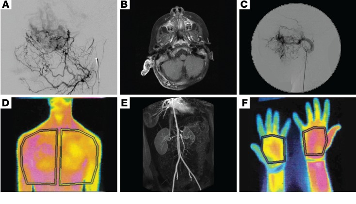
Background: Sporadic vascular malformations (VMs) are complex congenital anomalies of blood vessels that lead to stroke, life-threatening bleeds, disfigurement, overgrowth, and/or pain. Therapeutic options are severely limited, and multidisciplinary management remains challenging, particularly for high-flow arteriovenous malformations (AVM).

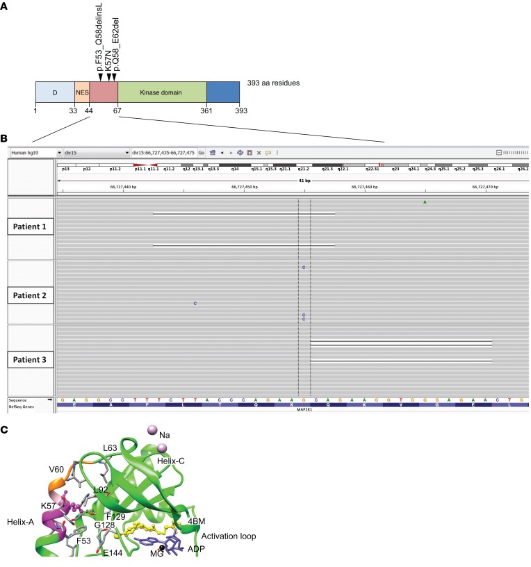
Methods: To investigate the pathogenesis of sporadic intracranial and extracranial VMs in 160 children in which known genetic causes had been excluded, we sequenced DNA from affected tissue and optimized analysis for detection of low mutant allele frequency.

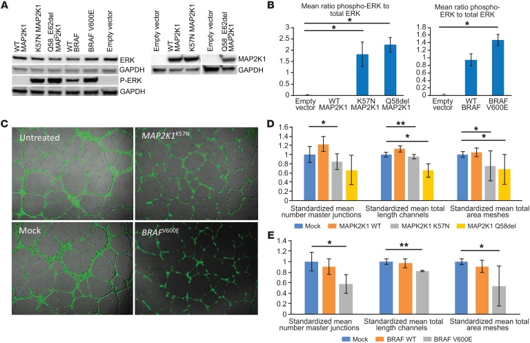
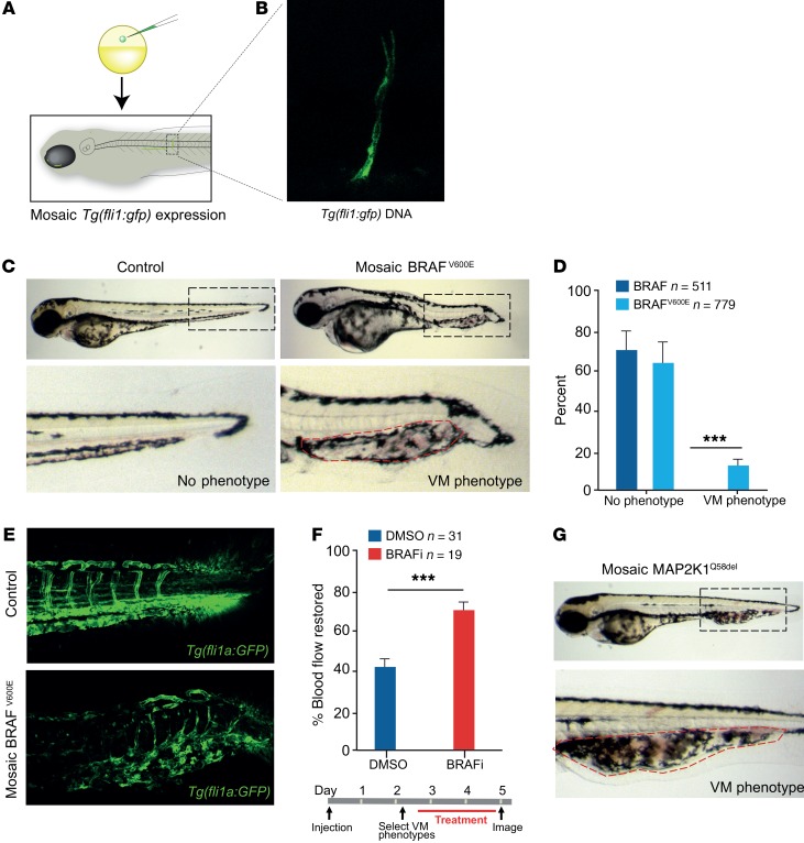
Results: We discovered multiple mosaic-activating variants in 4 genes of the RAS/MAPK pathway, KRAS, NRAS, BRAF, and MAP2K1, a pathway commonly activated in cancer and responsible for the germline RAS-opathies. These variants were more frequent in high-flow than low-flow VMs. In vitro characterization and 2 transgenic zebrafish AVM models that recapitulated the human phenotype validated the pathogenesis of the mutant alleles. Importantly, treatment of AVM-BRAF mutant zebrafish with the BRAF inhibitor vemurafinib restored blood flow in AVM.

Conclusion: Our findings uncover a major cause of sporadic VMs of different clinical types and thereby offer the potential of personalized medical treatment by repurposing existing licensed cancer therapies.

Funding: This work was funded or supported by grants from the AVM Butterfly Charity, the Wellcome Trust (UK), the Medical Research Council (UK), the UK National Institute for Health Research, the L'Oreal-Melanoma Research Alliance, the European Research Council, and the National Human Genome Research Institute (US).

Keywords: Drug therapy; Molecular genetics; Signal transduction; Therapeutics; Vascular Biology.


Conflict of interest statement

Conflict of interest: LGB is an uncompensated advisor to Illumina and receives royalties from Genentech and honoraria from Wiley-Blackwell.

Figures

Figure 1
Figure 1. A broad clinical spectrum of VMs in somatic RAS/MAPK mutations.
Inexorable enlargement of high-flow AVMs with age, affecting the face and leading to loss of vision in the right eye (AD), and affecting the right ear helix and posterior auricular soft tissues leading to eventual resection of the helix (EG). Varied clinical examples of the spectrum of high-flow VMs (AVMs) of the temple, left leg/buttock, and left face (H and I). Segmental overgrowth of the left arm, chest wall, and breast with a colocalized low-flow VM, detectable by a uniform brownish-pink macular capillary malformation and superimposed scattered telangiectasia, with clear midline demarcation (JL). Segmental overgrowth of the right arm and hand, with a colocalized uniform brownish-pink capillary malformation with superimposed scattered telangiectasia (M and N).
Figure 2
Figure 2. Imaging of sporadic VMs secondary to mutations in MAPK pathway genes demonstrating involvement of all blood vessel sizes.
(A) Lateral image from a digitally subtracted angiogram showing a leash of small, abnormal vessels shunting through a dense capillary bed to early filling veins. (B) Axial contrast-enhanced fat-saturated T1 weighted MRI image showing a grossly enlarged right pinna and thickened posterior auricular soft tissues. The abnormal tissue is filled with multiple signal voids, representing enlarged abnormal vessels. The pinna enhances avidly. (C) Lateral image from a digitally subtracted angiogram, with the catheter tip in the grossly enlarged left internal maxillary artery, which supplies a leash of abnormal high-flow vessels in the face. (D) Thermography of low-flow VMs with overgrowth of the left chest wall and arm demonstrates increased temperature (shown in magenta) compared with the right-sided structures (E), and in the right forearm and thumb compared with the left (F). 3D reconstruction of an abdominal CT angiogram demonstrating multifocal vascular disease, with severe stenoses of the descending aorta, coeliac axis, superior mesenteric artery origin, and right renal artery (E).
Figure 3
Figure 3. Somatic variants in MAP2K1 cluster in exon 2 and are predicted to destabilize the 3D structure of the inactive form of the kinase.
(A) Schematic representation of clustered somatic mutations in exon 2 of MAP2K1. (B) Low allele frequency mutations on IGV visualization of deep next-generation sequencing data from VM tissue samples of 3 patients. (C) 3D structural modelling of an inhibitor-bound (4BM) form (PDB ID: 3EQG) of MAP2K1 demonstrating the mutated residues. Deletions are highlighted in magenta (residues 53–58) and orange (residues 58–62, with residue 58 common to both in pink). Critical residue K57, which is substituted as a result of the missense mutation, is shown using ball and stick representation, with the dashed line indicating a hydrogen-bond interaction with the β-sheet of the kinase. Residues involved in interaction between helix A and the core kinase domain are also shown.
Figure 4
Figure 4. Mutations in MAPK pathway–encoding genes lead to activation of downstream signaling and disruption of vascular endothelial tube formation in vitro.
(A and B) Expression of mutant BRAFV600E, MAP2K1K57N, and MAP2K1Q58_E62del in HEK293T cells leads to significantly increased phosphorylation of ERK detected by immunoblotting (representative blot shown from duplicate biological replicates), compared with WT gene overexpression and controls. Data are shown as mean ± SD. *P < 0.05, 1-way ANOVA. (C) Expression of mutant BRAFV600E and MAP2K1K57N in HUVECs seeded onto Geltrex Matrix leads to visible disruption of endothelial vascular tube formation compared with controls. Original magnification, ×50. (D and E) Significant reductions in mean number of master junctions, total length of tubes, and total mesh area are indicated by asterisks. Data are shown as mean ± SD. Means were taken from triple technical replicates for each of the duplicate biological replicates, standardized to within-replicate controls, and analyzed by 1-way ANOVA with correction for multiple testing (*P < 0.0167; **P < 0.001).
Figure 5
Figure 5. BRAF and MAP2K mutations induce VM phenotypes in zebrafish that respond to targeted therapy.
(A) Schematic of zebrafish embryos injected with Tg (fli1a:GFP) at the 1-cell stage generate zebrafish larvae that are mosaic for the transgene integration and expression. (B) Image of mosaic expression of Tg (fli1a:GFP) in a vessel. (C) Images of zebrafish expressing WT or mutant BRAFV600E in a mosaic fashion from a fli1a promoter in endothelial cells. Accumulation of blood is visible at the caudal vein vascular plexus in the BRAFV600E–expressing zebrafish and outlined with a dashed red line. (D) Quantification of VM phenotype in BRAFWT-expressing (n = 511) and BRAFV600E-expressing (n = 779) zebrafish. (E) BRAFWT and BRAFV600E mosaic expression in stable Tg (fli1a:GFP) larvae to visualize all vessels in the zebrafish in the VM lesion. Increased numbers of vascular channels and disorganized architecture of VM lesions are clearly detectable. (F) Schematic of VM treatment protocol and quantification of the percentage of VM BRAFV600E zebrafish with improved blood flow following treatment with vemurafenib. VM BRAFV600E zebrafish were randomized prior to DMSO (n = 31) or vemurafenib (n = 19) treatment and blind scored. (G) Images of zebrafish expressing MAPK2K1Q58del in a mosaic fashion from a fli1a promoter in endothelial cells (n = 5/37). Zebrafish larvae in D were analyzed by an unpaired parametric t test with Welch’s correction and in F by a paired t test comparing matched pairs (before and after treatment). Data are shown as SEM. ***P < 0.001.
Figure 6
Figure 6. Schematic summary of the known and newly identified signaling proteins affected by mosaic mutations that lead to a VM phenotype.
Key signaling pathways PI3K/AKT/MTOR and RAS/RAF/MEK/ERK control cellular growth, apoptosis, and differentiation through complex transcriptional regulation. Multiple receptor types feed into one or both pathways. In addition, there is crosstalk between the 2 pathways at multiple levels (not shown). Proteins affected by the genetic mutations presented in this paper are shown in red, with previously identified sites shown in blue. Key classes of potential targeted therapeutics are shown in green boxes. RTK, receptor tyrosine kinase; GPCR, G protein–coupled receptor.

References

    1. Wassef M, et al. Vascular anomalies classification: recommendations from the International Society for the Study of Vascular Anomalies. Pediatrics. 2015;136(1):e203–e214. doi: 10.1542/peds.2014-3673. - DOI - PubMed
    1. Lindhurst MJ, et al. A mosaic activating mutation in AKT1 associated with the Proteus syndrome. N Engl J Med. 2011;365(7):611–619. doi: 10.1056/NEJMoa1104017. - DOI - PMC - PubMed
    1. Shirley MD, et al. Sturge-Weber syndrome and port-wine stains caused by somatic mutation in GNAQ. N Engl J Med. 2013;368(21):1971–1979. doi: 10.1056/NEJMoa1213507. - DOI - PMC - PubMed
    1. Thomas AC, et al. Mosaic activating mutations in GNA11 and GNAQ are associated with phakomatosis pigmentovascularis and extensive dermal melanocytosis. J Invest Dermatol. 2016;136(4):770–778. doi: 10.1016/j.jid.2015.11.027. - DOI - PMC - PubMed
    1. Rivière JB, et al. De novo germline and postzygotic mutations in AKT3, PIK3R2 and PIK3CA cause a spectrum of related megalencephaly syndromes. Nat Genet. 2012;44(8):934–940. doi: 10.1038/ng.2331. - DOI - PMC - PubMed

Publication types

MeSH terms