. 2018 May 1;128(5):1913-1918.
doi: 10.1172/JCI98680. Epub 2018 Apr 3.

CYP3A4 mutation causes vitamin D-dependent rickets type 3

Affiliations

CYP3A4 mutation causes vitamin D-dependent rickets type 3

Jeffrey D Roizen et al. J Clin Invest. .

Abstract

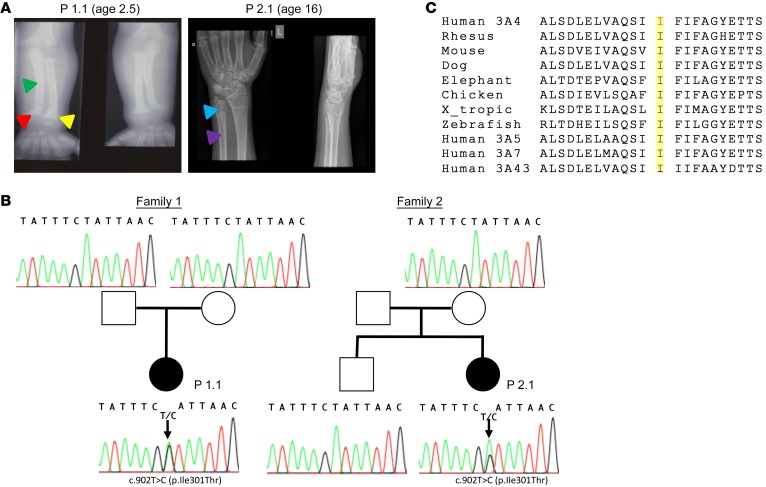
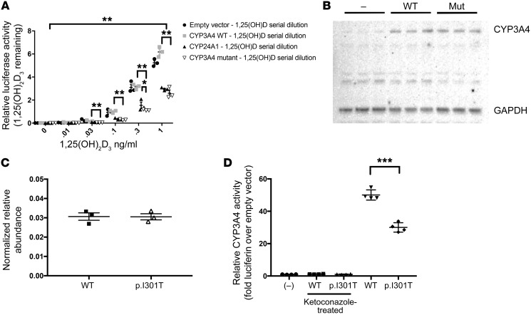
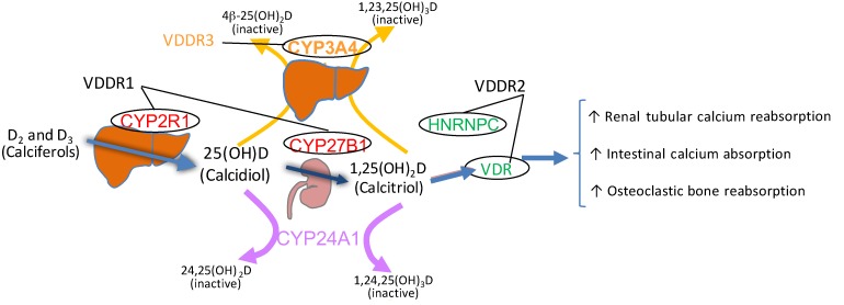
Genetic forms of vitamin D-dependent rickets (VDDRs) are due to mutations impairing activation of vitamin D or decreasing vitamin D receptor responsiveness. Here we describe two unrelated patients with early-onset rickets, reduced serum levels of the vitamin D metabolites 25-hydroxyvitamin D and 1,25-dihydroxyvitamin D, and deficient responsiveness to parent and activated forms of vitamin D. Neither patient had a mutation in any genes known to cause VDDR; however, using whole exome sequencing analysis, we identified a recurrent de novo missense mutation, c.902T>C (p.I301T), in CYP3A4 in both subjects that alters the conformation of substrate recognition site 4 (SRS-4). In vitro, the mutant CYP3A4 oxidized 1,25-dihydroxyvitamin D with 10-fold greater activity than WT CYP3A4 and 2-fold greater activity than CYP24A1, the principal inactivator of vitamin D metabolites. As CYP3A4 mutations have not previously been linked to rickets, these findings provide insight into vitamin D metabolism and demonstrate that accelerated inactivation of vitamin D metabolites represents a mechanism for vitamin D deficiency.

Keywords: Bone Biology; Bone disease; Genetic diseases; Genetics.


Conflict of interest statement

Conflict of interest: The authors have declared that no conflict of interest exists.

Figures

Figure 1
Figure 1. CYP3A4 mutation causes VDDR.
(A) Wrist radiographs of the 2 probands are consistent with those of untreated rickets despite high-dose supplementation. Wrist radiographs from these patients show the typical features of rickets: younger growing patients have bowing (green triangle), metaphyseal splaying (yellow triangle), and cupping (red triangle), as in proband 1.1 (P 1.1); older patients have metaphyseal lucency (blue triangle) and osteopenia (purple triangle), as in proband 2.1; features change as the role of calcification changes with age (supporting growth vs. maintaining bone integrity). (B) Family pedigrees are consistent with p.I301T mutations arising de novo. Family pedigrees of the 2 probands with Sanger sequencing results of the mutation region in CYP3A4 from all available family members. (C) Alignment of CYP3A4 protein sequence surrounding p.I301T mutation reveals high conservation of this residue across species. Isoleucine 301 is highly conserved in CYP3A4 across evolution and within the human 3A protein family.
Figure 2
Figure 2. CYP3A4 (p.I301T) mutant has increased vitamin D degradative activity but decreased activity for other substrates.
(A) CYP3A4 (p.I301T) has increased vitamin D degradative activity. CYP3A4 p.I301T had increased inactivation of calcitriol relative to WT (**P < 0.01 by 2-way ANOVA for curve), and post hoc multiple comparison–adjusted analyses confirmed significant differences at 0.03, 0.1, and 0.3 ng/ml (**P < 0.01), and relative to CYP24A1 at 0.3 ng/ml (*P < 0.05) (n = 3 for each). (B and C) There is no difference in the relative abundance of the WT and p.I301T CYP3A4 (n = 3 for each treatment). (B) Immunoblot of cell lysates for CYP3A4 and GAPDH. (C) Quantification of the relative abundance of CYP3A4 and GAPDH. No significant differences were observed between the abundance of the WT and the p.I301T mutant. (D) CYP3A4 (p.I301T) does not have increased catalytic activity for non–vitamin D substrates. The p.I301T mutant had significantly decreased activity for luciferin IPA relative to the WT enzyme (30.0 ± 1.6 vs. 50.1 ± 1.6, n = 4 for each, ***P < 0.001). **P < 0.05. Data are presented as mean ± SEM.
Figure 3
Figure 3. Pathogenesis of VDDR.
VDDR-1s are caused by mutations in genes encoding proteins that activate vitamin D: CYP2R1 and CYP27B1. VDDR-2s are caused by mutations in genes encoding signal transducing proteins: VDR and HNRNPC. Type 3 is due to gain-of-function mutations in a gene encoding a vitamin D–degrading enzyme: CYP3A4.

References

    1. Terushkin V, Bender A, Psaty EL, Engelsen O, Wang SQ, Halpern AC. Estimated equivalency of vitamin D production from natural sun exposure versus oral vitamin D supplementation across seasons at two US latitudes. J Am Acad Dermatol. 2010;62(6):929.e1–929.e9. - PubMed
    1. Kitanaka S, et al. Inactivating mutations in the 25-hydroxyvitamin D3 1alpha-hydroxylase gene in patients with pseudovitamin D-deficiency rickets. N Engl J Med. 1998;338(10):653–661. doi: 10.1056/NEJM199803053381004. - DOI - PubMed
    1. Cheng JB, Levine MA, Bell NH, Mangelsdorf DJ, Russell DW. Genetic evidence that the human CYP2R1 enzyme is a key vitamin D 25-hydroxylase. Proc Natl Acad Sci U S A. 2004;101(20):7711–7715. doi: 10.1073/pnas.0402490101. - DOI - PMC - PubMed
    1. Thacher TD, Fischer PR, Singh RJ, Roizen J, Levine MA. CYP2R1 mutations impair generation of 25-hydroxyvitamin D and cause an atypical form of vitamin D deficiency. J Clin Endocrinol Metab. 2015;100(7):E1005–E1013. doi: 10.1210/jc.2015-1746. - DOI - PMC - PubMed
    1. Chen H, Hewison M, Hu B, Adams JS. Heterogeneous nuclear ribonucleoprotein (hnRNP) binding to hormone response elements: a cause of vitamin D resistance. Proc Natl Acad Sci U S A. 2003;100(10):6109–6114. doi: 10.1073/pnas.1031395100. - DOI - PMC - PubMed

Publication types

MeSH terms