Review
. 2018 Aug;129(2):343-366.
doi: 10.1097/ALN.0000000000002130.

Neuroinflammation and Central Sensitization in Chronic and Widespread Pain

Affiliations
Review

Neuroinflammation and Central Sensitization in Chronic and Widespread Pain

Ru-Rong Ji et al. Anesthesiology. 2018 Aug.

Abstract

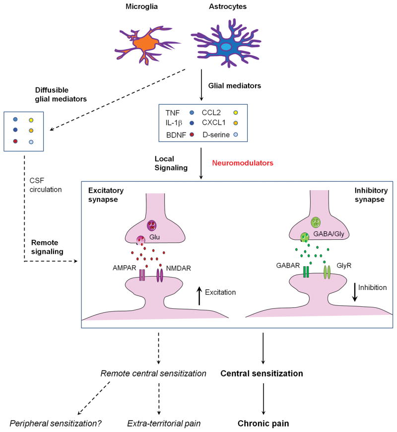
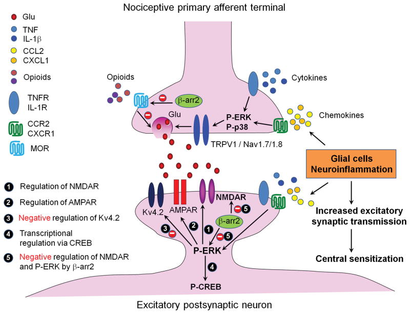
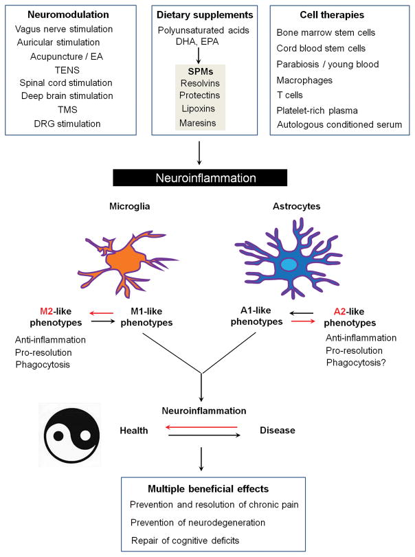
Chronic pain is maintained in part by central sensitization, a phenomenon of synaptic plasticity, and increased neuronal responsiveness in central pain pathways after painful insults. Accumulating evidence suggests that central sensitization is also driven by neuroinflammation in the peripheral and central nervous system. A characteristic feature of neuroinflammation is the activation of glial cells, such as microglia and astrocytes, in the spinal cord and brain, leading to the release of proinflammatory cytokines and chemokines. Recent studies suggest that central cytokines and chemokines are powerful neuromodulators and play a sufficient role in inducing hyperalgesia and allodynia after central nervous system administration. Sustained increase of cytokines and chemokines in the central nervous system also promotes chronic widespread pain that affects multiple body sites. Thus, neuroinflammation drives widespread chronic pain via central sensitization. We also discuss sex-dependent glial/immune signaling in chronic pain and new therapeutic approaches that control neuroinflammation for the resolution of chronic pain.


Conflict of interest statement

Conflict of interest: The authors have no financial interests in this study.

Figures

Figure 1
Figure 1
A PubMed Search on September 30, 2017 shows the number of publications on “neuroinflammation and pain” and “microglia and pain” in last 10 years. The numbers on the bottom show all time publications in PubMed.
Figure 2
Figure 2
Neuroinflammation is associated with various insults that evoke painful sensations. These insults include but not limited to trauma, major surgeries, drug treatments, autoimmune disease conditions, and other painful insults and tissue damage. Some of these insults such as major surgeries (breast surgery, amputation, thoracotomy), chemotherapy, and anti-viral treatment will cause nerve injury, as highlighted in red. Others will cause immune activation (highlighted in purple) and tissue injury (highlighted in light blue). Neuroinflammation results in several adverse effects, such as chronic pain and neurodegenerative diseases including Alzheimer’s disease (AD), Parkinson disease (PD), multiple sclerosis (MS), and stroke. Neuroinflammation is also associated with chronic overlapping pain conditions. After priming of nociceptive circuit by previous injury, stress, or existing genomic, environmental, and psychological factors, acute insult may cause transition from acute pain to chronic pain.
Figure 3
Figure 3
Distinct and time-dependent activation of microglia and astrocytes in the spinal cord after nerve injury. (A, B) Microglia activation revealed by increased CX3CR1 expression in the spinal cord 10 days (A) and 21 days (B) after nerve injury in Cx3cr1-GFP mice. Scale, 100 μm. (C) Phosphorylation of p38 MAP kinase (P-p38) in CD11b+ microglia in the spinal cord dorsal horn 7 days after nerve injury. Scale, 20 μm. (D, E) Astroctye activation revealed by increased GFAP expression in the spinal cord 10 days (D) and 21 days (E) after nerve injury in mice. Scale, 100 μm. (F) Expression of Cx43 in GFAP+ astrocytes in the spinal cord dorsal horn 21 days after nerve injury. Scale, 20 μm. All the images have not been published.
Figure 4
Figure 4
Schematic illustration of local and remote central sensitization induced by glial activation and neuroinflammation in the spinal cord. Activation of spinal microglia and astrocytes by painful insults results in secretion of glial mediators such as TNF, IL-1β, CCL2, CXCL1, BDNF, D-serine, which can act as neuromodulators to induce local central sensitization in surrounding excitatory synapses (facilitation) and inhibitory synapses (dis-inhibition). During neuroinflammation these glial mediators are also present in the CSF and affect synapses in different spinal segments to cause remote central sensitization and extra-territorial and widespread pain beyond the initial injury site.
Figure 5
Figure 5
Molecular mechanisms of central sensitization in first-order excitatory synapses in the spinal cord dorsal horn pain circuit and induction of central sensitization by proinflammatory cytokines and chemokines (e.g., TNF, IL-1β, CCL2, CXCL1) that are produced by glial cells. At presynaptic sites, i.e. central terminals of nociceptive primary afferents, activation of receptors of cytokine and chemokine receptors results in phosphorylation and activation of ERK and p38 (P-ERK, P-p38), leading to glutamate (Glu) release from synaptic vesicles, via activation of ion channels TRPV1, Nav1.7, and Nav1.8. At postsynaptic sties, increased release of neurotransmitters (e.g., Glu) also induces P-ERK, which can induce central sensitization by positive modulation of NMDA receptor (NMDAR, Step-1). AMPA receptor (AMPAR, Step-2) and negative modulation of potassium channel subunit Kv4.2 (Step-3). P-ERK also maintains central sensitization via inducing CREB phosphorylation (P-CREB, Step-4). Opioids such as morphine inhibit neurotransmitter release via mu opioid receptors (MOR) and N-type calcium channels. The scaffold protein β-arrestin-2 (βarr2) inhibits MOR signaling by desensitization and degradation of GPCRs, leading to enhanced acute opioid analgesia in βarr2 knockout mice. Paradoxically, βarr2 also inhibits NMDAR and ERK signaling, leading to a transition from acute pain to chronic pain.
Figure 6
Figure 6
Non-pharmacological approaches that can control neuroinflammation and produce multiple beneficial effects including prevention and resolution of chronic pain, prevention of neurodegeneration, and repair of cognitive function deficits. Abbreviations: DHA, docosahexaenoic acid; EA, electroacupuncture; EPA, eicosapentaenoic acid; VNS, vagus nerve stimulation; SPMs, specialized pro-resolution mediators; TENS, transcutaneous electrical nerve stimulation; TMS, transcranial magnetic stimulation.

References

    1. Pizzo PA, Clark NM. Alleviating suffering 101--pain relief in the United States. N Engl J Med. 2012;366:197–199. - PubMed
    1. Gereau RWt, Sluka KA, Maixner W, Savage SR, Price TJ, Murinson BB, Sullivan MD, Fillingim RB. A pain research agenda for the 21st century. J Pain. 2014;15:1203–14. - PMC - PubMed
    1. Basbaum AI, Bautista DM, Scherrer G, Julius D. Cellular and molecular mechanisms of pain. Cell. 2009;139:267–284. - PMC - PubMed
    1. Hucho T, Levine JD. Signaling pathways in sensitization: toward a nociceptor cell biology. Neuron. 2007;55:365–376. - PubMed
    1. Gold MS, Gebhart GF. Nociceptor sensitization in pain pathogenesis. Nat Med. 2010;16:1248–1257. - PMC - PubMed

Publication types

MeSH terms