. 2018 Feb 20;14(2):e1007233.
doi: 10.1371/journal.pgen.1007233. eCollection 2018 Feb.

RNF8 and SCML2 cooperate to regulate ubiquitination and H3K27 acetylation for escape gene activation on the sex chromosomes

Affiliations

RNF8 and SCML2 cooperate to regulate ubiquitination and H3K27 acetylation for escape gene activation on the sex chromosomes

Shannel R Adams et al. PLoS Genet. .

Abstract

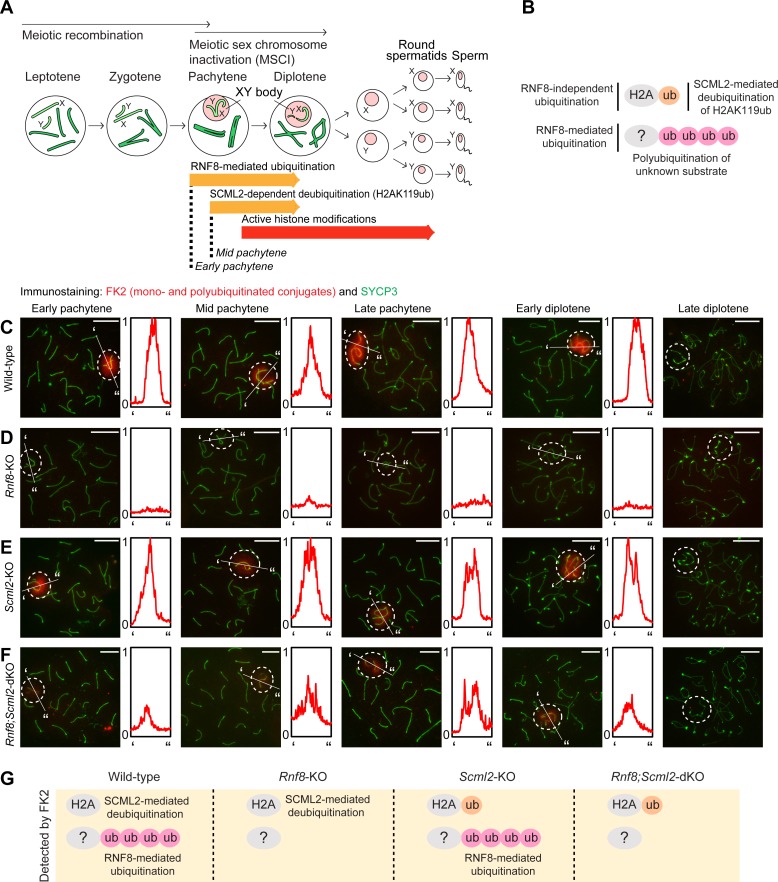
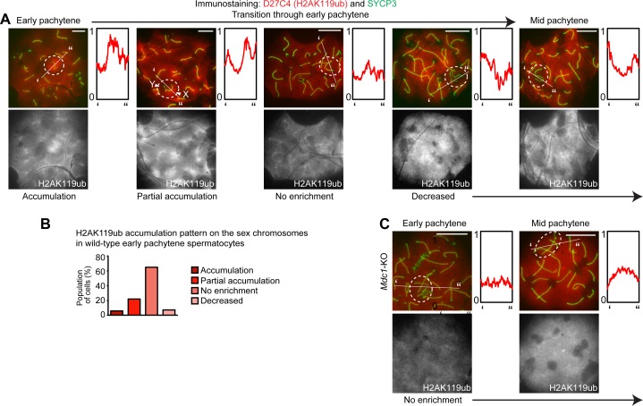
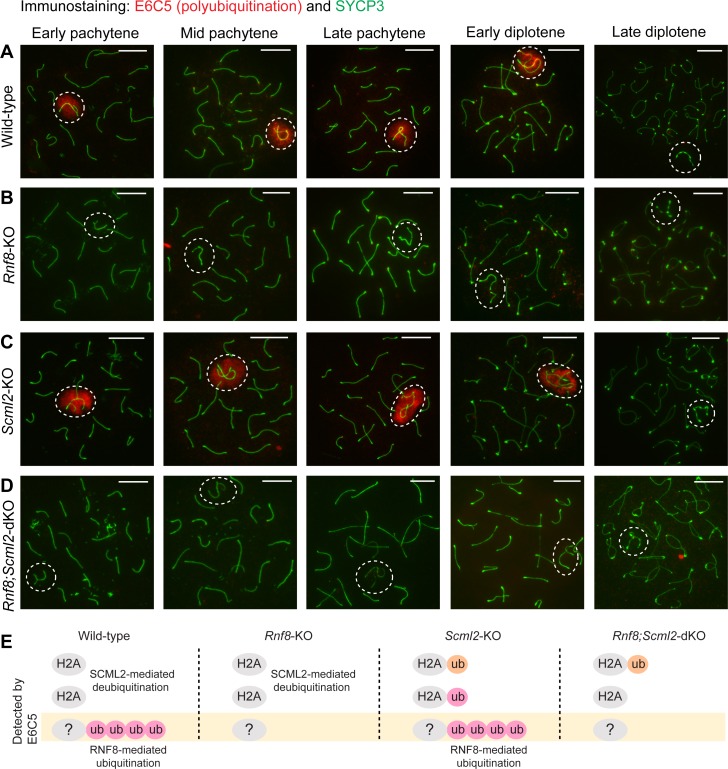
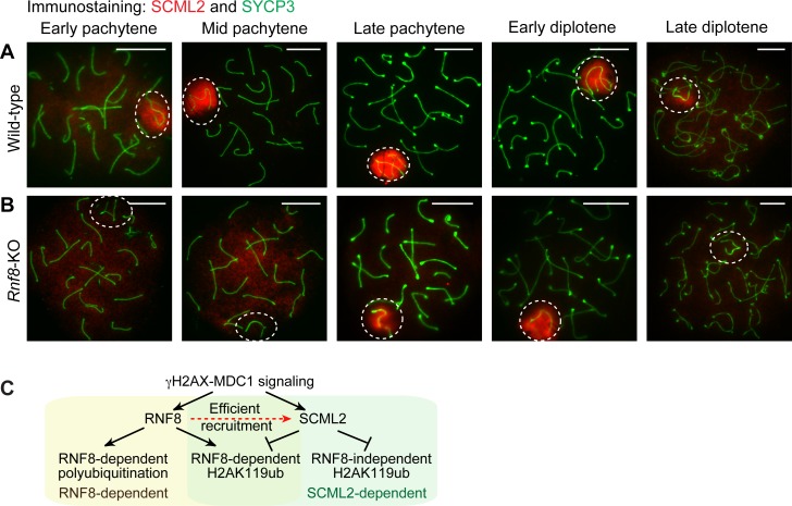
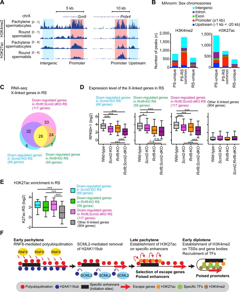
The sex chromosomes are enriched with germline genes that are activated during the late stages of spermatogenesis. Due to meiotic sex chromosome inactivation (MSCI), these sex chromosome-linked genes must escape silencing for activation in spermatids, thereby ensuring their functions for male reproduction. RNF8, a DNA damage response protein, and SCML2, a germline-specific Polycomb protein, are two major, known regulators of this process. Here, we show that RNF8 and SCML2 cooperate to regulate ubiquitination during meiosis, an early step to establish active histone modifications for subsequent gene activation. Double mutants of Rnf8 and Scml2 revealed that RNF8-dependent monoubiquitination of histone H2A at Lysine 119 (H2AK119ub) is deubiquitinated by SCML2, demonstrating interplay between RNF8 and SCML2 in ubiquitin regulation. Additionally, we identify distinct functions of RNF8 and SCML2 in the regulation of ubiquitination: SCML2 deubiquitinates RNF8-independent H2AK119ub but does not deubiquitinate RNF8-dependent polyubiquitination. RNF8-dependent polyubiquitination is required for the establishment of H3K27 acetylation, a marker of active enhancers, while persistent H2AK119ub inhibits establishment of H3K27 acetylation. Following the deposition of H3K27 acetylation, H3K4 dimethylation is established as an active mark on poised promoters. Together, we propose a model whereby regulation of ubiquitin leads to the organization of poised enhancers and promoters during meiosis, which induce subsequent gene activation from the otherwise silent sex chromosomes in postmeiotic spermatids.


Conflict of interest statement

AB is a co-founder of Datirium, LLC.

Figures

Fig 1
Fig 1. Distinct forms of ubiquitin regulation by RNF8 and SCML2: Immunostaining with FK2 antibody, which recognizes both mono- and polyubiquitinated conjugates.
(A) Schematic of spermatogenesis. (B) Model of distinct forms of regulation of ubiquitination by RNF8 and SCML2. (C-F) Immunostaining of SYCP3 and FK2 on meiotic chromosome spreads. Dotted circles: sex chromosomes. Scale bar: 10 μm. Representative images are shown for at least 30 spermatocytes from each substage, from at least 3 independent mice per mouse model. The intensity of immunostaining is quantified by densitometry across the indicated path (‘ to”) and plotted in a relative intensity range of 0–1, which is normalized among the samples at the same stage. (G) Schematic of ubiquitin targets recognized by the FK2 antibody in each mouse model. A pink circle denotes RNF8-dependent ubiquitination and an orange circle denotes events mediated by a different E3 ubiquitin ligase.
Fig 2
Fig 2. RNF8-dependent H2AK119ub is removed by SCML2: Immunostaining with D27C4 antibody, which recognizes H2AK119ub.
(A-D) Immunostaining of SYCP3 and D27C4 (H2AK119ub) on meiotic chromosome spreads. Dotted circles: sex chromosomes. Scale bar: 10 μm. Representative images are shown for at least 30 spermatocytes from each substage, from at least 3 independent mice per mouse model. The intensity of immunostaining is quantified by densitometry across the indicated path (‘ to”) and plotted in a relative intensity range of 0–1, which is normalized among all images in this figure, Fig 3 and S2 Fig. (E) Updated model of distinct forms of regulation of ubiquitination by RNF8 and SCML2, including RNF8-dependent H2AK119ub, which is removed by SCML2 based on data in this figure. (F) Schematic of ubiquitin targets recognized by the D27C4 (H2AK119ub) antibody in each mouse model.
Fig 3
Fig 3. MDC1-dependent H2AK119ub on the sex chromosomes at the onset of the early pachytene stage.
(A) Immunostaining of SYCP3 and D27C4 (H2AK119ub) on wild-type meiotic chromosome spreads. Dotted circles: sex chromosomes. Scale bar: 10 μm. Representative images are shown for each category through the early pachytene stage for spermatocytes from 9 independent mice. The intensity of immunostaining is quantified by densitometry across the indicated path (‘ to”) and plotted in a relative intensity range of 0–1, which is normalized among all images in this figure, Fig 2 and S2 Fig. (B) H2AK119ub accumulation patterns on the sex chromosomes in wild-type early pachytene spermatocytes. A total of 305 early pachytene spermatocytes were scored from 9 independent wild-type mice. (C) Immunostaining of SYCP3 and D27C4 (H2AK119ub) on Mdc1-KO meiotic chromosome spreads. H2AK119ub accumulation was not observed in Mdc1-KO spermatocytes. Dotted circles: sex chromosomes. Scale bar: 10 μm. Representative images are shown through the early pachytene stage for spermatocytes from 3 independent Mdc1-KO mice. The intensity of immunostaining is quantified by densitometry across the indicated path (‘ to”) and plotted in a relative intensity range of 0–1, which is normalized among all images in this figure, Fig 2 and S2 Fig.
Fig 4
Fig 4. RNF8 is required for polyubiquitination of the sex chromosomes: Immunostaining with E6C5 antibody, which recognizes polyubiquitination.
(A-D) Immunostaining of SYCP3 and E6C5 on meiotic chromosome spreads. Dotted circles: sex chromosomes. Scale bar: 10 μm. Representative images are shown for at least 30 spermatocytes from each substage, from at least 3 independent mice per mouse model. (E) Schematic of ubiquitin targets recognized by the E6C5 antibody in each mouse model.
Fig 5
Fig 5. RNF8 is involved in the efficient recruitment of SCML2 to the sex chromosomes.
(A, B) Immunostaining of SYCP3 and SCML2 on meiotic chromosome spreads. Dotted circles: sex chromosomes. Scale bar: 10 μm. Representative images are shown for at least 30 spermatocytes from each substage, from at least 3 independent mice per mouse model. (C) Model of the functions of RNF8 and SCML2 in the regulation of the ubiquitin network.
Fig 6
Fig 6. RNF8-dependent establishment of H3K4me2 and its inhibition by H2AK119ub.
(A-D) Immunostaining of SYCP3 and H3K4me2 on meiotic chromosome spreads. Dotted circles: sex chromosomes. Scale bar: 10 μm. Representative images are shown for at least 30 spermatocytes from each substage, from at least 3 independent mice per mouse model. The intensity of immunostaining is quantified by densitometry across the indicated path (‘ to”) and plotted in a relative intensity range of 0–1, which is normalized among all pictures in this figure. (E) Model of the regulation of H3K4me2. (F, G) Immunostaining of SYCP3 and H3K4me2 on wild-type and Scml2-KO meiotic chromosome spreads. Dotted circles: sex chromosomes. Scale bar: 10 μm. Representative images are shown for late pachytene spermatocytes from 6 independent wild-type mice and 3 independent Scml2-KO mice. The intensity of immunostaining is quantified by densitometry across the indicated path (‘ to”) and plotted in a relative intensity range of 0–1, which is normalized among all pictures in this figure. (H) H3K4me2 accumulation patterns on the sex chromosomes in late pachytene spermatocytes. A total of 244 late pachytene spermatocytes were scored from 6 independent wild-type mice, and a total 91 late pachytene spermatocytes were scored from 3 independent Scml2-KO mice.
Fig 7
Fig 7. RNF8-dependent establishment of H3K27ac and its inhibition by H2AK119ub.
(A-D) Immunostaining of SYCP3 and H3K27ac on meiotic chromosome spreads. Dotted circles: sex chromosomes. Scale bar: 10 μm. Representative images are shown for at least 30 spermatocytes from each substage, from at least 3 independent mice per mouse model. The intensity of immunostaining is quantified by densitometry across the indicated path (‘ to”) and plotted in a relative intensity range of 0–1, which is normalized among all pictures in A-D. (E) Model of the regulation of H3K27ac. (F) Immunostaining of SYCP3 and H3K27ac on wild-type and Rnf8-KO round spermatids using slides that preserve the relative nuclear organization of spermatogenic cells. Dotted circles: post-meiotic sex chromatin. Scale bar: 10 μm. Representative images are shown for at least 30 round spermatids from at least 3 independent mice.
Fig 8
Fig 8. Genomic distribution of H3K4me2 and H3K27ac on the sex chromosomes in late spermatogenesis.
(A) Track view of ChIP-seq data with biological replicates in pachytene spermatocytes (PS) and round spermatids (RS) of wild-type mice. (B) MAnorm analysis of ChIP-seq peaks of H3K4me2 and H3K27ac in PS and RS of wild-type mice. The genomic distribution of each peak is shown with colored bars. (C) The number of down-regulated genes in mutant round spermatids detected by RNA-seq (more than 1.5 fold change, expression in wild-type RS is at least 3 RPKM, and Padj is less than 0.05) between wild-type and each mutant (two biological replicates). (D) Expression levels of X-linked genes for each class in RS of the indicated genotypes. Other genes include all X-linked genes that are not classified into the three groups. * P < 0.01, *** P < 0.0001, paired t-test. (E) Enrichment analysis of H3K27ac (ChIP-seq) around ±2 kb from transcription starts sites for each class of genes. * P < 0.01, *** P < 0.0001, paired t-test. (F) Schematic model of gene selection mechanisms for gene activation. Specific enhancers serve as initiation sites by which escape genes can be selected.

References

    1. Silber SJ. A modern view of male infertility. Reprod Fertil Dev. 1994;6(1):93–103; discussion -4. Epub 1994/01/01. . - PubMed
    1. Krausz C, Escamilla AR, Chianese C. Genetics of male infertility: from research to clinic. Reproduction (Cambridge, England). 2015;150(5):R159–74. Epub 2015/10/09. doi: 10.1530/rep-15-0261 . - DOI - PubMed
    1. Hu YC, Namekawa SH. Functional significance of the sex chromosomes during spermatogenesis. Reproduction. 2015;149(6):R265–77. Epub 2015/05/08. doi: 10.1530/REP-14-0613 ; PubMed Central PMCID: PMCPMC4510947. - DOI - PMC - PubMed
    1. Luconi M, Cantini G, Baldi E, Forti G. Role of a-kinase anchoring proteins (AKAPs) in reproduction. Front Biosci. 2011;16:1315–30. Epub 2011/01/05. doi: 3791 [pii]. . - PubMed
    1. Lee S, Lee SH, Chung TG, Kim HJ, Yoon TK, Kwak IP, et al. Molecular and cytogenetic characterization of two azoospermic patients with X-autosome translocation. J Assist Reprod Genet. 2003;20(9):385–9. Epub 2003/10/09. doi: 10.1023/A:1025437329427 . - DOI - PMC - PubMed

Publication types