

Cell-free synthesis of functional antibody fragments to provide a structural basis for antibody-antigen interaction
- PMID: 29462206
- PMCID: PMC5819829
- DOI: 10.1371/journal.pone.0193158
Cell-free synthesis of functional antibody fragments to provide a structural basis for antibody-antigen interaction
Abstract
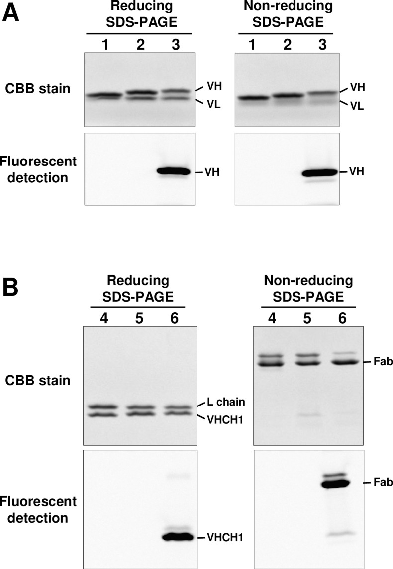
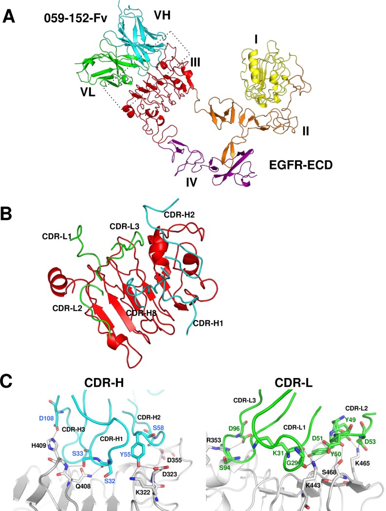
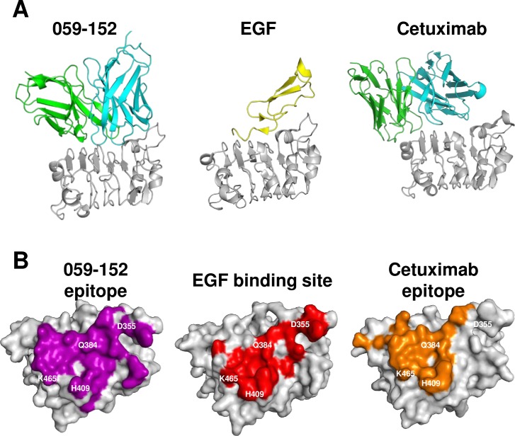
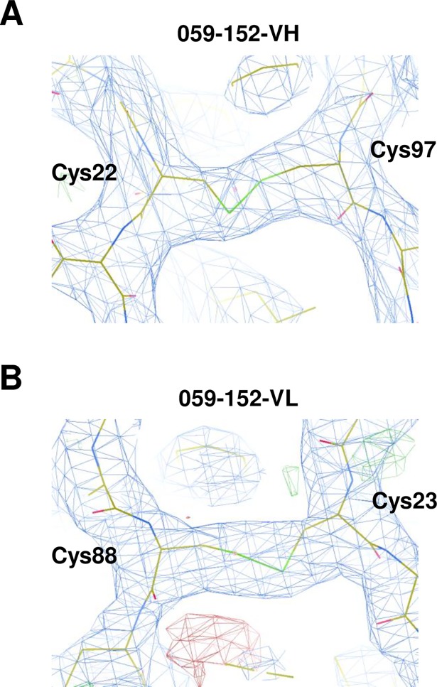
Growing numbers of therapeutic antibodies offer excellent treatment strategies for many diseases. Elucidation of the interaction between a potential therapeutic antibody and its target protein by structural analysis reveals the mechanism of action and offers useful information for developing rational antibody designs for improved affinity. Here, we developed a rapid, high-yield cell-free system using dialysis mode to synthesize antibody fragments for the structural analysis of antibody-antigen complexes. Optimal synthesis conditions of fragments (Fv and Fab) of the anti-EGFR antibody 059-152 were rapidly determined in a day by using a 30-μl-scale unit. The concentration of supplemented disulfide isomerase, DsbC, was critical to obtaining soluble antibody fragments. The optimal conditions were directly applicable to a 9-ml-scale reaction, with linear scalable yields of more than 1 mg/ml. Analyses of purified 059-152-Fv and Fab showed that the cell-free synthesized antibody fragments were disulfide-bridged, with antigen binding activity comparable to that of clinical antibodies. Examination of the crystal structure of cell-free synthesized 059-152-Fv in complex with the extracellular domain of human EGFR revealed that the epitope of 059-152-Fv broadly covers the EGF binding surface on domain III, including residues that formed critical hydrogen bonds with EGF (Asp355EGFR, Gln384EGFR, H409EGFR, and Lys465EGFR), so that the antibody inhibited EGFR activation. We further demonstrated the application of the cell-free system to site-specific integration of non-natural amino acids for antibody engineering, which would expand the availability of therapeutic antibodies based on structural information and rational design. This cell-free system could be an ideal antibody-fragment production platform for functional and structural analysis of potential therapeutic antibodies and for engineered antibody development.
Conflict of interest statement
Figures

References
-
- Chan AC, Carter PJ. Therapeutic antibodies for autoimmunity and inflammation. Nat Rev Immunol. 2010;10(5):301–16. doi: 10.1038/nri2761 - DOI - PubMed
-
- Carter PJ. Potent antibody therapeutics by design. Nat Rev Immunol. 2006;6(5):343–57. doi: 10.1038/nri1837 - DOI - PubMed
-
- Hynes NE, Lane HA. ERBB receptors and cancer: the complexity of targeted inhibitors. Nat Rev Cancer. 2005;5(5):341–54. doi: 10.1038/nrc1609 - DOI - PubMed
-
- Burgess AW, Cho HS, Eigenbrot C, Ferguson KM, Garrett TP, Leahy DJ, et al. An open-and-shut case? Recent insights into the activation of EGF/ErbB receptors. Mol Cell. 2003;12(3):541–52. doi: 10.1016/S1097-2765(03)00350-2 - DOI - PubMed
-
- Li S, Schmitz KR, Jeffrey PD, Wiltzius JJ, Kussie P, Ferguson KM. Structural basis for inhibition of the epidermal growth factor receptor by cetuximab. Cancer Cell. 2005;7(4):301–11. doi: 10.1016/j.ccr.2005.03.003 - DOI - PubMed
Publication types
MeSH terms
Substances
LinkOut - more resources
Full Text Sources
Other Literature Sources
Molecular Biology Databases
Research Materials
Miscellaneous
