

Mechanisms for the establishment and maintenance of pregnancy: synergies from scientific collaborations
- PMID: 29462279
- PMCID: PMC6044348
- DOI: 10.1093/biolre/ioy047
Mechanisms for the establishment and maintenance of pregnancy: synergies from scientific collaborations
Abstract
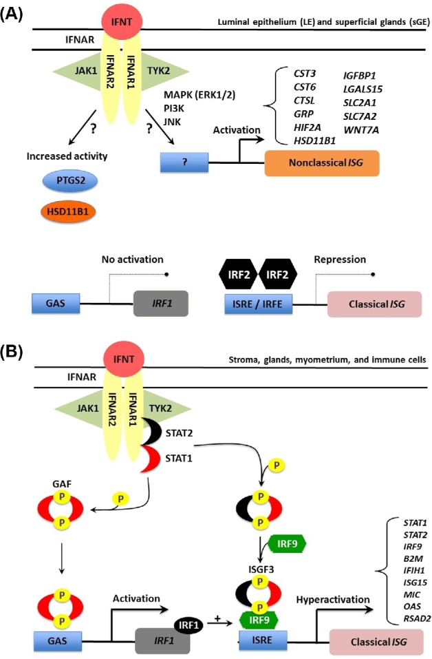
Research on the functions of interferon tau (IFNT) led to the theory of pregnancy recognition signaling in ruminant species. But IFNT does much more as it induces expression of interferon regulatory factor 2 (IRF2) in uterine luminal (LE), superficial glandular (sGE), but not glandular (GE) epithelia. First, IRF2 silences transcription of the estrogen receptor alpha gene and, indirectly, transcription of the oxytocin receptor gene to abrogate development of the luteolytic mechanism to prevent regression of the corpus luteum and its production of progesterone for establishing and maintaining pregnancy. Second, IRF2 silences expression of classical interferon-stimulated genes in uterine LE and sGE; however, uterine LE and sGE respond to progesterone (P4) and IFNT to increase expression of genes for transport of nutrients into the uterine lumen such as amino acids and glucose. Other genes expressed by uterine LE and sGE encode for adhesion molecules such as galectin 15, cathepsins, and cystatins for tissue remodeling, and hypoxia-inducible factor relevant to angiogenesis and survival of blastocysts in a hypoxic environment. IFNT is also key to a servomechanism that allows uterine epithelia, particularly GE, to proliferate and to express genes in response to placental lactogen and placental growth hormone in sheep. The roles of secreted phosphoprotein 1 are also discussed regarding its role in implantation in sheep and pigs, as well as its stimulation of expression of mechanistic target of rapamycin mRNA and protein which is central to proliferation, migration, and gene expression in the trophectoderm cells.
Figures
Similar articles
-
Interferons and progesterone for establishment and maintenance of pregnancy: interactions among novel cell signaling pathways.Reprod Biol. 2008 Nov;8(3):179-211. doi: 10.1016/s1642-431x(12)60012-6. Reprod Biol. 2008. PMID: 19092983 Review.
-
Biology of progesterone action during pregnancy recognition and maintenance of pregnancy.Front Biosci. 2002 Sep 1;7:d1879-98. doi: 10.2741/spencer. Front Biosci. 2002. PMID: 12161340 Review.
-
Conceptus signals for establishment and maintenance of pregnancy.Reprod Biol Endocrinol. 2004 Jul 5;2:49. doi: 10.1186/1477-7827-2-49. Reprod Biol Endocrinol. 2004. PMID: 15236653 Free PMC article. Review.
-
Fetal-maternal interactions during the establishment of pregnancy in ruminants.Soc Reprod Fertil Suppl. 2007;64:379-96. doi: 10.5661/rdr-vi-379. Soc Reprod Fertil Suppl. 2007. PMID: 17491160
-
Conceptus signals for establishment and maintenance of pregnancy.Anim Reprod Sci. 2004 Jul;82-83:537-50. doi: 10.1016/j.anireprosci.2004.04.014. Anim Reprod Sci. 2004. PMID: 15271478
Cited by
-
YPEL3 Negatively Regulates Endometrial Function via the Wnt/β-Catenin Pathways during Early Pregnancy in Goats.Animals (Basel). 2022 Oct 28;12(21):2973. doi: 10.3390/ani12212973. Animals (Basel). 2022. PMID: 36359097 Free PMC article.
-
Pre-implantation exogenous progesterone and pregnancy in sheep. II. Effects on fetal-placental development and nutrient transporters in late pregnancy.J Anim Sci Biotechnol. 2021 Apr 8;12(1):46. doi: 10.1186/s40104-021-00567-1. J Anim Sci Biotechnol. 2021. PMID: 33827696 Free PMC article.
-
Induced endometrial inflammation compromises conceptus development in dairy cattle†.Biol Reprod. 2023 Oct 13;109(4):415-431. doi: 10.1093/biolre/ioad088. Biol Reprod. 2023. PMID: 37540198 Free PMC article.
-
Dietary L-arginine supplementation during days 14-25 of gestation enhances aquaporin expression in the placentae and endometria of gestating gilts.Amino Acids. 2021 Aug;53(8):1287-1295. doi: 10.1007/s00726-021-03038-z. Epub 2021 Jul 9. Amino Acids. 2021. PMID: 34241695
-
Differences in Microbial Community Composition between Uterine Horns Ipsilateral and Contralateral to the Corpus Luteum in Beef Cows on Day 15 of the Estrous Cycle.Microorganisms. 2023 Aug 20;11(8):2117. doi: 10.3390/microorganisms11082117. Microorganisms. 2023. PMID: 37630677 Free PMC article.
References
-
- Schramm W, Bovaird L, Glew ME, Schramm G, McCracken JA. Corpus luteum regression induced by ultra-low pulses of prostaglandin F2 alpha. Prostaglandins 1983; 26:347–364. - PubMed
-
- Ott TL, Zhou Y, Mirando MA, Stevens C, Harney JP, Ogle TF, Bazer FW. Changes in progesterone and oestrogen receptor mRNA and protein during maternal recognition of pregnancy and luteolysis in ewes. J Mol Endocrinol 1993; 10:171–183. - PubMed
-
- Spencer TE, Bazer FW. Temporal and spatial alterations in uterine estrogen receptor and progesterone receptor gene expression during the estrous cycle and early pregnancy in the ewe. Biol Reprod 1995; 53:1527–1543. - PubMed
-
- Fincher KB, Bazer FW, Hansen PJ, Thatcher WW, Roberts RM. Proteins secreted by the sheep conceptus suppress induction of uterine prostaglandin F2a release by oestradiol and oxytocin. Reproduction 1986; 76:425–433. - PubMed
Publication types
MeSH terms
Substances
Grants and funding
LinkOut - more resources
Full Text Sources
Other Literature Sources
Research Materials
