

Left frontal hub connectivity delays cognitive impairment in autosomal-dominant and sporadic Alzheimer's disease
- PMID: 29462334
- PMCID: PMC5888938
- DOI: 10.1093/brain/awy008
Left frontal hub connectivity delays cognitive impairment in autosomal-dominant and sporadic Alzheimer's disease
Abstract
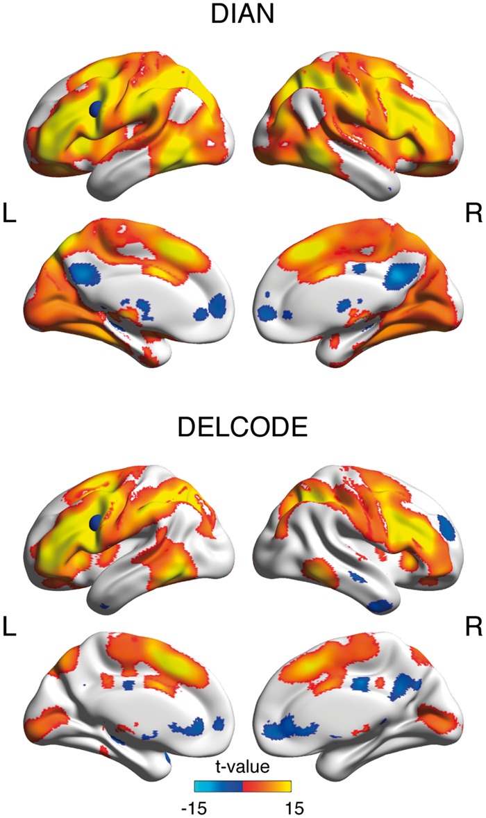
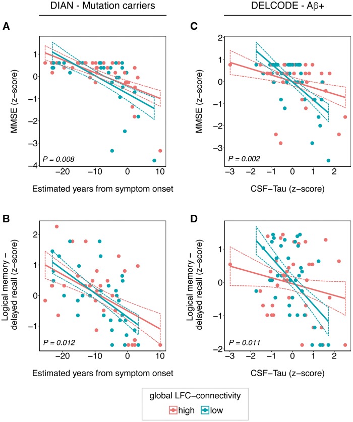
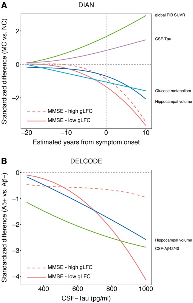
Patients with Alzheimer's disease vary in their ability to sustain cognitive abilities in the presence of brain pathology. A major open question is which brain mechanisms may support higher reserve capacity, i.e. relatively high cognitive performance at a given level of Alzheimer's pathology. Higher functional MRI-assessed functional connectivity of a hub in the left frontal cortex is a core candidate brain mechanism underlying reserve as it is associated with education (i.e. a protective factor often associated with higher reserve) and attenuated cognitive impairment in prodromal Alzheimer's disease. However, no study has yet assessed whether such hub connectivity of the left frontal cortex supports reserve throughout the evolution of pathological brain changes in Alzheimer's disease, including the presymptomatic stage when cognitive decline is subtle. To address this research gap, we obtained cross-sectional resting state functional MRI in 74 participants with autosomal dominant Alzheimer's disease, 55 controls from the Dominantly Inherited Alzheimer's Network and 75 amyloid-positive elderly participants, as well as 41 amyloid-negative cognitively normal elderly subjects from the German Center of Neurodegenerative Diseases multicentre study on biomarkers in sporadic Alzheimer's disease. For each participant, global left frontal cortex connectivity was computed as the average resting state functional connectivity between the left frontal cortex (seed) and each voxel in the grey matter. As a marker of disease stage, we applied estimated years from symptom onset in autosomal dominantly inherited Alzheimer's disease and cerebrospinal fluid tau levels in sporadic Alzheimer's disease cases. In both autosomal dominant and sporadic Alzheimer's disease patients, higher levels of left frontal cortex connectivity were correlated with greater education. For autosomal dominant Alzheimer's disease, a significant left frontal cortex connectivity × estimated years of onset interaction was found, indicating slower decline of memory and global cognition at higher levels of connectivity. Similarly, in sporadic amyloid-positive elderly subjects, the effect of tau on cognition was attenuated at higher levels of left frontal cortex connectivity. Polynomial regression analysis showed that the trajectory of cognitive decline was shifted towards a later stage of Alzheimer's disease in patients with higher levels of left frontal cortex connectivity. Together, our findings suggest that higher resilience against the development of cognitive impairment throughout the early stages of Alzheimer's disease is at least partially attributable to higher left frontal cortex-hub connectivity.
Figures
References
-
- Arenaza-Urquijo EM, Molinuevo JL, Sala-Llonch R, Sole-Padulles C, Balasa M, Bosch B, et al. Cognitive reserve proxies relate to gray matter loss in cognitively healthy elderly with abnormal cerebrospinal fluid amyloid-beta levels. J Alzheimers Dis 2013; 35: 715–26. - PubMed
-
- Ashburner J. A fast diffeomorphic image registration algorithm. NeuroImage 2007; 38: 95–113. - PubMed
Publication types
MeSH terms
Substances
Grants and funding
- U01 AG032438/AG/NIA NIH HHS/United States
- U24 AG021886/AG/NIA NIH HHS/United States
- P50 AG005681/AG/NIA NIH HHS/United States
- P01 AG026276/AG/NIA NIH HHS/United States
- MR/009076/1/MRC_/Medical Research Council/United Kingdom
- MR/L023784/1/MRC_/Medical Research Council/United Kingdom
- P30 NS048056/NS/NINDS NIH HHS/United States
- UF1 AG032438/AG/NIA NIH HHS/United States
- MR/L023784/2/MRC_/Medical Research Council/United Kingdom
- U19 AG032438/AG/NIA NIH HHS/United States
- R01 EB009352/EB/NIBIB NIH HHS/United States
- P30 NS098577/NS/NINDS NIH HHS/United States
LinkOut - more resources
Full Text Sources
Other Literature Sources
Medical
Research Materials
Miscellaneous
