. 2018 May 1;27(9):1556-1564.
doi: 10.1093/hmg/ddy062.

The beta-adrenergic agonist salbutamol modulates neuromuscular junction formation in zebrafish models of human myasthenic syndromes

Affiliations

The beta-adrenergic agonist salbutamol modulates neuromuscular junction formation in zebrafish models of human myasthenic syndromes

Grace McMacken et al. Hum Mol Genet. .

Abstract

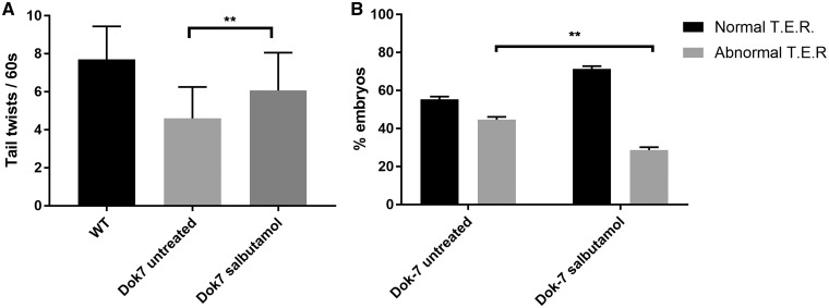
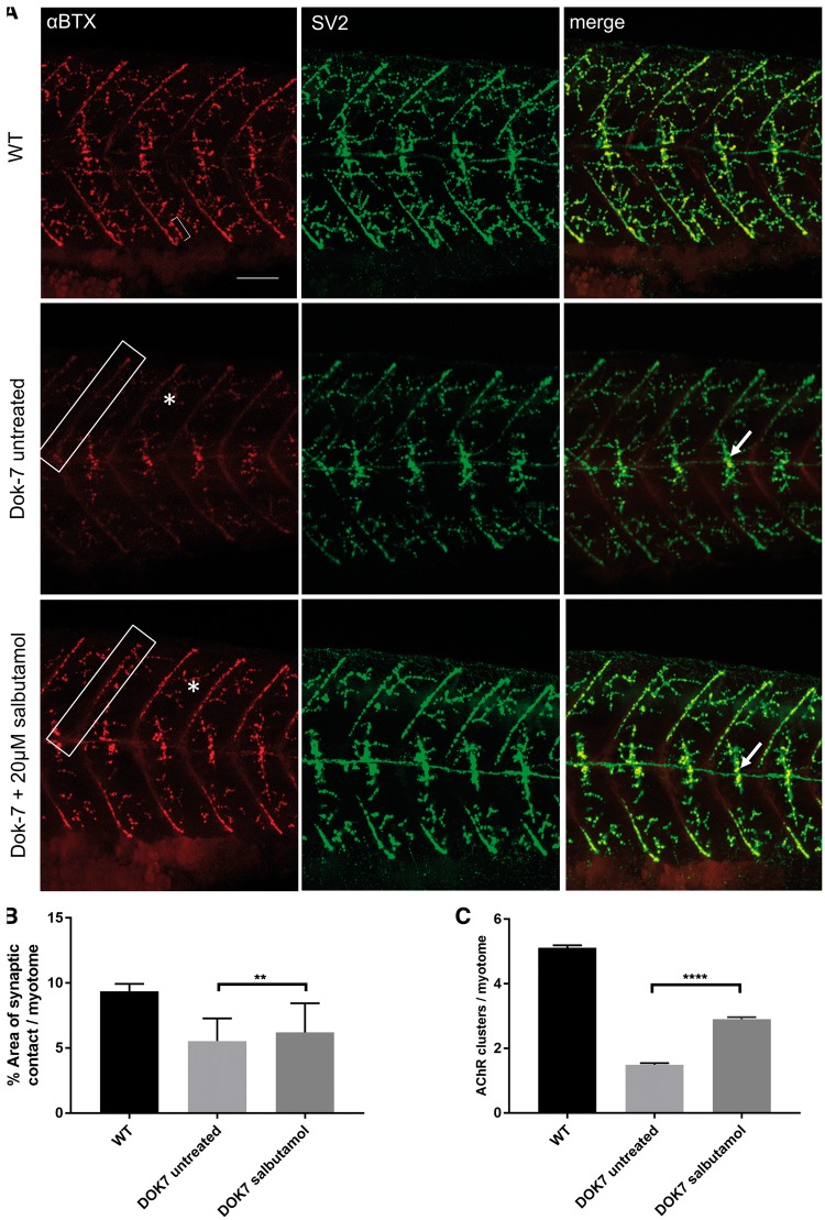
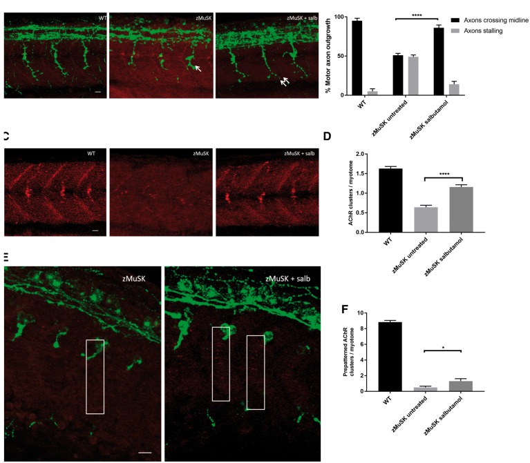
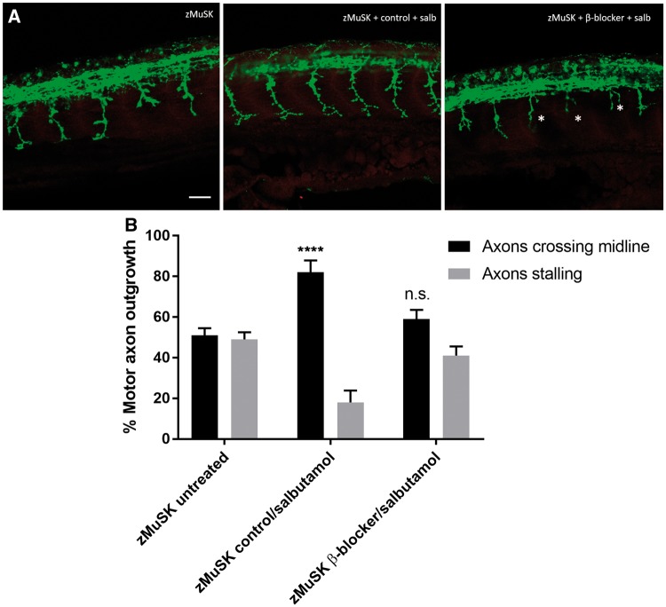
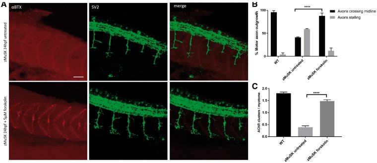
Inherited defects of the neuromuscular junction (NMJ) comprise an increasingly diverse range of disorders, termed congenital myasthenic syndromes (CMS). Therapies acting on the sympathetic nervous system, including the selective β2 adrenergic agonist salbutamol and the α and β adrenergic agonist ephedrine, have become standard treatment for several types of CMS. However, the mechanism of the therapeutic effect of sympathomimetics in these disorders is not understood. Here, we examined the effect of salbutamol on NMJ development using zebrafish with deficiency of the key postsynaptic proteins Dok-7 and MuSK. Treatment with salbutamol reduced motility defects in zebrafish embryos and larvae. In addition, salbutamol lead to morphological improvement of postsynaptic acetycholine receptor (AChR) clustering and size of synaptic contacts in Dok-7-deficient zebrafish. In MuSK-deficient zebrafish, salbutamol treatment reduced motor axon pathfinding defects and partially restored the formation of aneural prepatterned AChRs. In addition, the effects of salbutamol treatment were prevented by pre-treatment with a selective β2 antagonist. Treatment with the cyclic adenosine monophosphate (cAMP) activator forskolin, replicated the effects of salbutamol treatment. These results suggest that sympathomimetics exert a direct effect on neuromuscular synaptogenesis and do so via β2 adrenoceptors and via a cAMP-dependent pathway.


Figures

Figure 1.
Figure 1.
Dok7 morphant zebrafish demonstrate improved motility in the presence of salbutamol. (A) When monitored at 24 hpf, Dok7 MO-injected embryos are able to perform tail twisting movements in their chorion but do so at lower frequency than WT embryos. Following salbutamol treatment, Dok7 embryos performed tail twists at an increased frequency (mean 6.1 per 60 s) compared with untreated embryos (mean 4.6 per 60 s). A total of 30 embryos were observed for each category. ** indicates P < 0.01, Student’s t-test, n = 30 treated and 30 untreated embyros observed. (B) Dok7 embryos demonstrate equal percentages of normal and abnormal touch-evoke response (TER) to stimuli at 48 hpf. Embryos treated with salbutamol demonstrate an increased percentage of normal swim behaviour. ** indicates P < 0.01, chi-square test, n = 3 tests on 30 embryos were performed for each category. All error bars depict S.E.M.
Figure 2.
Figure 2.
Salbutamol treatment of Dok-7 MO-injected zebrafish improves NMJ morphology. Lateral views of 48 hpf embryos with neuromuscular synapses labelled with antibodies against SV2 (green, presynaptic vesicles) and αBTX (red, postsynaptic AChRs). Scale bar = 50 µm. (A) In Dok7 MO-injected embryos, the focal innervation point along the horizontal myoseptum is reduced in size compared with wild type, but is increased following treatment with 20 µm salbutamol (arrows). In addition, the morphology of myoseptal (boxes) and myotomal (asterisks) AChR clusters is partially rescued with salbutamol treatment. A representative 20 μm2 AChR cluster is labelled with a bracket. (B) Following salbutamol treatment, the area of synaptic contact on the horizontal myoseptum is significantly increased (** indicates P < 0.01, Student’s t-test, n = 160 treated and 160 untreated myotomal segments examined). (C) Salbutamol treatment caused a significant increase in the number of AChR clusters >20 μm2 per myotome in Dok7 zebrafish embryos (**** indicates P < 0.0001, Student’s t-test, n = 100 treated and 100 untreated myotomal segments examined).
Figure 3.
Figure 3.
Salbutamol treatment of zMuSK MO-injected embryos improves motor axon pathfinding defects and AChR clustering. Lateral views of embryos with neuromuscular synapses labelled with antibodies against SV2 (green, presynaptic vesicles) and αBTX (red, postsynaptic AChRs). Scale bars = 10 µm. (A) zMuSK MO-injected embryos at 24 hpf demonstrate characteristic stalling of the outgrowing motor axon at the choice point on the horizontal midline (arrow). Following salbutamol treatment more axons cross the horizontal midline and extend to the periphery of the myotome (double arrow). (B) Quantification of the improvement of motor axon growth following salbutamol treatment. (**** indicates P < 0.0001, chi-square test, n = 240 treated and 240 untreated myotomal segments examined). (C) zMuSK embryos at 24 hpf display almost complete absence of AChR clustering. In salbutamol-treated zMuSK embryos, AChR clustering is restored. (D) Quantification of the improvement in AChR clustering following salbutamol treatment of zMuSK embryos. AChR clustering is significantly increased with the mean number of AChR clusters >20 μm2 per myotome 1.16 in salbutamol treated embryos compared with 0.64 in untreated embryos (**** indicates P < 0.0001, Student’s t-test, n = 100 treated and 100 untreated myotomal segments examined). (E) zMuSK MO-injected embryos at 17 hpf. Untreated embryos lack elongated, diffuse prepatterned clusters on the myotomal surface prior to the arrival of the motor growth cone (boxed areas), but following salbutamol treatment some zMuSK embryos display prepatterned clusters. (F) Quantification of the effect of salbutamol on prepatterned AChR clustering in zMuSK embryos. Following salbutamol treatment, the number of prepatterned AChR clusters >3 μm2 in zMuSK embryos increased, with a mean of 0.5 prepatterned clusters per myotome in untreated embryos, compared with 1.3 in salbutamol-treated embryos (* indicates P < 0.05, Student’s t-test, n = 30 untreated and 30 untreated myotomal segments examined).
Figure 4.
Figure 4.
Pre-treatment with a selective β2 antagonist blocks the rescue of NMJ development by salbutamol. Lateral views of 24 hpf embryos with neuromuscular synapses labelled with antibodies against SV2 (green, presynaptic vesicles) and αBTX (red, postsynaptic AChRs). Scale bar= 50 µm. zMuSK embryos were treated with 5 µm of the selective β2 antagonist ICI118, 551 or control (water) added to E3 medium for 3 h prior to the addition of 20 µm salbutamol. (A) In zebrafish embryos pre-treated with ICI118, 551 no rescue of the axonal pathfinding defects was seen following 21 h of salbutamol treatment, with stalling and branching of motor growth cones (asterisks). (B) Quantification of the effect of pre-treatment of ICI118, 551 on motor axon growth. Following pre-treatment with 5 µm ICI118, 551, salbutamol treatment did not have an effect on axon pathfinding (n.s. P > 0.05, ****P < 0.0001, chi-square test, n = 240 treated and 240 untreated myotomal segments examined).
Figure 5.
Figure 5.
The effects of β2 receptor agonist activation on NMJ morphology are replicated by cAMP activation. Lateral views of 24 hpf embryos with neuromuscular synapses labelled with antibodies against SV2 (green, presynaptic vesicles) and αBTX (red, postsynaptic AChRs). Scale bar = 50 µm. (A) zMuSK embryos at 24 hpf demonstrate improvement in AChR clustering and axon pathfinding defects following forskolin treatment. (B) Quantification of motor axon guidance defects. About 88% (n = 28/240) of imaged somites of zMuSK embryos treated with forskolin displayed no axon guidance defects, compared with 41% (n = 142/240) of those in untreated zMuSK embryos. **** indicates P < 0.0001, chi-square test. (C) The number of AChR clusters larger than 20 μm2 on myotomal muscle fibres increased with forskolin treatment, with mean number of clusters 0.39 per myotome in untreated embryos, and 1.48 in treated zMuSK embryos. **** indicates P < 0.0001, Student’s t-test, n = 100 treated and 100 untreated myotomes examined.

References

    1. Cohen S.A., Fischbach G.D. (1977) Clusters of acetylcholine receptors located at identified nerve-muscle synapses in vitro. Dev. Biol., 59, 24–38. - PubMed
    1. Kummer T.T., Misgeld T., Sanes J.R. (2006) Assembly of the postsynaptic membrane at the neuromuscular junction: paradigm lost. Curr. Opin. Neurobiol., 16, 74–82. - PubMed
    1. Kim N., Burden S.J. (2008) MuSK controls where motor axons grow and form synapses. Nat. Neurosci., 11, 19–27. - PMC - PubMed
    1. Weatherbee S.D., Anderson K.V., Niswander L.A. (2006) LDL-receptor-related protein 4 is crucial for formation of the neuromuscular junction. Dev. Camb. Engl., 133, 4993–5000. - PubMed
    1. Flanagan-Steet H., Fox M.A., Meyer D., Sanes J.R. (2005) Neuromuscular synapses can form in vivo by incorporation of initially aneural postsynaptic specializations. Development, 132, 4471–4481. - PubMed

Publication types