Review
. 2018 Feb 15;18(2):599.
doi: 10.3390/s18020599.

Responsive Polydiacetylene Vesicles for Biosensing Microorganisms

Affiliations
Review

Responsive Polydiacetylene Vesicles for Biosensing Microorganisms

Estelle Lebègue et al. Sensors (Basel). .

Abstract

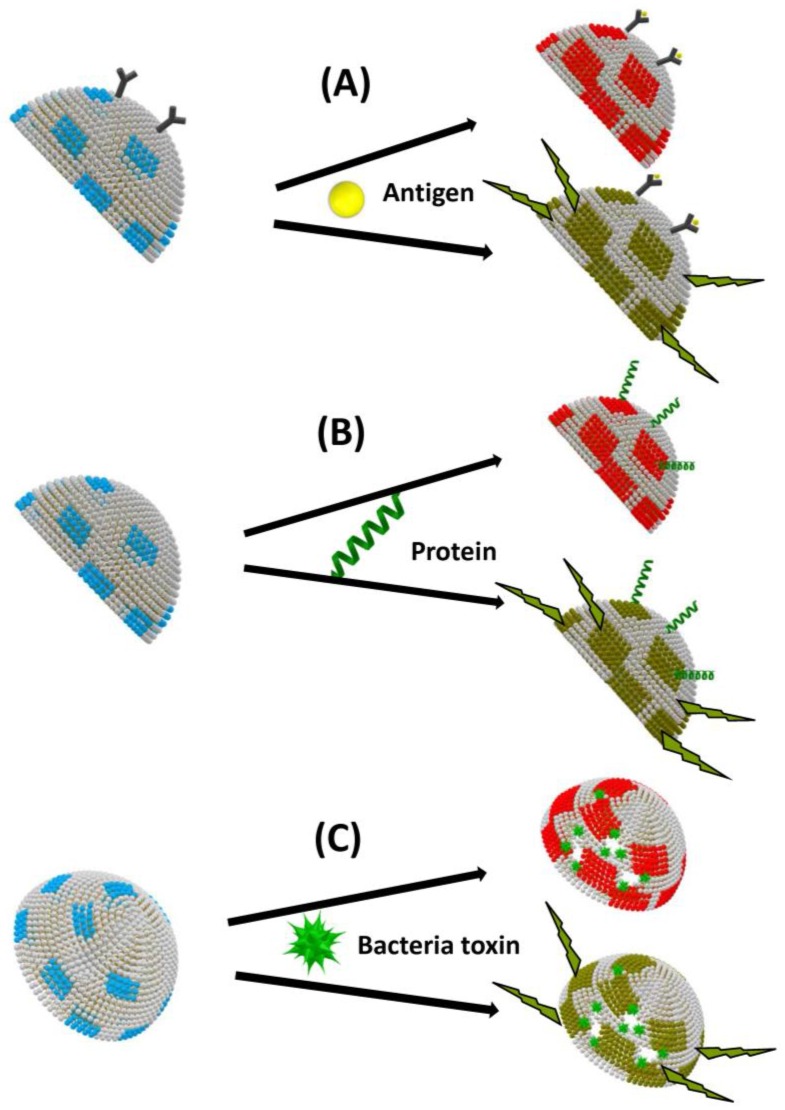
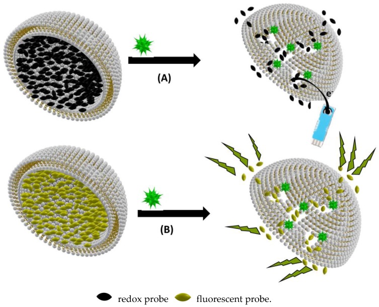
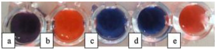
Polydiacetylene (PDA) inserted in films or in vesicles has received increasing attention due to its property to undergo a blue-to-red colorimetric transition along with a change from non-fluorescent to fluorescent upon application of various stimuli. In this review paper, the principle for the detection of various microorganisms (bacteria, directly detected or detected through the emitted toxins or through their DNA, and viruses) and of antibacterial and antiviral peptides based on these responsive PDA vesicles are detailed. The analytical performances obtained, when vesicles are in suspension or immobilized, are given and compared to those of the responsive vesicles mainly based on the vesicle encapsulation method. Many future challenges are then discussed.

Keywords: bacteria; biosensing; peptides; polydiacetylene; toxins; vesicles; virus.


Conflict of interest statement

The authors declare no conflict of interest.

Figures

Figure 1
Figure 1
Different structures of vesicles: SUV (small unilamellar vesicles), LUV (large unilamellar vesicles), GUV (giant unilamellar vesicles), and MLV (multilamellar large vesicles). Vesicles are presented as hemivesicle to show the inside.
Figure 2
Figure 2
TEM images of large unilamellar vesicles (LUV) (left) and multilamellar giant vesicles (MGV) (right), made of bilayers of synthetic double chain zwitterionic surfactants, taken after preparation by freeze-fracture and replication of the fracture section. LUV appear as small circles being either full or having an empty water pool inside depending on whether the fracture propagated across the vesicles or along their external surface. MGV appear as onion-like stacks of lipid bilayers. Such concentric bilayers fill the whole vesicle; the empty hole in the middle corresponds to part of the vesicle center that has been detached when fracturing the frozen sample.
Figure 3
Figure 3
Colorimetric (upper line) and fluorescence (lower line) biosensing based on biomimetic vesicles comprising polydiacetylene (PDA) (blue and red parts) induced by external stimuli: (A) surface binding; (B) insertion; and (C) pore formation. Vesicles are presented as hemivesicles.
Figure 4
Figure 4
(A) Amperometric biosensing based on biomimetic vesicle encapsulation of redox probes; and (B) fluorimetric biosensing based on biomimetic vesicle encapsulation of fluorescent probes. Vesicles are presented as hemivesicles to show the inside.
Figure 5
Figure 5
PCDA/SPH/Cholesterol/Lysine vesicles added to TSB (0.1%) and aqueous saline at pH 6.0 with: (a) E. coli; (b) P. aeruginosa; (c) S. aureus; (d) L. plantarum; and (e) S. Choleraesuis (108 CFU/mL). Reprinted with permission from [31]. Copyright 2017 Elsevier.
Figure 6
Figure 6
Colorimetric transition of mixed polyacetylene vesicles as a function of light irradiation time in the presence of TiO2 in E. coli K12 suspension. Reprinted with permission from [33]. Copyright 2005 Elsevier.
Figure 7
Figure 7
An intelligent hydrogel wound dressing based on fluorescent dye release from polydiacetylene vesicles. Reprinted with permission from [47]. Copyright 2016 American Chemical Society.
Figure 8
Figure 8
Color change of DMPE/TRCDA vesicles treated with 50 µL cell-free supernatant of 54 lactic acid bacteria strains. Reprinted with permission from [12]. Copyright 2017 Springer Nature.

References

    1. Ivnitski D., Abdel-Hamid I., Atanasov P., Wilkins E. Biosensors for detection of pathogenic bacteria. Biosens. Bioelectron. 1999;14:599–624. doi: 10.1016/S0956-5663(99)00039-1. - DOI - PubMed
    1. Okada S., Peng S., Spevak W., Charych D. Color and Chromism of Polydiacetylene Vesicles. Acc. Chem. Res. 1998;31:229–239. doi: 10.1021/ar970063v. - DOI
    1. Fendler J.H. Surfactant vesicles as membrane mimetic agents: Characterization and utilization. Acc. Chem. Res. 1980;13:7–13. doi: 10.1021/ar50145a002. - DOI
    1. Bally M., Bailey K., Sugihara K., Grieshaber D., Vörös J., Stâdler B. Liposome and lipid bilayer arrays towards biosensing applications. Small. 2010;22:2481–2497. doi: 10.1002/smll.201000644. - DOI - PubMed
    1. Liu Q., Boyd B.J. Liposomes in biosensors. Analyst. 2013;138:39–409. doi: 10.1039/C2AN36140J. - DOI - PubMed

LinkOut - more resources