. 2018 Feb 20;18(1):210.
doi: 10.1186/s12885-018-4126-y.

Inhibition of autophagy sensitizes cancer cells to Photofrin-based photodynamic therapy

Affiliations

Inhibition of autophagy sensitizes cancer cells to Photofrin-based photodynamic therapy

Antoni Domagala et al. BMC Cancer. .

Abstract

Background: Accumulating evidence suggest that autophagy plays a pivotal role in various anticancer therapies, including photodynamic therapy (PDT), acting as a pro-death or pro-survival mechanism in a context-dependent manner. Therefore, we aimed to determine the role of autophagy in Photofrin-based PDT.

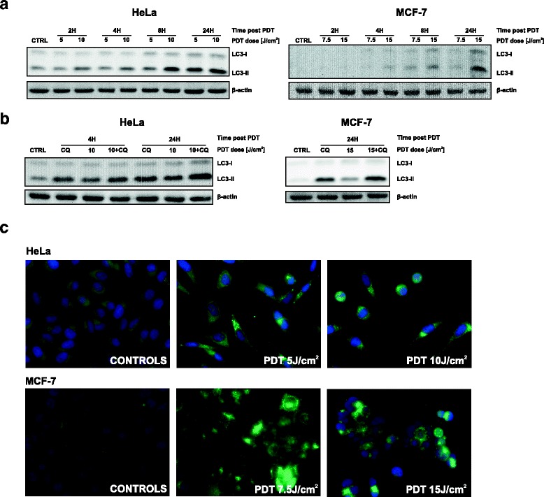
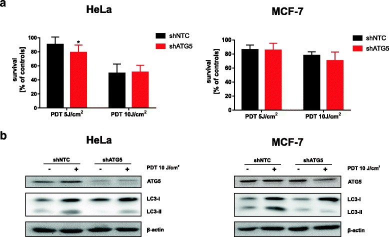
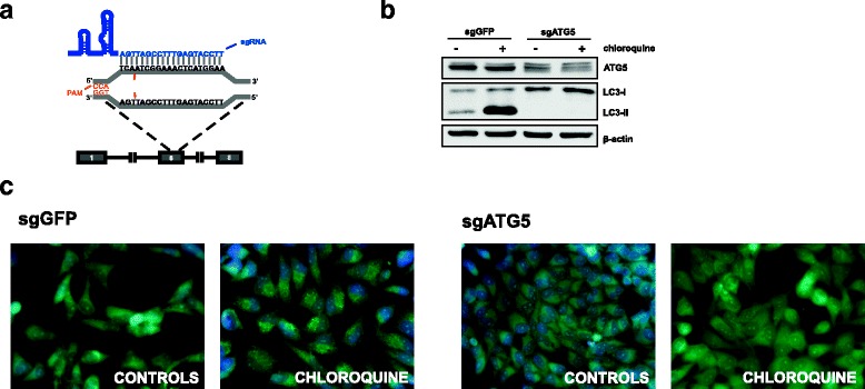
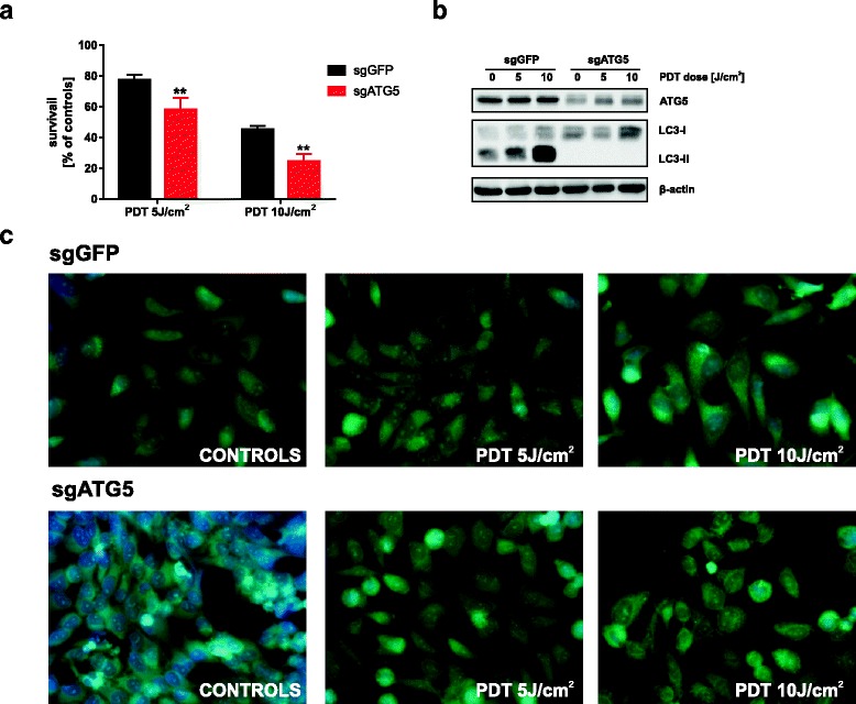
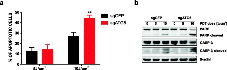
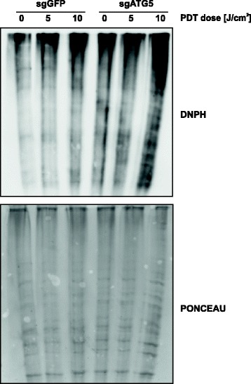
Methods: In vitro cytotoxic/cytostatic effects of PDT were evaluated with crystal violet cell viability assay. Autophagy induction was analyzed by immunoblotting and immunofluorescence using anti-LC3 antibody. Autophagy was inhibited by shRNA-mediated ATG5 knockdown or CRISPR/Cas9-mediated ATG5 knockout. Apoptosis was assessed by flow cytometry analysis of propidium iodide and anexin V-positive cells as well as by detection of cleaved PARP and caspase 3 proteins using immunoblotting. Protein carbonylation was evaluated by the 2,4-dinitrophenylhydrazine (DNPH) method.

Results: Photofrin-PDT leads to robust autophagy induction in two cancer cell lines, Hela and MCF-7. shRNA-mediated knockdown of ATG5 only partially blocks autophagic response and only marginally affects the sensitivity of Hela and MCF-7 cells to PDT. ATG5 knockout in HeLa cell line utilizing CRISPR/Cas9 genome editing results in increased PDT-mediated cytotoxicity, which is accompanied by an enhanced apoptotic response and increased accumulation of carbonylated proteins.

Conclusions: Altogether, these observations imply that autophagy contributes to Photofrin-PDT resistance by enabling clearance of carbonylated and other damaged proteins. Therefore, autophagy inhibition may serve as a strategy to improve PDT efficacy.

Keywords: ATG5; Autophagy; CRISR/Cas-9; Photodynamic therapy; Photofrin.


Conflict of interest statement

Ethics approval and consent to participate

There was no research involving human or animal subjects in the study.

Consent for publication

Not applicable.

Competing interests

The authors declare that they have no competing interest.

Publisher’s Note

Springer Nature remains neutral with regard to jurisdictional claims in published maps and institutional affiliations.

Figures

Fig. 1
Fig. 1
Photofrin-PDT induces autophagy. a HeLa (left panel) and MCF-7 (right panel) cells were incubated with Photofrin for 24 h before exposure to different light fluencies. Whole-cell lysates were collected at indicated time points after PDT and analyzed by Western blotting for LC3 and β-actin (loading control) expression. b The experiment was performed as in (a), but, for indicated samples, 10 μM chloroquine (CQ) was added to the culture medium after PDT. c 16 h after irradiations, the cells were fixed and stained with anti-LC3 antibody to visualize autophagosomes by immunofluorescence
Fig. 2
Fig. 2
ShRNA-mediated ATG5 downregulation moderately affects autophagy and PDT efficacy. a HeLa and MCF-7 cells were infected with lentiviral particles containing shRNA targeting ATG5 (shATG5) or scrambled shRNA (shNTC) and subsequently incubated with puromycin to isolate stable cell lines. The stable cell lines were subjected to in-vitro PDT and cell survival was determined 24 h post-PDT by crystal violet staining. The bars represent survival in each experimental versus its own untreated control. Data show the mean values of 2 independent experiments ± S.D. (represented by error bars), and *P < 0.05 (Student’s t-test) (b) Whole-cell lysates from shATG5 and shNTC cell lines were collected 24-h post PDT and ATG5 and LC3 expression was evaluated by Western blotting. β-actin expression was assessed as protein loading control
Fig. 3
Fig. 3
Construction and characterization of HeLa cells with CRISPR/Cas9-mediated genomic KO of ATG5. a Schematic diagram showing ATG5 gene and the sgRNA target site at exon 6. Cas-9-mediated double-strand break near the PAM sequence are indicated by arrows. b HeLa-sgGFP and HeLa-sgATG5 cells were incubated for 24 h in the presence or absence of 10 μM chloroquine (CQ) to block lysosomal degradation. Whole-cell lysates were collected and ATG5 and LC3 expression was analyzed by Western blotting. c To confirm impaired autophagosome formation, HeLa-sgGFP and HeLa-sgATG5 cells were incubated as in (b) and subsequently stained with anti-LC3 antibody to visualize autophagosomes by immunofluorescence microscopy
Fig. 4
Fig. 4
Elimination of ATG5-dependent autophagy sensitizes cells to PDT. a HeLa-sgGFP and HeLa-sgATG cells were subjected to in vitro PDT and the cell survival was determined by crystal violet staining 24 h after treatment. The viability in each experimental group is calculated versus its own untreated control. Data represent the mean values of 2 independent experiments ± S.D. (represented by error bars), **p < 0.001 (Student’s t-test). b To confirm impaired autophagic flux, cells were treated as in (a) and the whole-cell lysates were collected and ATG5 and LC3 levels were analyzed by Western blotting. c Immunofluorescence microscopy was used to confirm abrogated autophagosome formation after PDT
Fig. 5
Fig. 5
Abrogation of ATG5-dependent autophagy potentiates apoptosis. HeLa-sgGFP and HeLa sgATG cells were subjected to PDT. Cells were collected 24 h after illumination and stained with Annexin V-FITC and propidium iodide. Subsequently, early and late apoptotic cells (annexin V-positive and annexin V as well as propidium iodide double positive, respectively) were quantified by flow cytometry. Data represent the mean values of 2 independent experiments ± S.D. (represented by error bars), **p < 0.001 (Student’s t-test). a The bars show mean value of annexin V or propidium iodide positive cells. b cells were treated as in (a) and whole-cell lysates were collected 24 h after PDT, and the levels of PARP and caspase-3 and their activated cleaved forms were analyzed by Western blotting
Fig. 6
Fig. 6
Abrogation of ATG5-dependent autophagy increases protein carbonylation. HeLa-sgGFP and HeLa-sgATG cells were subjected to PDT. Total cell lysates were prepared from tumor cells 24 h after illumination and protein carbonylation was assessed by the 2,4-dinitrophenylhydrazine (DNPH) method (upper panel). To ensure equal protein loading, the blot was stained with Ponceau (lower panel)

Similar articles

Cited by

References

    1. Klionsky DJ, Emr SD. Autophagy as a regulated pathway of cellular degradation. Science. 2000;290(5497):1717–1721. doi: 10.1126/science.290.5497.1717. - DOI - PMC - PubMed
    1. Todde V, Veenhuis M, van der Klei IJ. Autophagy: principles and significance in health and disease. Biochim Biophys Acta. 2009;1792(1):3–13. doi: 10.1016/j.bbadis.2008.10.016. - DOI - PubMed
    1. Kroemer G, Marino G, Levine B. Autophagy and the integrated stress response. Mol Cell. 2010;40(2):280–293. doi: 10.1016/j.molcel.2010.09.023. - DOI - PMC - PubMed
    1. Denton D, Xu T, Kumar S. Autophagy as a pro-death pathway. Immunol Cell Biol. 2015;93(1):35–42. doi: 10.1038/icb.2014.85. - DOI - PubMed
    1. Nakatogawa H, Suzuki K, Kamada Y, Ohsumi Y. Dynamics and diversity in autophagy mechanisms: lessons from yeast. Nat Rev Mol Cell Biol. 2009;10(7):458–467. doi: 10.1038/nrm2708. - DOI - PubMed

Publication types