. 2018 Feb 20;17(1):32.
doi: 10.1186/s12944-018-0675-y.

Fatty acids modulate the expression levels of key proteins for cholesterol absorption in Caco-2 monolayer

Affiliations

Fatty acids modulate the expression levels of key proteins for cholesterol absorption in Caco-2 monolayer

Fang Yang et al. Lipids Health Dis. .

Abstract

Background: Fatty acids have been shown to modulate intestinal cholesterol absorption in cells and animals, a process that is mediated by several transporter proteins. Of these proteins, Niemann-Pick C1-Like 1 (NPC1L1) is a major contributor to this process. The current study investigates the unknown mechanism by which fatty acids modulate cholesterol absorption.

Methods: We evaluated the effects of six fatty acids palmitic acid (PAM), oleic acid (OLA), linoleic acid (LNA), arachidonic acid (ARA), eicosapentaenoic acid (EPA) and docosahexaenoic acid (DHA) on cholesterol uptake and transport in human enterocytes Caco-2 cells, and on the mRNA expression levels of NPC1L1, others proteins (ABCG5, ABCG8, ABCA1, ACAT2, MTP, Caveolin 1, Annexin-2) involved in cholesterol absorption, and SREBP-1 and SREBP-2 that are responsible for lipid metabolism.

Results: The polyunsaturated fatty acids (PUFAs), especially for EPA and DHA, dose-dependently inhibited cholesterol uptake and transport in Caco-2 monolayer, while saturated fatty acids (SFAs) and monounsaturated fatty acids (MUFAs) had no inhibitory effects. EPA and DHA inhibited cholesterol absorption in Caco-2 monolayer might be caused by down-regulating NPC1L1 mRNA and protein levels, which were associated with inhibition of SREBP-1/- 2 mRNA expression levels.

Conclusion: Results from this study indicate that functional food containing high PUFAs may have potential therapeutic benefit to reduce cholesterol absorption. Further studies on this topic may provide approaches to control lipid metabolism and to promote health.

Keywords: Caco-2 monolayer transport; Fatty acids; Intestinal cholesterol absorption; NPC1L1.


Conflict of interest statement

Ethics approval and consent to participate

Not applicable.

Consent for publication

Not applicable.

Competing interests

All authors declare that they have no competing interests.

Publisher’s Note

Springer Nature remains neutral with regard to jurisdictional claims in published maps and institutional affiliations.

Figures
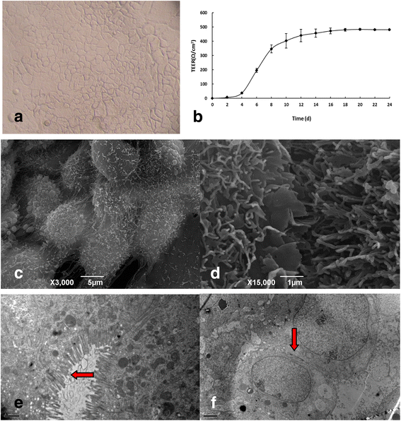
Fig. 1
Fig. 1
Morphology and characteristics of Caco-2 cell monolayer during and after differentiation. a Caco-2 monolayer morphology after the 21-day culture (Inverted microscope ×400). b Transepithelial electrical resistances (TEER) of Caco-2 monolayer at different time spots of the 24-day culture. The electrode of the Milicell ERS-2 resistance meter (Millipore, MA, USA) was immersed in 70% ethanol for 15 min, dried in air for 15 min, and then immersed in sterile electrolyte similar to the culture medium for 15 min. Then the two sides of electrodes were placed in two chambers of the TransWell™ culture plate, and the TEER of Caco-2 monolayer was detected, which was calculated as in Eq. (1) described in the Materials and Methods section. Results were presented as mean ± SD of three independent experiments. c Morphology of Caco-2 monolayer (C: × 3000) under scanning electron microscope. d Microvilli on the apical side of the Caco-2 monolayer (D: × 15,000) under scanning electron microscope. Cells after the 21-day culture were fixed in 2.5% glutaraldehyde treatment for 2 h, and then post-fixed for 30 min in 1% osmium tetroxide buffer and incubated in freshly made 1% carbohydrazide for 30 min. The fixed cells were rinsed three times with distilled water for over 15 min each time. The bottom of cell culture dishes was cut to fit the critical point dryer. Then, after a series of alcohol dehydration, dried in critical point dryer and sputter coat with 1-2 nm gold-palladium. Then, the cell ultrastructure was observed under scanning electron micrograph (JSM-6390/LV). e and f, the transmission electron micrographs of differentiated Caco-2 cells with microvilli (E: × 12,000) and tight junctions among cells (F: × 6000), respectively. Cells cultured for 21 days were fixed in 2.5% glutaraldehyde solution for 4 h and then post-fixed for 2 h in 1.33% osmium tetroxide buffered with 0.1 mol/L cacodylate. Then after a series of alcohol dehydration, propylene oxide/resin 2:1 infiltration, resin embedding and sectioning, the cell ultrastructure was observed under transmission electron microscopy (Hitachi H-7100)
Fig. 2
Fig. 2
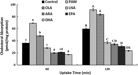
The effects of fatty acids on cholesterol uptake in Caco-2 monolayer. Caco-2 cells were incubated for 21 days, and the cholesterol uptake of 60 min and 120 min was analyzed as described under the Materials and Methods. Micellar solutions with 0.1% (v/v) DMSO were as follows: 2 μCi/mL [1,2-3H (N)]-cholesterol, 100 μmol/L cholesterol, 1 mmol/L OLA (for Control) or 0.5 mmol/L indicated fatty acid (PAM, OLA, LNA, ARA, EPA and DHA), 0.5 mmol/L monoolein, 6.6 mmol/L sodium taurocholate, and 0.1 mmol/L soy PtdCho in cultural medium containing the delipidized FBS. Data were represented as mean ± SD of three independent experiments (a > b > c > d, A > B > C > D > E, all p < 0.01). PAM: palmitic acid; OLA: oleic acid; LNA: linoleic acid; ARA: arachidonic acid; EPA: eicosapentaenoic acid; DHA: docosahexaenoic acid
Fig. 3
Fig. 3
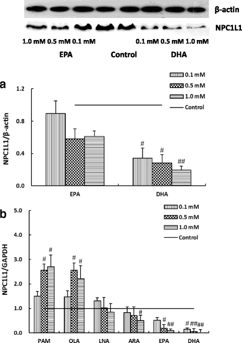
The effects of fatty acid treatments on the expression levels of Niemann-Pick C1-Like 1 (NPC1L1) protein (a) and its mRNA (b) in Caco-2 cells. Caco-2 cells were incubated in increasing concentrations of indicated fatty acids micellar solutions for 24 h, as described in the Methods and Materials section. Micellar solutions with 0.1% (v/v) DMSO were as follows: 2 μCi/mL [1,2-3H (N)]-cholesterol, 100 μmol/L cholesterol, 0 mmol/L (for control) or 0.1 / 0.5 / 1.0 mmol/L fatty acid (PAM, OLA, LNA, ARA, EPA or DHA), 0.5 mmol/L monoolein, 6.6 mmol/L sodium taurocholate, and 0.1 mmol/L soy PtdCho in cultural medium containing 20% delipidized FBS. Expression levels in A were normalized to that of the control β-actin protein (P < 0.05), and expression values in B were normalized to GAPDH mRNA, a housekeeping gene. The expression level of NPC1L1 protein or mRNA in the control group was set to 1. #p < 0.05, ##p < 0.01 were compared to that in the control cells (Caco-2 cells incubated with micellar solution without any fatty acid). Data were presented as mean ± SD of three independent experiments. PAM: palmitic acid; OLA: oleic acid; LNA: linoleic acid; ARA: arachidonic acid; EPA: eicosapentaenoic acid; DHA: docosahexaenoic acid
Fig. 4
Fig. 4
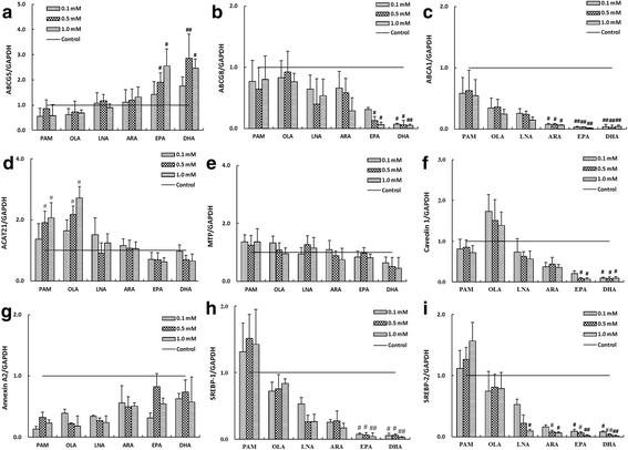
The effects of fatty acids on mRNA expression levels of ATP-binding cassette sub-family G member 5 (ABCG5) (a), ABCG8 (b), ABCA1 (c), acetyl-Coenzyme A acetyltransferase 2 (ACAT2) (d), microsomal triglyceride transfer protein (MTP) (e), Caveolin 1 (f), Annexin A2 (g), sterol regulatory element-binding transcription factor 1 (SREBP-1) (H) and SREBP-2 (I) in Caco-2 cells. Caco-2 cells were incubated in media with increasing concentrations of the indicated fatty acid micellar solutions for 24 h, as described in the Methods and Materials section. Micellar solutions with 0.1% (v/v) DMSO were as follows: 2 μCi/mL [1,2-3H (N)]-cholesterol, 100 μmol/L cholesterol, 0 mmol/L (for control) or 0.1 / 0.5 / 1.0 mmol/L fatty acids (PAM, OLA, LNA, ARA, EPA or DHA), 0.5 mmol/L monoolein, 6.6 mmol/L sodium taurocholate, and 0.1 mmol/L soy PtdCho in cultural medium containing 20% delipidized FBS. Values of the indicated genes were normalized to that of GAPDH, a housekeeping gene, which was set to 1. #p < 0.05, ##p < 0.01 were compared to control cells (Caco-2 cells incubated with micellar solution without fatty acid). Data were represented as mean ± SD of three independent experiments. PAM: palmitic acid; OLA: oleic acid; LNA: linoleic acid; ARA: arachidonic acid; EPA: eicosapentaenoic acid; DHA: docosahexaenoic acid

Similar articles

Cited by

References

    1. National Cholesterol Education Program (NCEP) Expert panel on detection, evaluation, and treatment of high blood cholesterol in adults (adult treatment panel III). Third report of the National Cholesterol Education Program (NCEP) expert panel on detection, evaluation, and treatment of high blood cholesterol in adults (adult treatment panel III) final report. Circulation. 2002;106(25):3143-3121. - PubMed
    1. Ahrens EH, Insull W, Blomstrand R, Hirsch J, Tsaltas TT, Peterson ML. The influence of dietary fats on serum-lipid levels in man. Lancet. 1957;272:943–953. doi: 10.1016/S0140-6736(57)91280-1. - DOI - PubMed
    1. Mensink RP, Katan MB. Effect of dietary fatty acids on serum lipids and lipoproteins. A meta-analysis of 27 trials. Arterioscler Thromb. 1992;12:911–919. doi: 10.1161/01.ATV.12.8.911. - DOI - PubMed
    1. Kris-Etherton PM, Yu S. Individual fatty acid effects on plasma lipids and lipoproteins: human studies. Am J Clin Nutr. 1997;65:1628S–1644S. doi: 10.1093/ajcn/65.5.1628S. - DOI - PubMed
    1. Rivellese AA, Maffettone A, Vessby B, Uusitupa M, Hermansen K, Berglund L, et al. Effects of dietary saturated, monounsaturated and n-3 fatty acids on fasting lipoproteins, LDL size and post-prandial lipid metabolism in healthy subjects. Atherosclerosis. 2003;167:149–158. doi: 10.1016/S0021-9150(02)00424-0. - DOI - PubMed

MeSH terms

Substances