

Complex Interplay between Sphingolipid and Sterol Metabolism Revealed by Perturbations to the Leishmania Metabolome Caused by Miltefosine
- PMID: 29463533
- PMCID: PMC5923112
- DOI: 10.1128/AAC.02095-17
Complex Interplay between Sphingolipid and Sterol Metabolism Revealed by Perturbations to the Leishmania Metabolome Caused by Miltefosine
Abstract
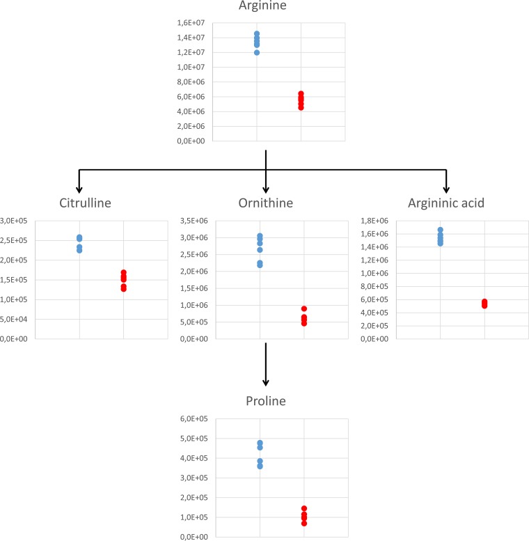
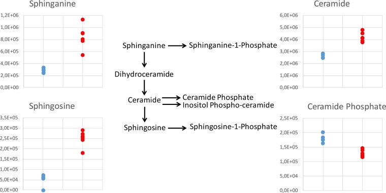
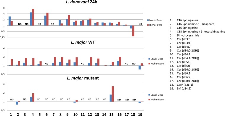
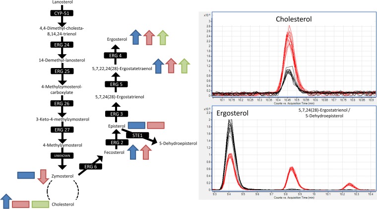
With the World Health Organization reporting over 30,000 deaths and 200,000 to 400,000 new cases annually, visceral leishmaniasis is a serious disease affecting some of the world's poorest people. As drug resistance continues to rise, there is a huge unmet need to improve treatment. Miltefosine remains one of the main treatments for leishmaniasis, yet its mode of action (MoA) is still unknown. Understanding the MoA of this drug and parasite response to treatment could help pave the way for new and more successful treatments for leishmaniasis. A novel method has been devised to study the metabolome and lipidome of Leishmania donovani axenic amastigotes treated with miltefosine. Miltefosine caused a dramatic decrease in many membrane phospholipids (PLs), in addition to amino acid pools, while sphingolipids (SLs) and sterols increased. Leishmania major promastigotes devoid of SL biosynthesis through loss of the serine palmitoyl transferase gene (ΔLCB2) were 3-fold less sensitive to miltefosine than wild-type (WT) parasites. Changes in the metabolome and lipidome of miltefosine-treated L. major mirrored those of L. donovani A lack of SLs in the ΔLCB2 mutant was matched by substantial alterations in sterol content. Together, these data indicate that SLs and ergosterol are important for miltefosine sensitivity and, perhaps, MoA.
Keywords: Leishmania; lipid metabolism; mechanisms of action; metabolomics; miltefosine.
Copyright © 2018 Armitage et al.
Figures
References
-
- Peña I, Pilar Manzano M, Cantizani J, Kessler A, Alonso-Padilla J, Bardera AI, Alvarez E, Colmenarejo G, Cotillo I, Roquero I, de Dios-Anton F, Barroso V, Rodriguez A, Gray DW, Navarro M, Kumar V, Sherstnev A, Drewry DH, Brown JR, Fiandor JM, Julio Martin J. 2015. New compound sets identified from high throughput phenotypic screening against three kinetoplastid parasites: an open resource. Sci Rep 5:8771. doi:10.1038/srep08771. - DOI - PMC - PubMed
-
- Pedrique B, Strub-Wourgaft N, Some C, Olliaro P, Trouiller P, Ford N, Pécoul B, Bradol JH. 2013. The drug and vaccine landscape for neglected diseases (2000–11): a systematic assessment. Lancet Glob Health 1:371–379. - PubMed
-
- Saunders EC, Ng WW, Chambers JM, Ng M, Naderer T, Kro JO, Likic VA, McConville MJ. 2011. Isotopomer profiling of Leishmania mexicana promastigotes reveals important roles for succinate fermentation and aspartate uptake in tricarboxylic acid cycle (TCA) anaplerosis, glutamate synthesis, and growth. J Biol Chem 286:27706–27717. doi:10.1074/jbc.M110.213553. - DOI - PMC - PubMed
Publication types
MeSH terms
Substances
Grants and funding
LinkOut - more resources
Full Text Sources
Other Literature Sources
Research Materials
