. 2018 Apr 19;131(16):1858-1869.
doi: 10.1182/blood-2017-10-812891. Epub 2018 Feb 20.

Neutrophils provide cellular communication between ileum and mesenteric lymph nodes at graft-versus-host disease onset

Affiliations

Neutrophils provide cellular communication between ileum and mesenteric lymph nodes at graft-versus-host disease onset

Jan Hülsdünker et al. Blood. .

Abstract

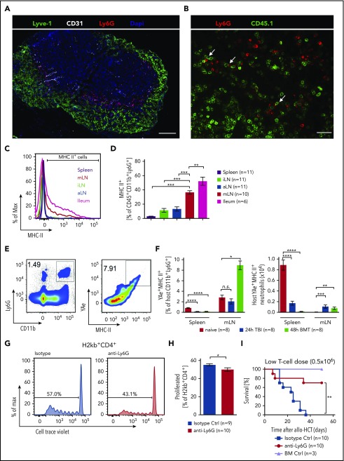
Conditioning-induced damage of the intestinal tract plays a critical role during the onset of acute graft-versus-host disease (GVHD). Therapeutic interference with these early events of GVHD is difficult, and currently used immunosuppressive drugs mainly target donor T cells. However, not donor T cells but neutrophils reach the sites of tissue injury first, and therefore could be a potential target for GVHD prevention. A detailed analysis of neutrophil fate during acute GVHD and the effect on T cells is difficult because of the short lifespan of this cell type. By using a novel photoconverter reporter system, we show that neutrophils that had been photoconverted in the ileum postconditioning later migrated to mesenteric lymph nodes (mLN). This neutrophil migration was dependent on the intestinal microflora. In the mLN, neutrophils colocalized with T cells and presented antigen on major histocompatibility complex (MHC)-II, thereby affecting T cell expansion. Pharmacological JAK1/JAK2 inhibition reduced neutrophil influx into the mLN and MHC-II expression, thereby interfering with an early event in acute GVHD pathogenesis. In agreement with this finding, neutrophil depletion reduced acute GVHD. We conclude that neutrophils are attracted to the ileum, where the intestinal barrier is disrupted, and then migrate to the mLN, where they participate in alloantigen presentation. JAK1/JAK2-inhibition can interfere with this process, which provides a potential therapeutic strategy to prevent early events of tissue damage-related innate immune cell activation and, ultimately, GVHD.


Conflict of interest statement

Conflict-of-interest disclosure: The authors declare no competing financial interests.

Figures

None
Graphical abstract
Figure 1.
Figure 1.
Neutrophil granulocytes selectively form clusters in the ileum after TBI. (A-B) Leukocytes were isolated from duodenum, ileum, and colon from untreated C57BL/6 mice, as well as after TBI, and analyzed by flow cytometry. Neutrophils are defined as viable CD45+CD11b+Ly6G+ cells. (A) Representative plots of neutrophil numbers from untreated samples and 48 hours after TBI. (B) Pooled data from 2 separate experiments showing the percentages of CD11b+Ly6G+ cells of all leukocytes (CD45+) at different points after TBI (n = 6-8). (C) C57BL/6 mice underwent allo-HCT (BALB/c into C57BL/6; 5 × 10⁶ BM + 106 CD4+/CD8+ T cells). Leukocytes were isolated at different points after allo-HCT, and the percentages of CD11b+Ly6G+ cells of all leukocytes (CD45+) are shown (n = 4). (D) Three-dimensional light sheet fluorescence microscopy of ileum 48 hours after TBI showed cluster formation of neutrophils (Ly6G, red; CD11b, green; tissue autofluorescence, blue). (E) Immunofluorescence staining of ileal cross-section 48 hours after TBI showed localization of neutrophils adjacent to intestinal crypts (Collagen IV, green; Ly6G, red; scale bar, 50 µm). (F) Bacterial load in lamina propria of duodenum, ileum, and colon of untreated C57BL/6 mice and C57BL/6 mice 48 hours after TBI was analyzed by quantitative reverse transcription-polymerase chain reaction. 16S rDNA content was normalized to mouse genomic DNA (glyceraldehyde-3-phosphate dehydrogenase). Pooled data from 2 individual experiments are shown (n = 6).
Figure 2.
Figure 2.
Neutrophils migrate from the ileum to the mesenteric lymph node. Mice transgenic for the photoconvertible green-to-red protein Dendra2 in all cells underwent TBI. After 10 or 34 hours, the ileum was exposed to 405-nm light during surgery to convert Dendra2 from its green to the red fluorescent form. Mice were sacrificed 10-14 hours after illumination and the amount of photoconverted cells that had trafficked from ileum to spleen, mLN, ndLN (nondraining lymph node, pooled iLN and aLN), blood, and BM was determined. (A) Illustration of experimental setup. (B) Representative fluorescence image showing successful photoconversion selectively in the ileum after surgery. (C) Representative FACS plots of photoconverted CD45+CD11b+Ly6G+ cells 48 hours after TBI in different organs. Pixel size of dots was increased for better visibility. (D-E) Percentage of photoconverted CD11b+Ly6G+ neutrophils is shown. (D) Photoconversion on day 0, 10 hours post-TBI, and analysis on day 1, 24 hours post-TBI, according to setup A1 (n = 6 for each organ from 2 independent experiments). (E) Photoconversion on day 1, 34 hours post-TBI, and analysis on day 2, 48 hours after TBI according to setup A2 (n = 7 for each organ from 2 independent experiments). (F-G) Neutrophils from the BM of mice transgenic for the photoconvertible green-to-red fluorescence protein Dendra2 were isolated and enriched by MACS purification. The cells were photoconverted ex vivo, and 107 neutrophils were adoptively transferred into C57BL/6 mice 34 hours after they underwent TBI. Fourteen hours after injection and 48 hours after TBI, mice were sacrificed, and photoconverted Dendra2 cells in spleen, mLN, ileum, and blood was analyzed by flow cytometry. (F) Representative FACS plots of photoconverted CD45+CD11b+Ly6G+ cells derived from the spleen. (G) Percentage of photoconverted CD11b+Ly6G+ neutrophils in different organs is shown (n = 8).
Figure 3.
Figure 3.
Neutrophil infiltration in lymphatic organs. (A) Neutrophil counts in spleen, mLN, iLN, and aLN at different points after TBI were analyzed by flow cytometry. Fold change of absolute numbers with regard to untreated mice (day 0) is presented (n = 6 for each point and organ from 2 independent experiments). (B) Giemsa-Wright staining of mLN-derived CD45+CD11b+Ly6G+ neutrophils after FACS sorting and cytospin. (C) Correlation of neutrophil frequency in mLN and ileum from WT or solvent control-treated mice (n = 57). (D) Bacterial load in mLN of untreated C57BL/6 and 48 hours after TBI were analyzed by quantitative reverse transcription-polymerase chain reaction. 16S rDNA content was normalized to genomic DNA (glyceraldehyde-3-phosphate dehydrogenase). (E) Fold change of neutrophil numbers on day 2 after TBI normalized day o d0 (no TBI) in C57BL/6 mice with and without gut decontamination is shown (Control treatment, n = 10; gut decontamination, n = 6 each point and organ from 2-3 independent experiments). (F) C57BL/6 mice were treated daily by subcutaneous injection of aztreonam (75 mg/kg/d) or solvent control from day −7 until day +1 and underwent TBI on day 0. Fold change of neutrophil numbers on day 2 post TBI normalized to mean of solvent controls is shown (n = 11 pooled from 2 independent experiments). (G) BALB/c mice were treated daily by subcutaneous injection of aztreonam (75mg/kg/day) or solvent control from day −7 until day +1 and received allo-HCT (C57BL/6 into BALB/c; 5 × 10⁶ BM cells + 0.3 × 10⁶ CD4+/CD8+ T cells) on day 0, and survival was monitored (n = 20 for each group pooled from 2 independent experiments). (H) C57BL/6 mice received 20 mg/kg doxorubicin on day 0 or were left untreated. The mLN were analyzed on day 3 by flow cytometry to assess neutrophils counts (CD45+CD11b+Ly6G+). Fold change of neutrophil numbers normalized to the mean of untreated samples is shown (n = 10 for each group pooled from 2 independent experiments).
Figure 4.
Figure 4.
Antigen presentation by neutrophils. (A) Immunofluorescence staining of a mLN 48 hours after TBI showing ring-shaped localization of Ly6G+ cells (red) around lymphocytes next to lymphatic vessels (Lyve-1, green; 4′,6-diamidino-2-phenylindole, blue; blood vessels (CD31), white; scale bar, 200 µm). (B) MLN 48 hours after allo-HSCT showing neutrophils (Ly6G+ = red) close to transplanted CD45.1+ T cells (green, scale bar = 150 µm). (C) Representative histogram of MHC-II expressing CD45+CD11b+Ly6G+ neutrophils in spleen, iLN, aLN, mLN, and ileum in C57BL/6 mice 48 hours after TBI. (D) Frequency of MHC-II+ (percentage of CD45+CD11b+Ly6G+) neutrophils in different organs in CD57Bl/6 mice 48 hours after TBI. (E-F) MHC-II expression and peptide presentation (YAe+) on CD11b+Ly6G+ neutrophils in B6D2F1 mice were assessed by flow cytometry in untreated mice, 24 hours after TBI and 48 hours after BM transplantation. (E) Representative FACS plots showing gating strategy. (F, left) Relative numbers of YAe+MHC-II+ cells of host neutrophils. (Right) Absolute numbers of host YAe+MHC-II+ neutrophils. (G-H) For neutrophil depletion, BALB/c mice were injected with either anti-Ly6G (0.5 mg intraperitoneally) or an isotype control (0.5 mg) on day −1 and transplanted after TBI on day 0 with 1 × 10⁷ purified and CellTrace Violet-labeled CD4+/CD8+ T cells from C57BL/6 mice. Mice were sacrificed on day 3 and mLN analyzed for donor-derived (H2kb+) T cells that had proliferated. (G) Representative flow cytometry plots of CellTrace Violet-dilution of H2kb+CD4+ T cells. (H) Pooled data from 2 independent experiments showing proliferated H2kb+CD4+ T cells after anti-Ly6G neutrophils depletion. (I) BALB/c mice were treated on day −1 by anti-Ly6G (0.5 mg) or isotype control antibody (0.5 mg) injection and underwent allo-HCT (FVB into BALB/c; 5 × 10⁶ BM cells + 0.5 × 10⁶ CD4+/CD8+ T cells). BM controls were treated on day 0 by isotype control antibody (0.5 mg) injection and underwent transplantation using BM alone (FVB 5 × 10⁶ cells).
Figure 5.
Figure 5.
JAK1/JAK2 inhibition reduces neutrophils and MHC-II expression. C57BL/6 mice were treated twice daily with 30 mg/kg ruxolitinib starting on day −1 before TBI. On day 2 after TBI, mice were sacrificed and analyzed by flow cytometry. (A) Absolute numbers of neutrophils (CD45+CD11b+Ly6G+) in mLN and ileum. (B) Representative histogram of MHC-II expression on CD45+CD11b+Ly6G+ neutrophils in ileum after ruxolitinib or solvent control treatment. (C) Frequency of MHC-II+ (% of CD45+CD11b+Ly6G+) neutrophils in ileum and mLN after ruxolitinib or solvent control treatment. (D) Fold change of MFI for MHC-II on MHC-II+CD11b+Ly6G+ cells isolated from mLN or ileum. (E) Fold change in absolute MHC-II+CD45+CD11b+Ly6G+ numbers by ruxolitinib treatment normalized to solvent control. (F) Correlation of MHC-II+CD45+CD11b+Ly6G+ neutrophil numbers in mLN vs ileum after ruxolitinib or solvent control treatment.

Comment in

References

    1. Forman SJ, Negrin RS, Antin JH, Appelbaum FR, eds. Thomas’ Hematopoietic Cell Transplantation. 5th ed. Hoboken, NJ: Wiley-Blackwell; 2016.
    1. Gratwohl A, Pasquini MC, Aljurf M, et al. ; Worldwide Network for Blood and Marrow Transplantation (WBMT). One million haemopoietic stem-cell transplants: a retrospective observational study. Lancet Haematol. 2015;2(3):e91-e100. - PubMed
    1. Zeiser R, Blazar BR. Acute Graft-versus-host disease - Biologic process, prevention, and therapy. N Engl J Med. 2017;377(22):2167-2179. - PMC - PubMed
    1. Pasquini MC, Zhu X Current use and outcome of hematopoietic stem cell transplantation. Milwaukee, WI: 2015 CIBMTR Summary Slides; 2014.
    1. Arai S, Arora M, Wang T, et al. ; Graft-vs-Host Disease Working Committee of the CIBMTR. Increasing incidence of chronic graft-versus-host disease in allogeneic transplantation: a report from the Center for International Blood and Marrow Transplant Research. Biol Blood Marrow Transplant. 2015;21(2):266-274. - PMC - PubMed

Publication types

MeSH terms