

Published Erratum
doi: 10.1084/jem.2016107002142018c.
Epub 2018 Feb 20.
Correction: Enzymatic lipid oxidation by eosinophils propagates coagulation, hemostasis, and thrombotic disease
- PMID: 29463570
- PMCID: PMC5839750
- DOI: 10.1084/jem.2016107002142018c
Item in Clipboard
Published Erratum
Correction: Enzymatic lipid oxidation by eosinophils propagates coagulation, hemostasis, and thrombotic disease
J Exp Med.
.
No abstract available
Figures
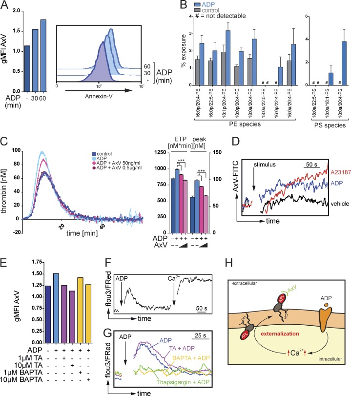
Ca2+-dependent exposure of aminophospholipids by eosinophils promote thrombin generation. (A) Flow cytometry analysis of the binding of annexin V (AxV) to aminophospholipids on the surface of resting or ADP-stimulated mouse eosinophils. Histograms show representative annexin V stainings, and bar graphs show mean geometric fluorescence intensities (gMFI). (B) LC/MS/MS-based quantification of the exposure of the aminophospholipids PE and PS in mouse eosinophils in response to ADP stimulation. (C, left) Calibrated thrombin generation assays with resting or ADP-stimulated mouse eosinophils in the presence of annexin V. (Right) Bar graphs show endogenous thrombin potential (ETP; nM*min) and peak of thrombin generation (peak; nM). (D) Flow cytometry analysis of annexin V binding on mouse eosinophils over time in the presence of calcium ionophore A23187, ADP, or vehicle. (E) Flow cytometry analysis of annexin V binding on mouse eosinophils in the presence of tannic acid (TA) or intracellular Ca2+-chelator BAPTA/AM. Bar graphs show geometric mean fluorescence intensity. (F) Flow cytometry–based analysis of intracellular Ca2+ signaling, indicated by Fluo3/FuraRed ratio, over time in a Ca2+-free environment. Where indicated (arrow and Ca2+), CaCl2 at a final concentration of 1 mM was added. (G) Flow cytometry–based analysis of intracellular Ca2+ signaling, indicated by Fluo3/FuraRed ratio, over time in a Ca2+-free environment. (H) Postulated mechanism of a sequential generation and Ca2+-dependent externalization of aminophospholipids (APL) at the surface of eosinophils. OH indicates hydroxyl group. Data are representative of at least three independent experiments. Error bars represent SEM. *, P < 0.05; ***, P < 0.001.
Erratum for
-
Enzymatic lipid oxidation by eosinophils propagates coagulation, hemostasis, and thrombotic disease.J Exp Med. 2017 Jul 3;214(7):2121-2138. doi: 10.1084/jem.20161070. Epub 2017 May 31. J Exp Med. 2017. PMID: 28566277 Free PMC article.
Publication types
Grants and funding
LinkOut - more resources
Full Text Sources
Other Literature Sources
