. 2018 Apr;59(4):658-669.
doi: 10.1194/jlr.M082651. Epub 2018 Feb 20.

Reduced atherosclerosis lesion size, inflammatory response in miR-150 knockout mice via macrophage effects

Affiliations

Reduced atherosclerosis lesion size, inflammatory response in miR-150 knockout mice via macrophage effects

Fu-Han Gong et al. J Lipid Res. 2018 Apr.

Abstract

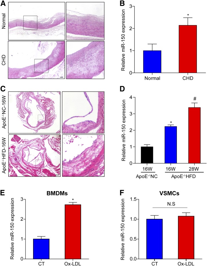
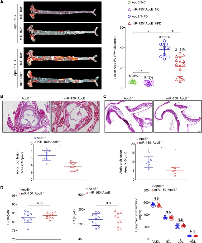
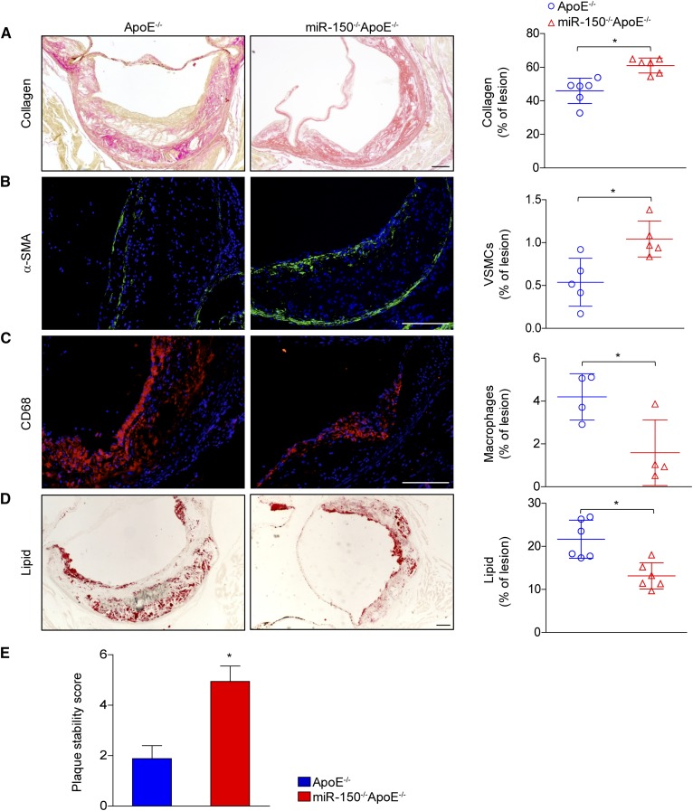
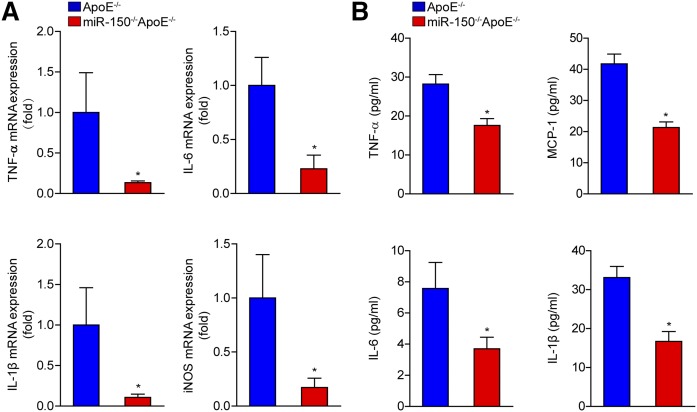
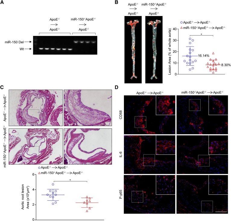
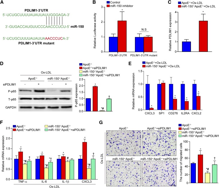
Atherosclerosis is considered to be a chronic inflammatory disease that can lead to severe clinically important cardiovascular events. miR-150 is a small noncoding RNA that significantly enhances inflammatory responses by upregulating endothelial cell proliferation and migration, as well as intravascular environmental homeostasis. However, the exact role of miR-150 in atherosclerosis remains unknown. Here, we investigated the effect of miR-150 deficiency on atherosclerosis development. Using double-knockout (miR-150-/- and ApoE-/-) mice, we measured atherosclerotic lesion size and stability. Meanwhile, we conducted in vivo bone marrow transplantation to identify cellular-level components of the inflammatory response. Compared with mice deficient only in ApoE, the double-knockout mice had significantly smaller atherosclerotic lesions and displayed an attenuated inflammatory response. Moreover, miR-150 ablation promoted plaque stabilization via increases in smooth muscle cell and collagen content and decreased macrophage infiltration and lipid accumulation. The in vitro experiments indicated that an inflammatory response with miR-150 deficiency in atherosclerosis results directly from upregulated expression of the cytoskeletal protein, PDZ and LIM domain 1 (PDLIM1), in macrophages. More importantly, the decreases in phosphorylated p65 expression and inflammatory cytokine secretion induced by miR-150 ablation were reversed by PDLIM1 knockdown. These findings suggest that miR-150 is a promising target for the management of atherosclerosis.

Keywords: PDZ and LIM domain 1; inflammation; microRNA-150.


Conflict of interest statement

The authors declare no conflicts of interest.

Figures

Fig. 1.
Fig. 1.
miR-150 expression is increased in atherosclerotic plaques and macrophages A: H&E-stained right coronary arteries from a donor and a patient with CHD. Scale bar = 200 μm. B: qPCR analysis of miR-150 mRNA expression in human artery tissue samples (n = 4). *P < 0.05 versus normal group. C: H&E-stained aortic roots from ApoE−/− mice treated with NC or a HFD for 16 weeks (16W). Scale bar = 200 μm. D: qPCR analysis of miR-150 mRNA expression in the entire aorta in ApoE−/− mice treated with NC for 16 weeks or a HFD for 16 or 28 weeks (28W) (n = 3). *P < 0.05 versus ApoE−/− with 16W-NC treatment group; #P < 0.05 versus ApoE−/− with 16W-HFD treatment group. E, F: miR-150 mRNA expression in BMDMs or VSMCs stimulated with Ox-LDL. *P < 0.05 versus control (CT) group; N.S indicates not significant.
Fig. 2.
Fig. 2.
miR-150 ablation protects mice from atherosclerosis. A: Oil Red O staining of atherosclerotic lesions in the entire aorta in ApoE−/− and miR-150−/−ApoE−/− mice treated with NC or a HFD for 28 weeks. Quantitative lesion data are shown in the right panel (n = 10–16). *P < 0.05 versus ApoE−/− NC group; #P < 0.05 versus ApoE−/− HFD group. B, C: H&E staining for the morphology of the plaques in the aortic roots (B) and ascending aortic arches (C) of ApoE−/− and miR-150−/−ApoE−/− mice treated with a HFD for 28 weeks (n = 6–12). Scale bar = 200 μm. *P < 0.05 versus ApoE−/− group. D: The lipid metabolism parameters of ApoE−/− and miR-150−/−ApoE−/− mice (n = 10 each group). Not significant (N.S) versus ApoE−/− littermates. TC, total cholesterol; TG, triglyceride.
Fig. 3.
Fig. 3.
miR-150 deficiency increases plaque stability. A–D: Cross-sections of aortic roots from ApoE−/− and miR-150−/−ApoE−/− mice were stained with picrosirius red to assess collagen deposition (A) and α-smooth muscle actin (α-SMA) expression (green) to determine SMC compositions (B). The tissues were also stained with CD68 (red) to assess macrophage infiltration (C) and Oil Red O to assess lipid accumulation (D). The quantitative results for each image are shown in the right panels. E: The assessment of plaque stability score in ApoE−/− and miR-150−/−ApoE−/− mice (n = 4–6). Scale bar = 100 μm.*P < 0.05 versus ApoE−/− group.
Fig. 4.
Fig. 4.
The inflammatory response is inhibited in the absence of miR-150. A: The mRNA expression levels of secreted cytokines, namely, TNF-α, IL-6, IL-1β, and iNOS, in the entire aortas of ApoE−/− and miR-150−/−ApoE−/− mice were measured by qPCR (n = 4). *P < 0.05 versus ApoE−/− group. B: The serum levels of secreted cytokines in ApoE−/− and miR-150−/−ApoE−/− mice (n = 6). *P < 0.05 versus ApoE−/− group.
Fig. 5.
Fig. 5.
miR-150 deficiency in marrow-derived cells attenuates atherosclerosis. A: The efficacy of bone marrow transplantation was determined with genomic DNA isolated from white blood cells after the chimera generation procedure. B: Oil Red O staining of atherosclerotic lesions in the entire aortas of ApoE−/−→ApoE−/− and miR-150−/−ApoE−/−→ApoE−/− mice treated with an HFD for 16 weeks. Quantitative data for the lesions are shown in the right panel (n = 13–17). *P < 0.05 versus ApoE−/−→ApoE−/− group. C: H&E-stained plaques in the aortic roots of ApoE−/−→ApoE−/− and miR-150−/−ApoE−/−→ApoE−/− mice (n = 8–10). Scale bar = 200 μm. D: The immunofluorescence staining of CD68, IL-6, and phosphorylated p65 (P-p65) (red). Scale bar = 100 μm. *P < 0.05 versus ApoE−/−→ApoE−/− group.
Fig. 6.
Fig. 6.
The inflammatory response and macrophage migration are attenuated by miR-150 ablation. A: qPCR analysis of the expression of inflammatory cytokines secreted by BMDMs from miR-150−/−ApoE−/− or ApoE−/− mice stimulated with Ox-LDL. *P < 0.05 versus the ApoE−/− group. B: Western blot analysis of the expression levels of phosphorylated (P)-IκBα and P-p65 in BMDMs from miR-150−/−ApoE−/− or ApoE−/− mice. *P < 0.05 versus the ApoE−/− group. C: MCP-1 mRNA expression in BMDMs from miR-150−/−ApoE−/− or ApoE−/− mice. *P < 0.05 versus the ApoE−/− group. D: Macrophage infiltration ability was tested with transwell experiments and crystal violet staining. Scale bar = 100 μm. *P < 0.05 versus the ApoE−/− group.
Fig. 7.
Fig. 7.
The upregulation of PDLIM1 expression by miR-150 deficiency. A: Potential target sites for miR-150 in the 3′UTR of murine PDLIM1 mRNA (blue). The sequence in the binding region that is highlighted in red was mutated. B: Luciferase reporter assays in HEK293 cells treated with a miR-150 inhibitor or negative control using a pEZX-MT01 vector containing the PDLIM1-3′UTR or the PDLIM1-3′UTR with mutations in the predicted miR-150 binding site. *P < 0.05 versus control group. C: qPCR analysis of PDLIM1 expression in BMDMs from miR-150−/−ApoE−/− or ApoE−/− mice stimulated with Ox-LDL. *P < 0.05 versus the ApoE−/− group. D: Western blot analysis of phosphorylated p65 (P-p65) expression levels in BMDMs from miR-150−/−ApoE−/− or ApoE−/− mice transfected with control or PDLIM1-specific siRNAs before Ox-LDL treatment. *P < 0.05 versus the ApoE−/− group; #P < 0.05 versus the ApoE−/− group treated with PDLIM1-specific siRNA; †P < 0.05 versus the miR-150−/−ApoE−/− group. E: qPCR analysis of the expression of potential target genes in BMDMs from miR-150−/−ApoE−/− or ApoE−/− mice stimulated with Ox-LDL. *P < 0.05 versus the ApoE−/− group. F: Inflammatory cytokine mRNA expression in BMDMs from miR-150−/−ApoE−/− or ApoE−/− mice pretreated with control or PDLIM1-specific siRNA before Ox-LDL treatment. *P < 0.05 versus the ApoE−/− group; #P < 0.05 versus the ApoE−/− group treated with PDLIM1-specific siRNA; †P < 0.05 versus the miR-150−/−ApoE−/− group. G: Macrophage infiltration ability was tested with transwell assays and crystal violet staining. *P < 0.05 versus the ApoE−/− group; #P < 0.05 versus the ApoE−/− group treated with PDLIM1-specific siRNA; †P < 0.05 versus the miR-150−/−ApoE−/− group. Scale bar = 100 μm.

References

    1. Benjamin E. J., Blaha M. J., Chiuve S. E., Cushman M., Das S. R., Deo R., de Ferranti S. D., Floyd J., Fornage M., Gillespie C., et al. 2017. Heart disease and stroke statistics-2017 update: a report from the American Heart Association. Circulation. 135: e146–e603. - PMC - PubMed
    1. Ross R. 1999. Atherosclerosis–an inflammatory disease. N. Engl. J. Med. 340: 115–126. - PubMed
    1. Kim C. H. J., Mitchell J. B., Bursill C. A., Sowers A. L., Thetford A., Cook J. A., van Reyk D. M., and Davies M. J.. 2015. The nitroxide radical TEMPOL prevents obesity, hyperlipidaemia, elevation of inflammatory cytokines, and modulates atherosclerotic plaque composition in apoE-/- mice. Atherosclerosis. 240: 234–241. - PMC - PubMed
    1. Weber C., and von Hundelshausen P.. 2017. CANTOS trial validates the inflammatory pathogenesis of atherosclerosis: setting the stage for a new chapter in therapeutic targeting. Circ. Res. 121: 1119–1121. - PubMed
    1. Kluiver J. L., and Chen C. Z.. 2012. MicroRNAs regulate B-cell receptor signaling-induced apoptosis. Genes Immun. 13: 239–244. - PubMed

Publication types