. 2018 Apr 16;38(9):e00672-17.
doi: 10.1128/MCB.00672-17. Print 2018 May 1.

β1-Integrin Impacts Rad51 Stability and DNA Double-Strand Break Repair by Homologous Recombination

Affiliations

β1-Integrin Impacts Rad51 Stability and DNA Double-Strand Break Repair by Homologous Recombination

Kazi Mokim Ahmed et al. Mol Cell Biol. .

Abstract

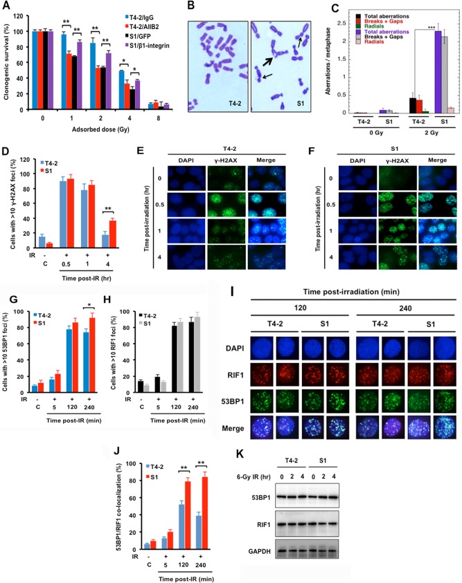
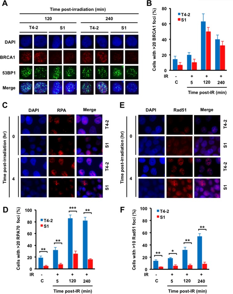
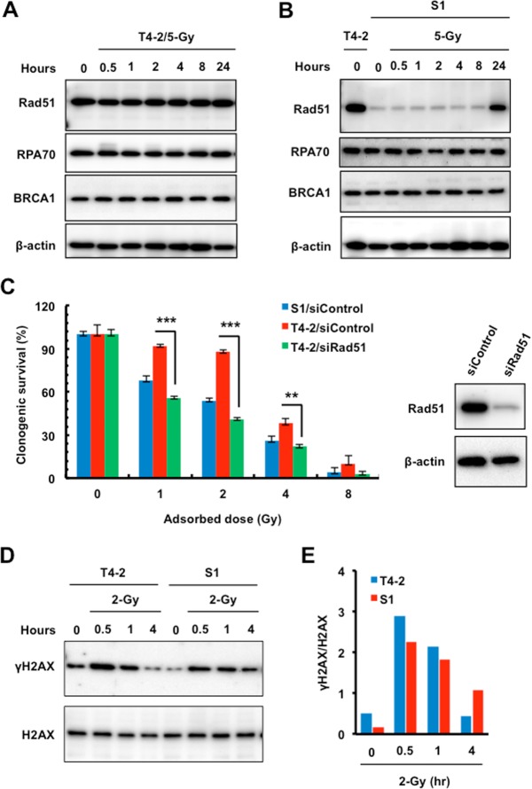
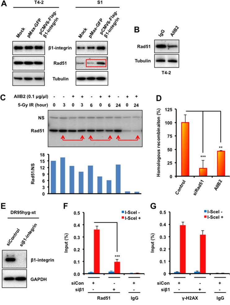
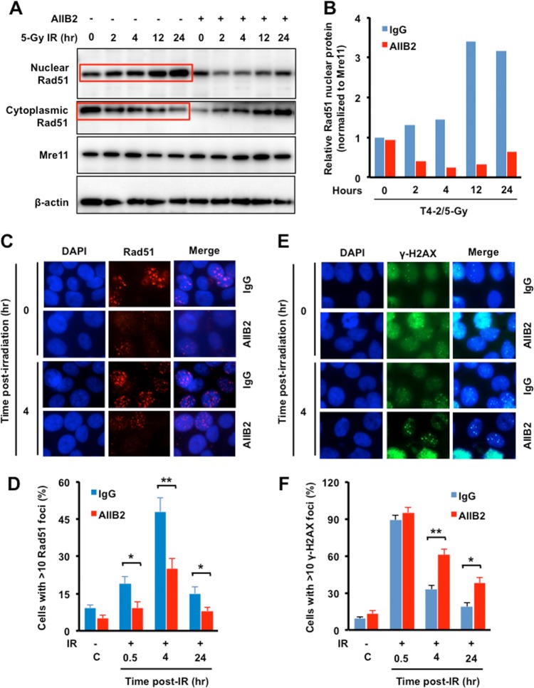
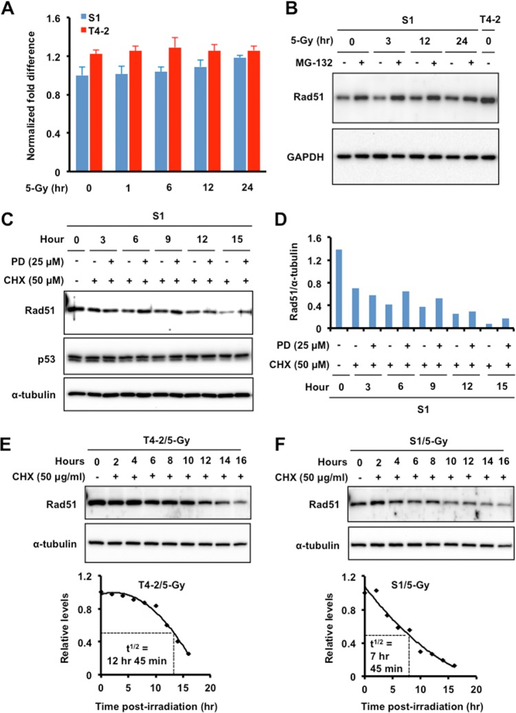
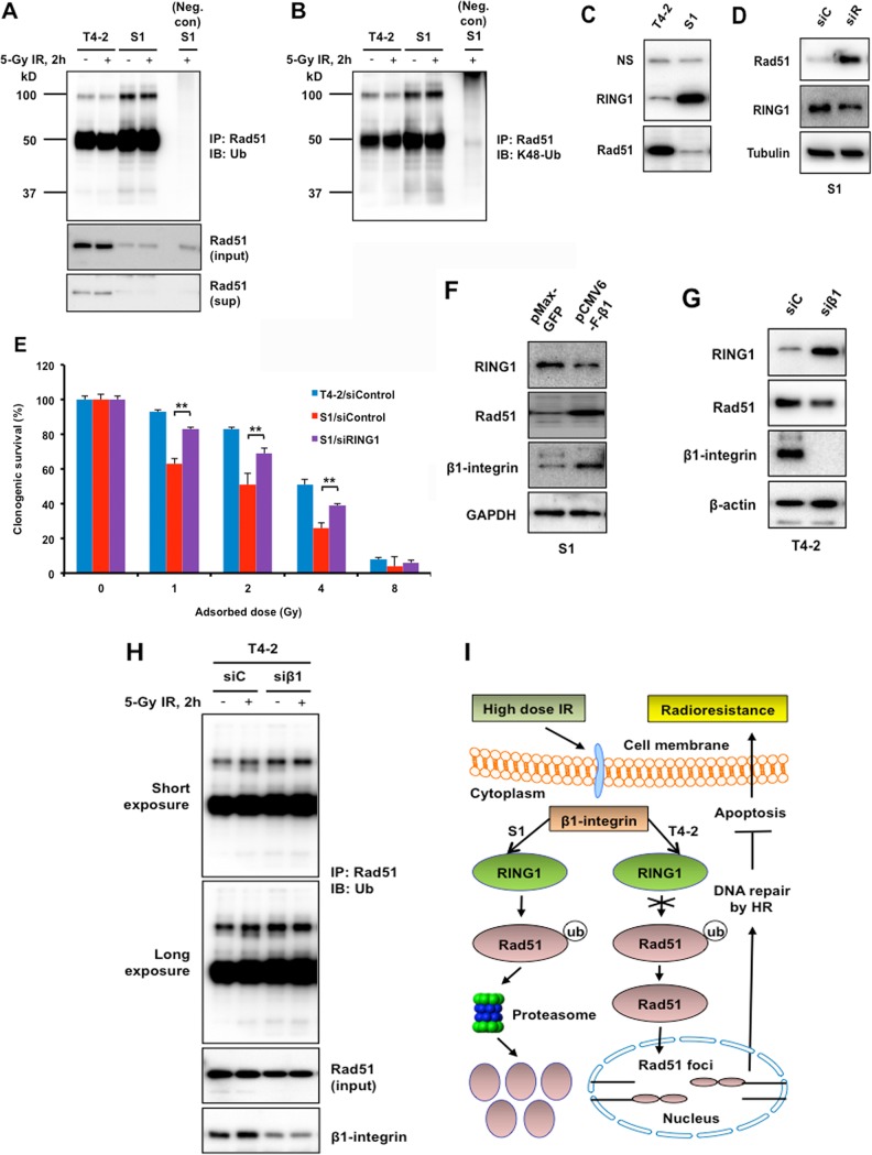
The molecular mechanisms underlying resistance to radiotherapy in breast cancer cells remain elusive. Previously, we reported that elevated β1-integrin is associated with enhanced breast cancer cell survival postirradiation, but how β1-integrin conferred radioresistance was unclear. Ionizing radiation (IR) induced cell killing correlates with the efficiency of DNA double-strand break (DSB) repair, and we found that nonmalignant breast epithelial (S1) cells with low β1-integrin expression have a higher frequency of S-phase-specific IR-induced chromosomal aberrations than the derivative malignant breast (T4-2) cells with high β1-integrin expression. In addition, there was an increased frequency of IR-induced homologous recombination (HR) repairosome focus formation in T4-2 cells compared with that of S1 cells. Cellular levels of Rad51 in T4-2 cells, a critical factor in HR-mediated DSB repair, were significantly higher. Blocking or depleting β1-integrin activity in T4-2 cells reduced Rad51 levels, while ectopic expression of β1-integrin in S1 cells correspondingly increased Rad51 levels, suggesting that Rad51 is regulated by β1-integrin. The low level of Rad51 protein in S1 cells was found to be due to rapid degradation by the ubiquitin proteasome pathway (UPP). Furthermore, the E3 ubiquitin ligase RING1 was highly upregulated in S1 cells compared to T4-2 cells. Ectopic β1-integrin expression in S1 cells reduced RING1 levels and increased Rad51 accumulation. In contrast, β1-integrin depletion in T4-2 cells significantly increased RING1 protein levels and potentiated Rad51 ubiquitination. These data suggest for the first time that elevated levels of the extracellular matrix receptor β1-integrin can increase tumor cell radioresistance by decreasing Rad51 degradation through a RING1-mediated proteasomal pathway.

Keywords: HR; Rad51; breast cancer; homologous recombination; radioresistance; β1-integrin.


Figures

FIG 1
FIG 1
Ionizing radiation (IR)-induced chromosome aberrations and 53BP1/RIF1 cofoci are increased in S1 cells compared to T4-2 cells after IR. (A) Inhibition of β1-integrin in T4-2 cells or ectopic expression of β1-integrin in S1 cells increased or decreased radiosensitivity, respectively. Malignant breast T4-2 cells, derived from the nonmalignant S1 breast epithelial cell line, were left untreated, T4-2 cells were treated with β1-integrin inhibitory antibody AIIB2 (0.1 μg/μl), or S1 cells were transiently transfected with expression vector for β1-integrin (pCMV6-Flag-β1-integrin) or control (pMax-GFP) before exposure to 1, 2, 4, or 8 Gy X rays. Clonogenic survival was measured 14 days after IR. Colonies consisting of more than 50 cells were scored as surviving colonies and normalized against nonirradiated clones. (B and C) Higher frequencies of chromosome aberrations at metaphase post-IR occurred in S1 cells than in T4-2 cells. Metaphase chromosome aberrations were determined in S phase of the cell cycle in cells exposed to 2 Gy X rays. (B) Thick arrow, breaks and gaps; thin arrows, radials. (C) Histogram of S-phase aberrations in T4-2 and S1 cells sham irradiated or exposed to 2 Gy of IR. (D to F) Delayed disappearance of γ-H2AX foci post-IR in S1 cells. Exponentially growing T4-2 and S1 cells were treated with 2 Gy X rays, fixed post-IR, and immunostained for γ-H2AX (histogram of >10 γ-H2AX foci). DAPI, 4′,6-diamidino-2-phenylindole. (G to J) Recruitment of IR-induced 53BP1/RIF1 foci is reduced in T4-2 cells but not in S1 cells. (G to I) Cells were treated with 6 Gy X rays, fixed post-IR, and immunostained for 53BP1 and RIF1. (I and J) Coimmunostaining for 53BP1 and RIF1 was done for fixed cells post-IR. 53BP1/RIF1 foci were counted for 3 sets of 30 cells, and the percentage of colocalized 53BP1/RIF1 foci was calculated relative to the total number of foci, i.e., 53BP1 plus RIF1 foci. (K) Western analysis of 53BP1 and RIF1 in whole-cell lysates prepared from T4-2 and S1 cells sham irradiated or exposed to 6 Gy X rays (GAPDH as a loading control). (A, C, D, G, H, and J) Columns represent the means (n = 3), and bars represent the SDs; *, P < 0.05; **, P < 0.01; ***, P < 0.001.
FIG 2
FIG 2
HR is increased in T4-2 malignant breast epithelial cells compared to that in S1 cells. (A and B) T4-2 and S1 cells were irradiated with 6 Gy X rays, fixed post-IR, and coimmunostained with 53BP1 and BRCA1 antibodies. (A) 53BP1 and BRCA1 staining; (B) histogram of BRCA1 foci. (C and E) Cells were treated with 6 Gy X rays, fixed post-IR, and immunostained for RPA70 (C) and Rad51 (E) antibodies. (D and F) Quantification of focus formation. (D) Histogram of >20 RPA70 foci; (F) histogram of >10 Rad51 foci. (B, D, and F) Columns represent the means (n = 3), and bars represent the SDs; *, P < 0.05; **, P < 0.01; ***, P < 0.001.
FIG 3
FIG 3
Inhibition of Rad51 increased malignant breast cancer T4-2 cell radiosensitivity. (A and B) Western analysis of Rad51, BRCA1, and RPA70 using whole-cell lysates prepared from T4-2 and S1 cells sham irradiated or exposed to 5 Gy X rays (β-actin as a loading control). (C) Radiosensitivity assay of T4-2 and S1 cells treated with control siRNA or T4-2 cells treated with Rad51 siRNA before exposure to 1, 2, 4, or 8 Gy X rays. Clonogenic survival was measured 14 days after IR. Colonies consisting of more than 50 cells were scored as surviving colonies and normalized against nonirradiated clones (n = 3, mean ± SD; **, P < 0.01; ***, P < 0.001). Right panel, Western blot analysis of the expression of Rad51 using whole-cell lysates prepared from T4-2 cells electroporated with control or Rad51 siRNA. β-Actin served as an internal loading control. (D and E) Western analysis of γ-H2AX in T4-2 and S1 cells sham irradiated or exposed to 2 Gy X rays. (D) The lysates were also blotted for H2AX as an internal loading control. (E) Relative expression levels of γ-H2AX normalized to the expression levels of H2AX.
FIG 4
FIG 4
β1-Integrin regulates Rad51 protein levels and recruitment to DSB sites and promotes HR. (A) T4-2 and S1 cells were electroporated with mammalian expression vectors encoding Flag-tagged β1-integrin or with GFP as a control. Cells were lysed 48 h following electroporation and subjected to immunoblotting with β1-integrin and Rad51 antibodies. Tubulin served as an internal loading control. (B and C) Western blot analysis of Rad51 in whole-cell lysates prepared from malignant breast cancer T4-2 cells treated with the β1-integrin inhibitory monoclonal antibody AIIB2 (0.1 μg/μl) or nonspecific rat IgG before exposure to IR (tubulin served as a loading control). (D) HR was assessed using a DR-GFP reporter assay, as described in Materials and Methods, using T4-2 cells electroporated with control siRNA or Rad51 siRNA or treated with AIIB2 or IgG (“control” represents both control siRNA and IgG). (E) DR95hyg-xt cells were electroporated with either control siRNA or β1-integrin siRNA and harvested after 48 h. Cell lysates were immunoblotted for the indicated proteins. (F and G) ChIP analysis of Rad51 (F) and γ-H2AX (G) on a unique DSB induced by I-SceI in vivo in DR95hyg-xt cells treated with control siRNA (siCon) or β1-integrin siRNA (siβ1), before and after DSB induction by I-SceI transfection. Real-time PCR on ChIP samples used primers directed at nucleotides 94 to 378 from the DSB. The enrichment of Rad51 and γ-H2AX after induction of the DSB was compared with that of an IgG control. (D, F, and G) Columns represent the means (n = 3), and bars represent the SDs; **, P < 0.01; ***, P < 0.001.
FIG 5
FIG 5
β1-Integrin promotes IR-induced nuclear accumulation of Rad51 and focus formation in irradiated cells. (A and B) Increased nuclear Rad51 levels in irradiated cells. (A) Western blotting was performed with nuclear and cytosolic Rad51 of T4-2 cells treated with AIIB2 for 16 h before exposure to 5 Gy X rays (Mre11 and β-actin are markers for nuclear and cytosolic fractions). (B) Relative amounts of nuclear Rad51 shown in panel A were estimated by densitometry. (C and E) Decreases in Rad51 foci by AIIB2 treatment were associated with an increase in γ-H2AX foci in irradiated cells. T4-2 cells were treated with AIIB2 before exposure to 6 Gy X rays, fixed post-IR, and immunostained for Rad51 (C) and γ-H2AX (E) antibodies. (D and F) The percentages of cells with more than 10 Rad51 (D) or γ-H2AX (F) foci are represented. (B, D, and F) Columns represent the means (n = 3), and bars represent the SDs; *, P < 0.05; **, P < 0.01.
FIG 6
FIG 6
Regulation of Rad51 protein levels is predominantly through the ubiquitin proteasomal pathway. (A) Quantitative reverse transcription-PCR analysis of β1-integrin mRNA expression in T4-2 and S1 cells exposed to 5 Gy X rays. GAPDH served as an internal control. (B) Proteasomal inhibitor MG-132 (carbobenzoxy-Leu-Leu-leucinal) blocks the degradation of Rad51. Western blot analysis of the expression of Rad51 was done using whole-cell lysate prepared from S1 cells treated with MG-132 (20 μM) or vehicle (dimethyl sulfoxide [DMSO]; GAPDH as a loading control and whole-cell lysate of T4-2 cells as a control for the expression level of Rad51). (C and D) A specific synthetic inhibitor of calpain, PD150606 (PD), blocks the degradation of Rad51. (C) Western blot analysis of Rad51 expression was performed using whole-cell lysate prepared from S1 cells treated with cycloheximide (CHX; 50 μg/ml) to inhibit protein synthesis or not treated with CHX and then treated with PD150606 or vehicle (DMSO; α-tubulin as a loading control and p53 as a negative or system control). (D) Relative expression levels of Rad51 normalized to the expression levels of α-tubulin. (E and F) Half-lives (t1/2) of Rad51 in T4-2 (E) and S1 (F) cells. Western blot analysis of Rad51 expression was performed using whole-cell lysate prepared from S1 cells treated with CHX (50 μg/ml) or not treated with CHX before exposure to 5 Gy X rays (α-tubulin as the loading control). Bottom panels, densitometric analysis of Rad51 normalized with α-tubulin.
FIG 7
FIG 7
β1-Integrin regulates Rad51 ubiquitination and Rad51 protein levels by E3 ubiquitin-protein ligase RING1. (A and B) T4-2 and S1 cells were left untreated or treated with 5 Gy of IR. Cells were lysed under denaturing conditions. Rad51 was immunoprecipitated (IP), and immunoblots (IB) were probed with panubiquitin (A) and K48-Ub (B) antibodies. Positions and sizes in kilodaltons of marker proteins are shown on the left. (A) Lower panels show the Rad51 before immunoprecipitation (input) and Rad51 in the supernatant postimmunoprecipitation (sup). Neg. con, negative control. (C and D) Western blot analysis of RING1 and Rad51 expression using whole-cell lysate prepared from T4-2 and S1 cells (C) and from S1 cells electroporated with control siRNA (siC) or Rad51 siRNA (siR) (D). Nonspecific bands (NS) and α-tubulin served as loading controls. (E) Inhibition of RING1 increased radioresistance in S1 cells. Malignant breast T4-2 cells and the nonmalignant S1 breast epithelial cell counterparts were treated with control siRNA or the S1 cells were treated with RING1 siRNA before exposure to 1, 2, 4, or 8 Gy X rays. Clonogenic survival was measured 14 days post-IR. Colonies consisting of more than 50 cells were scored as surviving colonies and normalized against nonirradiated clones (n = 3, mean ± SD; **, P < 0.01). (F and G) Western blot analysis of RING1, Rad51, and β1-integrin expression using whole-cell lysate prepared from S1 cells electroporated with mammalian expression vector for β1-integrin or GFP (control) (F) and from T4-2 cells electroporated with control siRNA (siC) or β1-integrin siRNA (siβ1) (G). GAPDH and β-actin served as loading controls. (H) T4-2 cells, electroporated with either control siRNA (siC) or β1-integrin siRNA (siβ1), were left untreated or were treated with 5 Gy of IR. Cells were lysed under denaturing conditions. Rad51 was immunoprecipitated, and blots were probed with panubiquitin antibody. Lower panels show the Rad51 (input) and β1-integrin before immunoprecipitation. (I) Model of a novel strategy to enhance the efficacy of radiotherapy in breast cancer patients.

References

    1. Hynes RO. 2002. Integrins: bidirectional, allosteric signaling machines. Cell 110:673–687. doi: 10.1016/S0092-8674(02)00971-6. - DOI - PubMed
    1. Zahir N, Weaver VM. 2004. Death in the third dimension: apoptosis regulation and tissue architecture. Curr Opin Genet Dev 14:71–80. doi: 10.1016/j.gde.2003.12.005. - DOI - PubMed
    1. Maser RS, Choudhury B, Campbell PJ, Feng B, Wong KK, Protopopov A, O'Neil J, Gutierrez A, Ivanova E, Perna I, Lin E, Mani V, Jiang S, McNamara K, Zaghlul S, Edkins S, Stevens C, Brennan C, Martin ES, Wiedemeyer R, Kabbarah O, Nogueira C, Histen G, Aster J, Mansour M, Duke V, Foroni L, Fielding AK, Goldstone AH, Rowe JM, Wang YA, Look AT, Stratton MR, Chin L, Futreal PA, DePinho RA. 2007. Chromosomally unstable mouse tumours have genomic alterations similar to diverse human cancers. Nature 447:966–971. doi: 10.1038/nature05886. - DOI - PMC - PubMed
    1. Shibue T, Weinberg RA. 2009. Integrin beta1-focal adhesion kinase signaling directs the proliferation of metastatic cancer cells disseminated in the lungs. Proc Natl Acad Sci U S A 106:10290–10295. doi: 10.1073/pnas.0904227106. - DOI - PMC - PubMed
    1. Sethi T, Rintoul RC, Moore SM, MacKinnon AC, Salter D, Choo C, Chilvers ER, Dransfield I, Donnelly SC, Strieter R, Haslett C. 1999. Extracellular matrix proteins protect small cell lung cancer cells against apoptosis: a mechanism for small cell lung cancer growth and drug resistance in vivo. Nat Med 5:662–668. doi: 10.1038/9511. - DOI - PubMed

Publication types