. 2018 Apr 16;38(9):e00107-17.
doi: 10.1128/MCB.00107-17. Print 2018 May 1.

Ligand-Dependent Corepressor (LCoR) Is a Rexinoid-Inhibited Peroxisome Proliferator-Activated Receptor γ-Retinoid X Receptor α Coactivator

Affiliations

Ligand-Dependent Corepressor (LCoR) Is a Rexinoid-Inhibited Peroxisome Proliferator-Activated Receptor γ-Retinoid X Receptor α Coactivator

Tali Shalom-Barak et al. Mol Cell Biol. .

Abstract

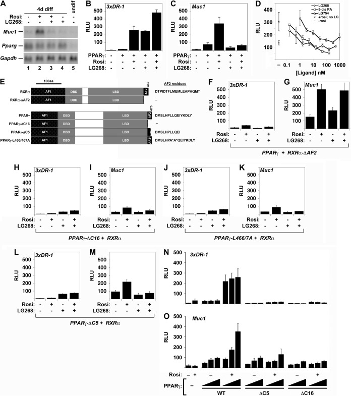
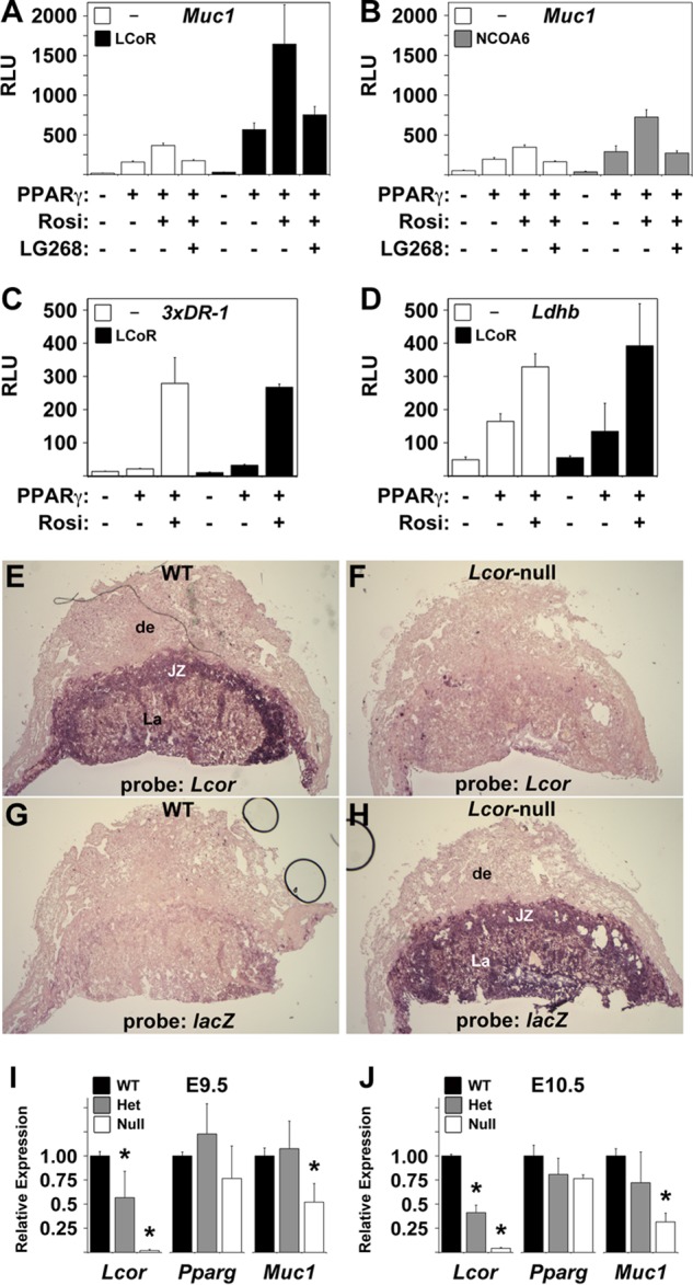
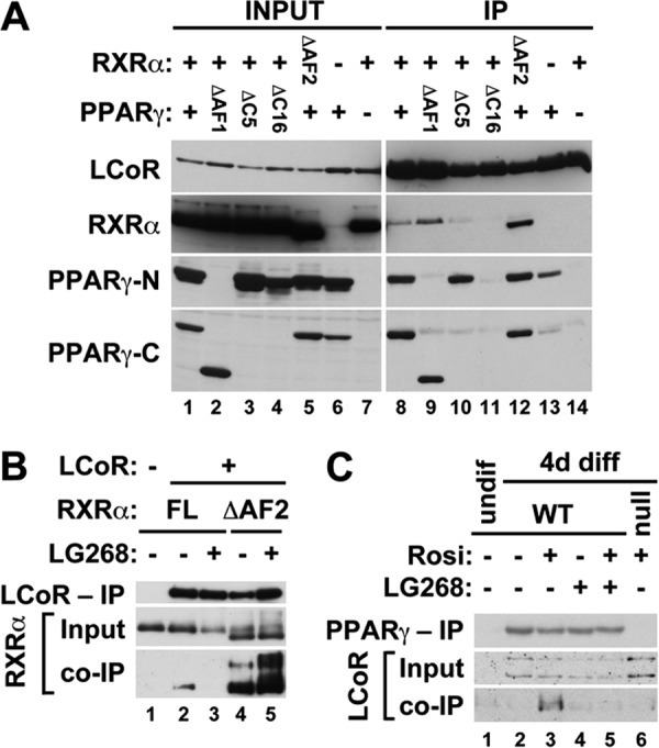
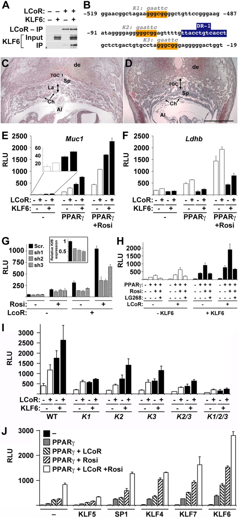
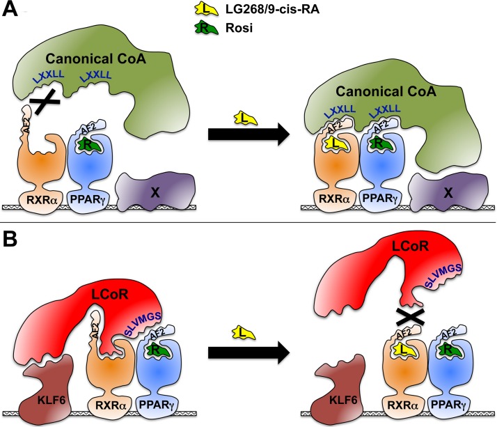
The nuclear receptor peroxisome proliferator-activated receptor gamma (PPARγ) is an essential regulator of placental development. To gain deeper insights into placental PPARγ signaling, we dissected its regulation of the Muc1 promoter. We find that, unlike prototypic target activation by heterodimeric receptors, which is either stimulated by or refractory to retinoid X receptor (RXR) ligands (rexinoids), the induction of Muc1 by liganded PPARγ requires RXRα but is inhibited by rexinoids. We demonstrate that this inhibition is mediated by the activation function 2 (AF2) domain of RXRα and that Muc1 activation entails altered AF2 structures of both PPARγ and RXRα. This unique regulation of Muc1 reflects specific coactivation of PPARγ-RXRα heterodimers by the transcription cofactor ligand-dependent corepressor (LCoR), corroborated by significant downregulation of Muc1 in Lcor-null placentas. LCoR interacts with PPARγ and RXRα in a synergistic fashion via adjacent noncanonical protein motifs, and the AF2 domain of ligand-bound RXRα inhibits this interaction. We further identify the transcription factor Krüppel-like factor 6 (KLF6) as a critical regulator of placental development and a component of Muc1 regulation in cooperation with PPARγ, RXRα, and LCoR. Combined, these studies reveal new principles and players in nuclear receptor function in general and placental PPARγ signaling in particular.

Keywords: KLF6; LCoR; PPARgamma; RXR; coactivators; rexinoids; transcription regulation.


Figures

FIG 1
FIG 1
Effects of rexinoids and RXRα and PPARγ AF2 domain mutations on Muc1 promoter activity. (A) Northern blot analysis of Muc1, Pparg, and Gapdh (normalization control) in cultures of the TSC line GFP-Trf differentiated for 4 days (4d diff) without treatment or in the presence of 1 μM Rosi, 1 μM LG268, or both, as well as an undifferentiated culture (undif), as indicated. (B) Relative light units (RLU) in extracts of CV1 cells transiently transfected with pCMX-βGAL, a 3xDR1-luciferase (3xDR1-luc) construct, and RXRα alone versus RXRα and PPARγ with the indicated combinations of Rosi and LG268, normalized to β-galactosidase activity. (C) Same as described for panel B, with a Muc1-luc construct instead of 3xDR1-luc. (D) Normalized RLU in CV1 cells transfected with pCMX-βGAL, Muc1-luc, RXRα, and PPARγ and treated with 1 μM Rosi and incremental concentrations of the distinct rexinoids LG268, 9-cis retinoic acid, and LG754, from 0.3 nM to 1 μM. Left, mean basal RLU ± standard error (SE) without RXR ligands: bottom, no Rosi (–); top, 1 μM Rosi (×). Right, mean RLU ± SE of experimental data. (E) Summary of RXRα and PPARγ AF2 domain mutants used. Exact AF2 residues present in each species are shown. (F to M) Normalized RLU in CV1 cells transfected with pCMX-βGAL, either 3xDR1-luc (F, H, J, L) or Muc1-luc (G, I, K, M), and the indicated RXRα or PPARγ combinations: full-length (FL)-PPARγ + RXRα-ΔAF2 (F, G), PPARγ-ΔC16 + FL-RXRα (H, I), PPARγ-L466A/L467A + FL-RXRα (J, K), and PPARγ-ΔC5 + WT RXRα (L, M). The results shown in panels B, C, and F to M are part of a transfection series performed side by side and are directly comparable. (N, O) Dose-dependent effects of FL-PPARγ, PPARγ-ΔC5, and PPARγ-ΔC16 on the Muc1 and DR1 reporters. Normalized RLU in CV1 cells transfected with pCMX-βGAL, RXRα, and 3xDR1-luc (N) or Muc1-luc (O) and one to three quanta of the indicated PPARγ AF2 domain configurations (WT, ΔC5, and ΔC16), incubated in the absence or presence of 1 μM Rosi, as indicated; a filler plasmid (pCMX-GAL4N) was used to equalize the DNA concentration in reaction mixtures containing less than three quanta of the PPARγ variants. Bars and error bars show mean values and SE.
FIG 2
FIG 2
LCoR is a Muc1-specific PPARγ coactivator. (A, B) LCoR strongly coactivates PPARγ on the Muc1 promoter. Normalized RLU in CV1 cells transfected with pCMX-βGAL, Muc1-luc, and RXRα, with or without PPARγ as indicated, and treated with combinations of Rosi and LG268 (1 μM each), as marked, with or without cotransfection of LCoR (A) or NCOA6 (B). (C, D) LCoR has no effect on a 3xDR1 reporter or the Ldhb promoter. Same as described for panel A, with 3xDR1-luc (C) or Ldhb(2.7Kb)-luc (D) instead of Muc1-luc. (E to H) Lcor is expressed abundantly in all mouse placental layers. ISH of E11.5 WT (E, G) and Lcor-null (F, H) placentas with antisense riboprobes for Lcor (E, F) or the null-specific lacZ knock-in (G, H). de, decidua; JZ, junctional zone; La, labyrinthine layer. A similar staining pattern is observed at E16.5 (not shown). (I, J) Muc1 is significantly downregulated in Lcor-null placentas. RT-qPCR of Lcor, Pparg, and Muc1 in three or four litter-matched pools of three WT, Lcor+/−, or Lcor-null placentas each at E9.5 (I) or three or four individual placentas of each genotype at E10.5 (J). *, values that differ from the respective WT placentas in a statistically significant manner (P < 0.05). Bars and error bars show mean values and SE.
FIG 3
FIG 3
Effects of AF2 domain mutations and ligands on physical interactions of PPARγ and RXRα with LCoR. (A) Extracts of 293T cells cotransfected with N-terminally Flag-tagged LCoR and the indicated RXRα and/or PPARγ mutant combinations were resolved without IP (INPUT) or after IP with anti-Flag Ab beads (IP) and probed using Abs against Flag, RXRα, and the N or C terminus of PPARγ; the last two were used to detect PPARγ species lacking the opposite termini. (B) CV1 cells were grown in stripped serum and cotransfected with full-length (FL) RXRα alone (lane 1) or with Flag-LCoR and either FL-RXRα (lanes 2 and 3) or RXRα-ΔAF2 (lanes 4 and 5) in the absence (lanes 2 and 4) or presence (lanes 3 and 5) of LG268. Extracts were resolved without IP (Input) or after IP with anti-Flag Ab beads (top and bottom) and probed with anti-Flag (top) or anti-RXRα (middle, bottom) Abs. (C) TSC (gy11, WT, lanes 1 to 5, and gy9, Pparg null, lane 6) were cultured in the presence (lane 1) or absence (lanes 2 to 6) of fibroblast growth factor 4 (FGF4), heparin, and conditioned medium, along with combinations of Rosi and LG268, as indicated. After 4 days of differentiation, nuclear extracts were resolved without IP (Input, 5 μg) or after IP with anti-PPARγ Ab in the presence of the respective ligands (280 μg, top and bottom). Blots were probed with anti-PPARγ (top) or anti-LCoR (middle and bottom) Ab.
FIG 4
FIG 4
Mapping the PPARγ and RXRα interaction domains of LCoR. (A) Bar representation of the LCoR mutants used for the mapping. Locations of functional motifs are shown, including the canonical nuclear receptor box (NR), the tandem CtBP-binding motifs (CtBP), the putative helix-loop-helix sequence (HLH), and the deduced PPARγ- and RXRα-binding region (black box, aa 265 to 285). FL, full-length LCoR; NR*, NR box mutant; t355 to t203, mutants with truncation of all amino acids downstream from the indicated position. Internal deletions and point mutations are indicated. Amino acid sequences are spelled out in the corresponding locations of the NR and PPARγ/RXRα interaction boxes. (B to K) Extracts of 293T cells cotransfected with the indicated LCoR mutants along with either PPARγ (B to F) or RXRα-ΔAF2 (G to K) were resolved and blotted without IP (Input, middle) or after IP with anti-Flag Ab beads (top and bottom). Blots were probed with anti-Flag Ab (top), anti-PPARγ Ab (B to F, middle and bottom), or anti-RXRα Ab (G to K, middle and bottom). (B) Black arrowheads identify the presumptive unprocessed LCoR species translated from the respective constructs, a rightward white arrow points to the ∼270-aa-long cleavage products of LCoR-t319 and LCoR-t300, and a leftward white arrow points to its ∼380-aa-long deduced conjugate. Identically processed species or, where applicable, their internally deleted versions can be observed in the remaining panels, most robustly with C-terminal truncations of LCoR between aa 282 and 319. Additional LCoR breakdown products of other sizes appear in various extracts sporadically but inconsistently.
FIG 5
FIG 5
KLF6 regulates placental development and cooperates with PPARγ, RXRα, and LCoR in Muc1 induction. (A) Extracts of 293T cells cotransfected with the indicated combinations of Flag-LCoR and KLF6 were resolved without IP (Input, middle) or after IP with anti-Flag Ab beads (top, bottom) and probed using Abs against Flag (top) or KLF6 (middle, bottom). (B) Putative SP1/KLF-binding sites (orange boxes) within the minimal PPARγ-responsive sequences of the Muc1 promoter. Sequences designated K1, K2, and K3 delineate the alterations of the three mutant KLF/SP motifs analyzed in the experiments whose results are shown in panel H. The blue box marks the previously determined proximal PPRE (9). (C and D) Midsections of WT (C) and Klf6-null placentas at E9.5 (D) were stained with hematoxylin and eosin. Both WT and Klf6-null placentas have undergone allantoic (Al) fusion, but the Klf-null placenta is completely devoid of the vascular labyrinth (La in panel C; none in panel D) and exhibits aberrant expansion of the spongiotrophoblast (Sp) and, particularly, the trophoblast giant cell (TGC) layer. Ch, chorion; de, decidua. (E, F) Normalized RLU in CV1 cells transfected with pCMX-βGAL and RXRα (all), as well as PPARγ, LCoR, and/or KLF6 where indicated, and treated with 1 μM Rosi as marked, along with either Muc1-luc (E) or Ldhb-luc (F). (G) Normalized RLU in CV1 cells transfected with pCMX-βGAL, Muc1-luc, RXRα and PPARγ (all), with or without LCoR, as labeled, in the presence or absence of 1 μM Rosi, as marked, and a plasmid expressing the indicated control, scramble shRNA (Scr). or one of three CV1 Klf6-specific shRNA molecules. Inset, RT-qPCR analysis of endogenous Klf6 in CV1 cells transfected with a GFP-expressing vector along with a control or Klf6-specific shRNA construct, as indicated, and enriched to approximately 50 to 60% via 8 days of selection with puromycin. (H) Normalized RLU in CV1 cells transfected with pCMX-βGAL, Muc1-luc, and RXRα (all), as well as PPARγ and/or LCoR, as labeled, and treated with 1 μM Rosi, alone or with 1 μM LG268, as marked, in the absence or presence of KLF6. (I) Normalized RLU in CV1 cells transfected with pCMX-βGAL, RXRα, and PPARγ and treated with 1 μM Rosi (all), as well as LCoR and/or KLF6, as labeled, and Muc1 promoter variants carrying the indicated single and combined KLF/SP motif mutations. (J) Normalized RLU in CV1 cells transfected with pCMX-βGAL, Muc1-luc, and RXRα (all), as well as PPARγ and/or LCoR, as labeled, with or without 1 μM Rosi, as marked, and the indicated members of the SP/KLF family. Bars and error bars show mean values and SE.
FIG 6
FIG 6
Model of LCoR as a rexinoid-inhibited PPARγ/RXRα coactivator. A schematic comparison of rexinoid effects on the interactions of PPARγ/RXRα-occupied promoter complexes with canonical coactivators (A) versus LCoR (B). See Discussion for details.

References

    1. Kliewer SA, Umesono K, Noonan DJ, Heyman RA, Evans RM. 1992. Convergence of 9-cis retinoic acid and peroxisome proliferator signalling pathways through heterodimer formation of their receptors. Nature 358:771–774. doi: 10.1038/358771a0. - DOI - PMC - PubMed
    1. Forman BM, Tontonoz P, Chen J, Brun RP, Spiegelman BM, Evans RM. 1995. 15-Deoxy-δ 12,14-prostaglandin J2 is a ligand for the adipocyte determination factor PPARγ. Cell 83:803–812. doi: 10.1016/0092-8674(95)90193-0. - DOI - PubMed
    1. Lehmann JM, Moore LB, Smith-Oliver TA, Wilkison WO, Willson TM, Kliewer SA. 1995. An antidiabetic thiazolidinedione is a high affinity ligand for peroxisome proliferator-activated receptor gamma (PPAR gamma). J Biol Chem 270:12953–12956. doi: 10.1074/jbc.270.22.12953. - DOI - PubMed
    1. Tontonoz P, Hu E, Spiegelman BM. 1994. Stimulation of adipogenesis in fibroblasts by PPAR gamma 2, a lipid-activated transcription factor. Cell 79:1147–1156. doi: 10.1016/0092-8674(94)90006-X. - DOI - PubMed
    1. Barak Y, Nelson MC, Ong ES, Jones YZ, Ruiz-Lozano P, Chien KR, Koder A, Evans RM. 1999. PPARγ is required for placental, cardiac, and adipose tissue development. Mol Cell 4:585–595. doi: 10.1016/S1097-2765(00)80209-9. - DOI - PubMed

Publication types

MeSH terms