. 2018 Feb 20;9(1):e02419-17.
doi: 10.1128/mBio.02419-17.

Novel Segment- and Host-Specific Patterns of Enteroaggregative Escherichia coli Adherence to Human Intestinal Enteroids

Affiliations

Novel Segment- and Host-Specific Patterns of Enteroaggregative Escherichia coli Adherence to Human Intestinal Enteroids

Anubama Rajan et al. mBio. .

Abstract

Enteroaggregative Escherichia coli (EAEC) is an important diarrheal pathogen and a cause of both acute and chronic diarrhea. It is a common cause of pediatric bacterial diarrhea in developing countries. Despite its discovery in 1987, the intestinal tropism of the pathogen remains unknown. Cell lines used to study EAEC adherence include the HEp-2, T-84, and Caco-2 lines, but they exhibit abnormal metabolism and large variations in gene expression. Animal models either do not faithfully manifest human clinical symptoms or are cumbersome and expensive. Using human intestinal enteroids derived from all four segments of the human intestine, we find that EAEC demonstrates aggregative adherence to duodenal and ileal enteroids, with donor-driven differences driving a sheet-like and layered pattern. This contrasts with the colon, where segment-specific tropisms yielded a mesh-like adherence pattern dominated by interconnecting filaments. Very little to no aggregative adherence to jejunal enteroids was observed, regardless of the strain or donor, in contrast to a strong duodenal association across all donors and strains. These unique patterns of intestinal segment- or donor-specific adherence, but not the overall numbers of associated bacteria, were dependent on the major subunit protein of aggregative adherence fimbriae II (AafA), implying that the morphology of adherent clusters and the overall intestinal cell association of EAEC occur by different mechanisms. Our results suggest that we must give serious consideration to inter- and intrapatient variations in what is arguably the first step in pathogenesis, that of adherence, when considering the clinical manifestation of these infections.IMPORTANCE EAEC is a leading cause of pediatric bacterial diarrhea and a common cause of diarrhea among travelers and immunocompromised individuals. Heterogeneity in EAEC strains and lack of a good model system are major roadblocks to the understanding of its pathogenesis. Utilizing human intestinal enteroids to study the adherence of EAEC, we demonstrate that unique patterns of adherence are largely driven by unidentified factors present in different intestinal segments and from different donors. These patterns are also dependent on aggregative adherence fimbriae II encoded by EAEC. These results imply that we must also consider the contribution of the host to understand the pathogenesis of EAEC-induced inflammation and diarrhea.

Keywords: adherence; enteroaggregative E. coli; enteroid; fimbriae; intestine; tropism.


Figures

FIG 1
FIG 1
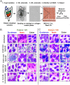
Patterns of EAEC adherence to HIEMs obtained from different donors and segments of the intestine. (A) Schematic representation of the methods used to assess adherence to enteroid monolayers. (B and C) Duodenal, ileal, and colon differentiated 2D HIEMs obtained from donors 103 and 109, respectively, were infected with EAEC 042 (a prototype strain), EAEC A2A (clinical isolate, typical EAEC), EAEC 5EA (clinical isolate, atypical EAEC), and E. coli HS (nonpathogenic control strain) at an MOI of 10 for 3 h. After infection, the cells were washed, fixed, stained with Giemsa-Wright stain, and imaged at ×100 to visualize the pattern of bacterial adherence. Technical replicates, three wells representative of 12 images; six biological replicates (two donors and three segments).
FIG 2
FIG 2
Definition and quantification of the different patterns of EAEC aggregative adherence on differentiated 2D HIEMs. (A to D) The insets show the occurrence of typical, sheet-like, microcolonies and mesh-like aggregative adherence (AA) exhibited by EAEC 042 across different donors and segments of intestine during infection as described in the legend to Fig. 1. Samples (D, duodenum [blue]; I, ileum [red]; C, colon [black]) were obtained from donors 103, 104, and 109, and the adherence pattern was quantified from an average of 12 different fields of 0.8 mm2 taken at ×100. Data represent the mean values of three independent experiments, and the error bars denote the standard error of the mean. Technical replicates, three wells representative of 12 images; eight biological replicates (three donors and three segments).
FIG 3
FIG 3
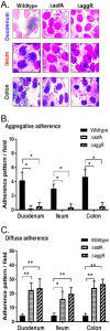
Adherence of wild-type EAEC (042) and ΔaafA and ΔaggR mutant strains to 2D differentiated HIEMs. (A) Wild-type EAEC and ΔaafA and ΔaggR mutants were added to duodenal, ileal, or colonic enteroids obtained from donor 103 as described in the legend to Fig. 1, and the adherence pattern was visualized by Giemsa-Wright staining. (B and C) The four adherence patterns from Fig. 2, classified collectively as aggregative adherence, were quantified versus that of diffuse adherence (see Materials and Methods). Data represent the mean values of three independent experiments, and the error bars denote the standard error of the mean, *, P < 0.05; **, P < 0.01. Technical replicates, three wells representative of 12 images; three biological replicates (one donor and three segments).
FIG 4
FIG 4
Total adherence of EAEC to three intestinal segments from three different donors. (A to C) 2D differentiated HIEMs obtained from duodenal, ileal, and colonic segments from donors 103, 104, and 109 were infected with EAEC 042 (A), A2A (B), or 5EA (C) at an MOI of 10 for 3 h. (D) Pooled adherence data from enteroids from all four intestinal segments from additional donors. Adherence was quantified as described in Materials and Methods. Data represent the mean values of three independent experiments, and the error bars denote the standard error of the mean. **, P < 0.01; ***, P < 0.001. Technical replicates, three wells; 9 (three donors and three segments) (A to C) and 17 (D) biological replicates.
FIG 5
FIG 5
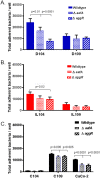
Total adherence of wild-type and mutant EAEC to HIEMs. (A, B) Wild-type EAEC and ΔaafA and ΔaggR mutants were incubated with 2D differentiated HIEMs from duodenum, ileum, and colon samples from donors 104 and 109, and adherence was quantified as described in Materials and Methods. Panel C shows the results of an adherence experiment with Caco-2 cells. Data represent the mean values of three independent experiments, and the error bars denote the standard error of the mean. Technical replicates, three wells; six biological replicates (two donors and three segments).
FIG 6
FIG 6
EAEC adherence to human intestinal enteroids. EAEC adheres to intestinal enteroids via five distinct aggregation patterns: sheet-like or microcolony adherence to duodenal (D) cells, diffuse adherence to jejunal (J) cells, typical stacked-brick adherence to ileal (I) cells, and a mesh-like adherence of interconnecting clusters to cells of the colon (C). While these patterns (as well as the strength of the interaction) are dependent on the large subunit of AAF/II, an unidentified host factor mediates adherence across different donors and different intestinal segments.

References

    1. Wanke CA, Schorling JB, Barrett LJ, Desouza MA, Guerrant RL. 1991. Potential role of adherence traits of Escherichia coli in persistent diarrhea in an urban Brazilian slum. Pediatr Infect Dis J 10:746–751. doi:10.1097/00006454-199110000-00006. - DOI - PubMed
    1. Steiner TS, Lima AA, Nataro JP, Guerrant RL. 1998. Enteroaggregative Escherichia coli produce intestinal inflammation and growth impairment and cause interleukin-8 release from intestinal epithelial cells. J Infect Dis 177:88–96. doi:10.1086/513809. - DOI - PubMed
    1. Adachi JA, Jiang ZD, Mathewson JJ, Verenkar MP, Thompson S, Martinez-Sandoval F, Steffen R, Ericsson CD, DuPont HL. 2001. Enteroaggregative Escherichia coli as a major etiologic agent in traveler’s diarrhea in 3 regions of the world. Clin Infect Dis 32:1706–1709. doi:10.1086/320756. - DOI - PubMed
    1. Glandt M, Adachi JA, Mathewson JJ, Jiang ZD, DiCesare D, Ashley D, Ericsson CD, DuPont HL. 1999. Enteroaggregative Escherichia coli as a cause of traveler’s diarrhea: clinical response to ciprofloxacin. Clin Infect Dis 29:335–338. doi:10.1086/520211. - DOI - PubMed
    1. Wanke CA, Mayer H, Weber R, Zbinden R, Watson DA, Acheson D. 1998. Enteroaggregative Escherichia coli as a potential cause of diarrheal disease in adults infected with human immunodeficiency virus. J Infect Dis 178:185–190. doi:10.1086/515595. - DOI - PubMed

Publication types

Substances

LinkOut - more resources