. 2018 Mar 6;115(10):E2302-E2310.
doi: 10.1073/pnas.1716747115. Epub 2018 Feb 20.

Carbon monoxide protects the kidney through the central circadian clock and CD39

Affiliations

Carbon monoxide protects the kidney through the central circadian clock and CD39

Matheus Correa-Costa et al. Proc Natl Acad Sci U S A. .

Abstract

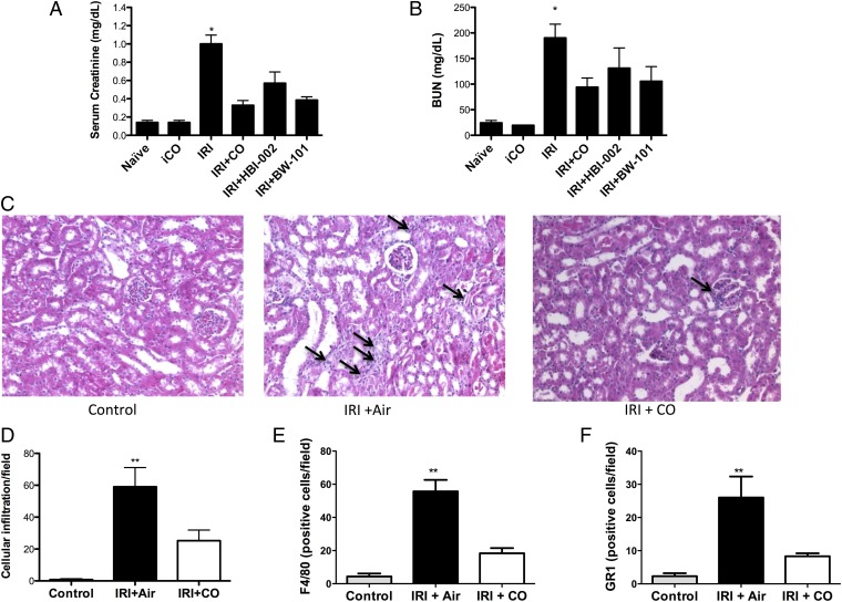
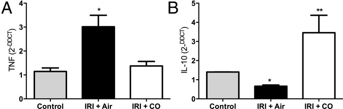
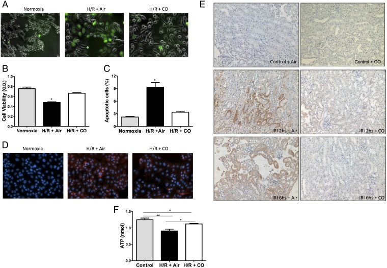
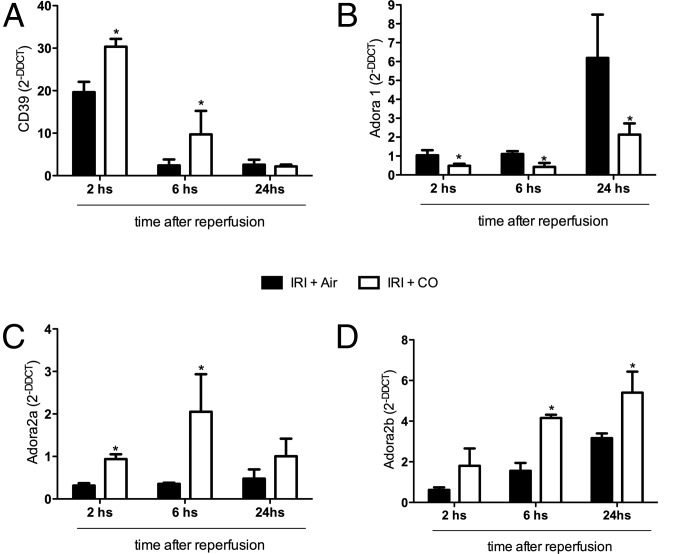
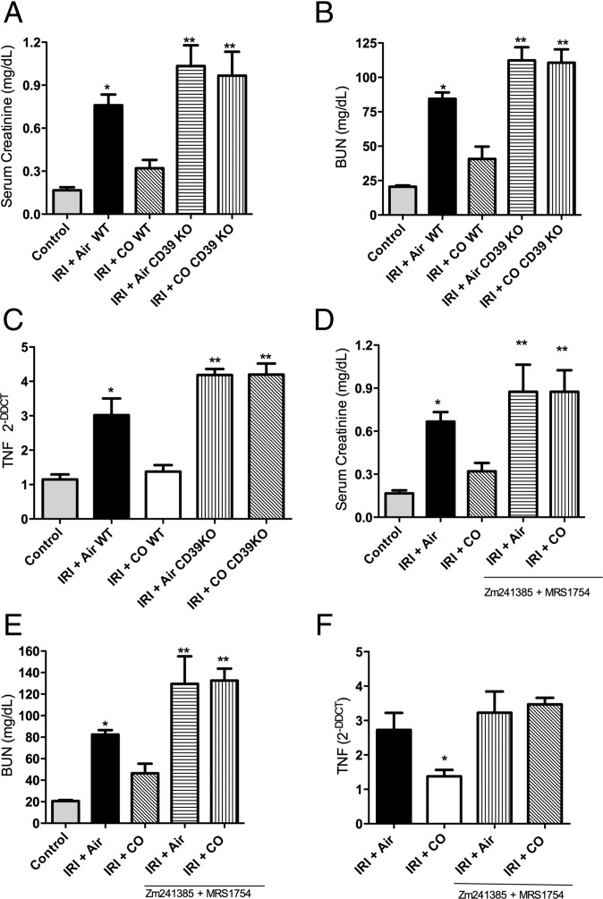
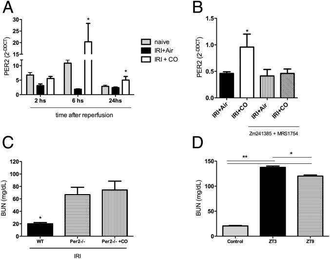
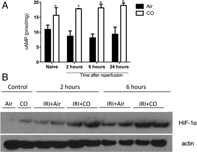
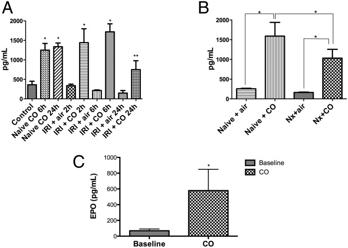
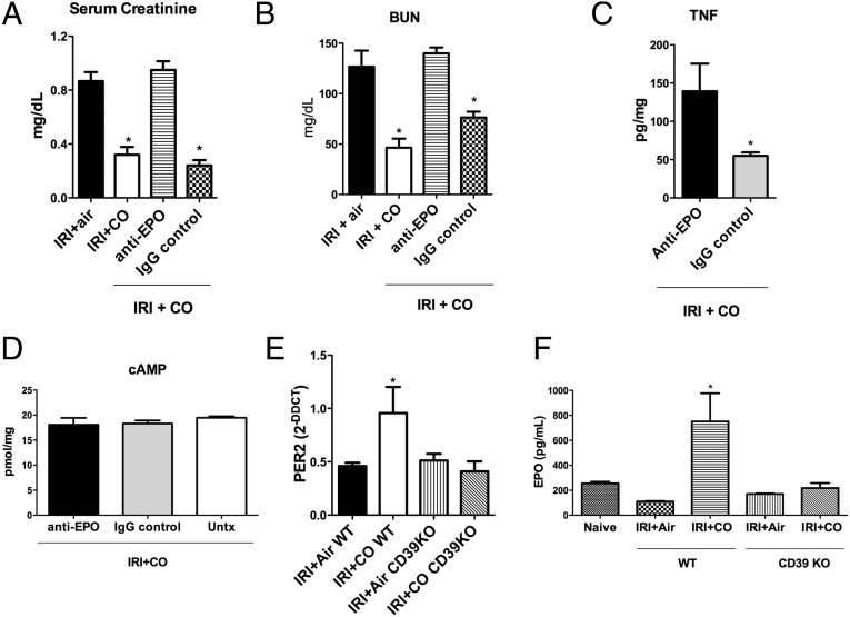
Ischemia reperfusion injury (IRI) is the predominant tissue insult associated with organ transplantation. Treatment with carbon monoxide (CO) modulates the innate immune response associated with IRI and accelerates tissue recovery. The mechanism has been primarily descriptive and ascribed to the ability of CO to influence inflammation, cell death, and repair. In a model of bilateral kidney IRI in mice, we elucidate an intricate relationship between CO and purinergic signaling involving increased CD39 ectonucleotidase expression, decreased expression of Adora1, with concomitant increased expression of Adora2a/2b. This response is linked to a >20-fold increase in expression of the circadian rhythm protein Period 2 (Per2) and a fivefold increase in serum erythropoietin (EPO), both of which contribute to abrogation of kidney IRI. CO is ineffective against IRI in Cd39-/- and Per2-/- mice or in the presence of a neutralizing antibody to EPO. Collectively, these data elucidate a cellular signaling mechanism whereby CO modulates purinergic responses and circadian rhythm to protect against injury. Moreover, these effects involve CD39- and adenosinergic-dependent stabilization of Per2. As CO also increases serum EPO levels in human volunteers, these findings continue to support therapeutic use of CO to treat IRI in association with organ transplantation, stroke, and myocardial infarction.

Keywords: DAMPS; adenosine; circadian rhythm; heme oxygenase; innate immunity.


Conflict of interest statement

Conflict of interest statement: L.E.O. is a scientific consultant for Hillhurst Biopharmaceuticals and has stock options. E.G. is a founder of Hillhurst Biopharmaceuticals and owns stock.

Figures

Fig. 1.
Fig. 1.
CO prevents kidney IRI in mice. (A and B) Mice were treated with inhaled CO (iCO), oral CO (HBI-002), or a CO-prodrug (BW-101) 1 h before a 45-min bilateral kidney ischemia. Serum creatinine (A) and BUN (B) were measured 24 h after reperfusion. Results represent mean ± SD of 5–10 mice per group. *P < 0.001 vs. iCO, P < 0.05 vs. HBI-002, and P < 0.01 vs. BW-101. (C) Representative H&E-stained sections from control kidney and from mice subjected to IRI treated with Air or iCO as above. Images are representative of 10 sections from five mice. Arrows indicate leukocyte infiltration. (DF) Quantitation of F4/80- and GR1-positive staining for macrophage and neutrophils, respectively. Results represent mean ± SD of 6–8 sections from five mice in each group. **P < 0.05 vs. CO.
Fig. 2.
Fig. 2.
CO modulates cytokine expression in the kidney after IRI. (A and B) Tissue expression of TNF and IL-10 mRNA in the kidney in naïve mice or kidneys from mice 24 h after IRI treated with either Air or CO. CO blocked TNF and simultaneously enhanced IL-10 expression. Results represent mean ± SD of 5 per group. *P < 0.05 vs. CO; **P < 0.01 vs. control and IRI + Air.
Fig. 3.
Fig. 3.
Effects of CO on promoting kidney epithelial cell bioenergetics and viability in response to H/R conditions. (A) Exposure to CO (250 ppm) prevented H/R-induced elevations in DCF fluorescence, as a marker for reactive oxygen species production. Images are representative of at least three independent experiments in triplicate. (B and C) LLCPK-1 kidney epithelial cell viability measured by crystal violet (B) or propidium iodine incorporation (C) was determined in response to H/R in the presence and absence of CO (250 ppm). Results represent mean ± SD of 4–6 samples in triplicate. *P < 0.02 vs. normoxia or H/R + CO. (D and E) CO reduced tissue hypoxia measured with the O2-sensitive hypoxyprobe. Cells and animals were treated with hypoxyprobe at 400 mM and 60 mg/kg 1 h before harvesting, and cells and tissue sections from H/R and IRI, respectively, were stained as described in Materials and Methods. (F) ATP levels in LLCPK-1 cells shows CO reversing an H/R-induced decrease in ATP at 24 h of H/R. Results represent mean ± SD of three independent experiments in triplicate. *P < 0.05, **P < 0.02.
Fig. 4.
Fig. 4.
Effects of IRI and CO on CD39 and A2 receptor expression. (AD) Expression levels of CD39, A1, A2a, and A2b were measured by PCR over time after reperfusion. Animals treated with CO increased expression of CD39 and the A2 receptors but inhibited IRI-induced increases in A1 receptor expression. Results represent mean ± SD of 4–6 mice per group at each time point. *P < 0.02 vs. Air.
Fig. 5.
Fig. 5.
CD39 activity and A2 receptor signaling are required for CO to prevent kidney IRI in mice. (AC) CO blocks IRI-induced elevations in creatinine and BUN and tissue levels of TNF, but these effects are lost in Cd39−/− mice. Results represent mean ± SD of 4–6 mice per group. Moreover, animals lacking CD39 and subjected to IRI showed enhanced severity of injury compared with WT mice with IRI regardless of CO. (DF) Similar to Cd39−/− mice, WT mice treated with selective inhibitors of A2a and A2b receptors, Zm241385 and MRS1754, respectively, were also nonresponsive to the protection afforded by CO against IRI. Serum creatinine, BUN, and tissue TNF levels were similar to that observed in Cd39−/− mice. Results represent mean ± SD of 4–6 mice per group. *P < 0.05 vs. control and CO-WT; **P < 0.001 vs. other groups.
Fig. 6.
Fig. 6.
Expression of the circadian rhythm gene Per2 is required for CO to protect the kidney against IRI and is dependent on A2 receptor activation. (A) Expression levels of Per2 over time in the kidney in response to IRI ± CO. CO markedly increases Per2 in conjunction with IRI versus no change observed in IRI+Air-treated mice. *P < 0.05 vs. Air. (B) Blockade of A2 receptors prevents the CO-induced increase in Per2 expression. *P < 0.05 vs. Air. (C) CO is unable to prevent IRI-induced elevations in serum BUN in Per2−/− mice. *P < 0.05 vs. other groups. (D) Since Per2 cycles throughout the day and peaks at ZT9, we tested susceptibility to kidney IRI compared with ZT3 when Per2 expression is low (Fig. S3). Mice that underwent IRI at ZT9 showed less severe IRI measured by elevations in BUN versus those subjected to IRI at ZT3. Results represent mean ± SD of 4–6 mice per group. *P < 0.05, **P < 0.001.
Fig. 7.
Fig. 7.
A2 receptor signaling is activated in CO-treated mice and associated with increased expression of HIF-1α. (A) Mice subjected to kidney IRI ± CO were killed at different times after reperfusion, and cAMP levels were measured by ELISA. *P < 0.01 vs. Air. (B) Kidney lysates were evaluated at different time points after reoxygenation for HIF-1α expression as indicated. Results represent mean ± SD of 4–6 mice per group.
Fig. 8.
Fig. 8.
CO exposure increases serum EPO levels. (A) Serum EPO levels were evaluated in naive, Air-, and CO-treated mice ± IRI. Blood samples were collected at different times after reoxygenation. *P < 0.05 vs. Air; **P < 0.001 vs. Air. (B) EPO levels were measured in naive and bilateral nephrectomized mice. *P < 0.05. (C) EPO levels were measured in per2−/− mice. *P < 0.05 vs. baseline. Results represent mean ± SD of 4–6 mice per group.
Fig. 9.
Fig. 9.
EPO is associated with a better outcome after CO treatment, and CD39 is an important mediator of the CO-induced protection in the IRI model. Mice were subjected to IRI ± CO in the presence or absence of anti-EPO blocking antibody or an IgG control antibody and were evaluated by Serum Creatinine (A), BUN (B), kidney TNF (C), and cAMP levels (D). *P < 0.05 vs. the other groups. (E and F) Per2 mRNA and serum EPO levels in WT and CD39 KO mice, subjected to IRI ± CO. *P < 0.02 vs. all of the other groups. Results represent mean ± SD of 5 mice per group.

References

    1. Eltzschig HK, Eckle T. Ischemia and reperfusion–From mechanism to translation. Nat Med. 2011;17:1391–1401. - PMC - PubMed
    1. Chertow GM, Burdick E, Honour M, Bonventre JV, Bates DW. Acute kidney injury, mortality, length of stay, and costs in hospitalized patients. J Am Soc Nephrol. 2005;16:3365–3370. - PubMed
    1. Jang HR, Ko GJ, Wasowska BA, Rabb H. The interaction between ischemia-reperfusion and immune responses in the kidney. J Mol Med (Berl) 2009;87:859–864. - PubMed
    1. Munshi R, Hsu C, Himmelfarb J. Advances in understanding ischemic acute kidney injury. BMC Med. 2011;9:11. - PMC - PubMed
    1. Bonventre JV, Weinberg JM. Recent advances in the pathophysiology of ischemic acute renal failure. J Am Soc Nephrol. 2003;14:2199–2210. - PubMed

Publication types

MeSH terms