. 2018 Feb 20;8(1):3363.
doi: 10.1038/s41598-018-21589-2.

Interleukin-33 is activated by allergen- and necrosis-associated proteolytic activities to regulate its alarmin activity during epithelial damage

Affiliations

Interleukin-33 is activated by allergen- and necrosis-associated proteolytic activities to regulate its alarmin activity during epithelial damage

Ian C Scott et al. Sci Rep. .

Abstract

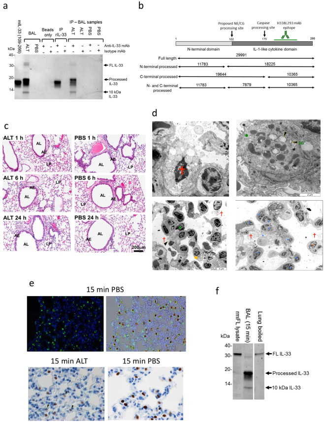
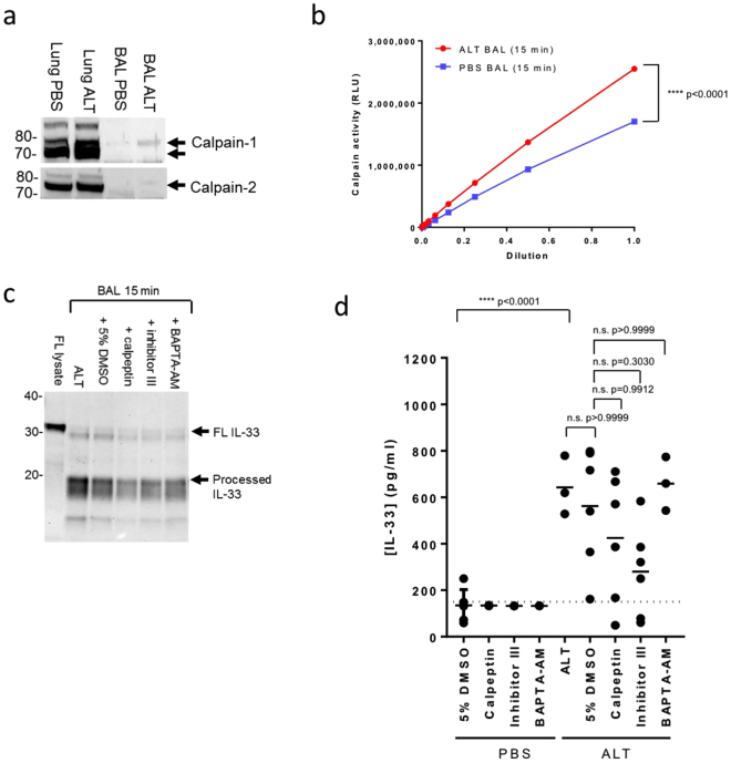
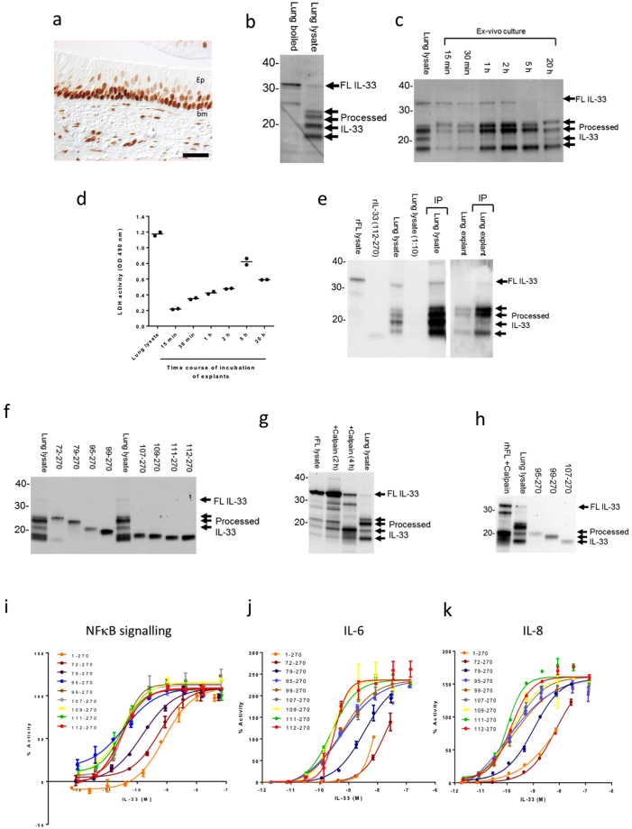
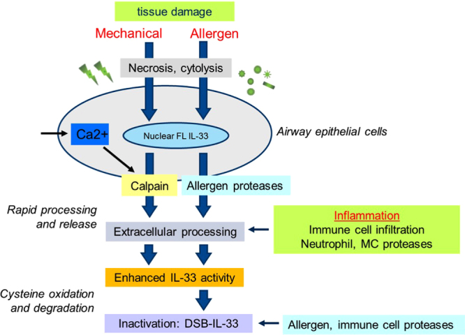
Interleukin (IL)-33 is an IL-1 family alarmin released from damaged epithelial and endothelial barriers to elicit immune responses and allergic inflammation via its receptor ST2. Serine proteases released from neutrophils, mast cells and cytotoxic lymphocytes have been proposed to process the N-terminus of IL-33 to enhance its activity. Here we report that processing of full length IL-33 can occur in mice deficient in these immune cell protease activities. We sought alternative mechanisms for the proteolytic activation of IL-33 and discovered that exogenous allergen proteases and endogenous calpains, from damaged airway epithelial cells, can process full length IL-33 and increase its alarmin activity up to ~60-fold. Processed forms of IL-33 of apparent molecular weights ~18, 20, 22 and 23 kDa, were detected in human lungs consistent with some, but not all, proposed processing sites. Furthermore, allergen proteases degraded processed forms of IL-33 after cysteine residue oxidation. We suggest that IL-33 can sense the proteolytic and oxidative microenvironment during tissue injury that facilitate its rapid activation and inactivation to regulate the duration of its alarmin function.


Conflict of interest statement

Authors that are employees of the AstraZeneca Group have stock/stock options in AstraZeneca.

Figures

Figure 1
Figure 1
IL-33 processing in ALT model is independent of immune cell proteases. DPP-1 (a), NE (b), and CG (c) enzyme activities in bone marrow (BM) lysates from wild type (WT) and Ctsc−/− mice 30 min after ALT or PBS challenge (n = 6–8/mice group). %Activity (relative to WT/PBS group) is shown above the data. (d) Concentration of IL-33 (pg/ml) in BAL from WT and Ctsc−/− mice 15 min to 24 h after i.n. ALT or PBS challenge measured by ELISA (n = 3–4 mice/group). Data points are mean ± SEM. (e) Western blot of IL-33 of BAL samples (n = 3–4 pooled /group). Controls are as follows: FL lysate, lysate of CHO cells transfected with full length mouse IL-33; in house (109–266), in house generated recombinant mouse IL-33; mouse R&D (109–266), R&D systems recombinant mouse IL-33 (109–266 aa). (f) Concentration of IL-5 (pg/ml) in BAL from WT and Ctsc−/− mice 15–1440 min after i.n. ALT or PBS challenge (n = 3–4 mice/group). Data points are mean ± SEM. Statistical analysis: two-way ANOVA test, Tukey’s post-test, F = 7.4, degrees of freedom = 9. n.s.: non-significant between WT/ALT and Ctsc−/−/ALT groups at 1440 min. (g) Western blot of IL-33 in BAL samples (n = 3–4 pooled /group) from WT and mast cell-deficient mice 30 min after ALT or PBS challenge. Controls: FL lysate, lysate of CHO cells transfected with full length mouse IL-33; 109–266-FH, recombinant mouse IL-33 109–266 aa with N-terminal Flag-His tag. Data is pooled from n = 3 independent studies (a–c). Data representative of n = 3 (d–f) and n = 2 (g) independent studies. Western blot images (e and g) have been cropped for clarity with full blots presented in Fig. S6.
Figure 2
Figure 2
ALT drives rapid N-terminal processing and release of IL-33 from damaged airway epithelium. (a) Immunoprecipitation (#H338L293 or NIP228 mAb) and western blot (AF3626 mAb) of IL-33 in BAL (pooled n = 3–4 mice/group) after ALT or PBS challenge. Controls: mIL-33 (109–266), recombinant mouse IL-33 (109–266 aa). Abbreviations: IP, immunoprecipitation. (b) Diagram of the mouse IL-33 structure, proposed CG/NE and caspase sites, epitope for H338L293 mAb and theoretical MW (Da) of proteins. (c) Haematoxylin and eosin staining of mouse lung sections 1, 6 and 24 h after i.n. 25μg ALT or PBS challenge. AL, airway lumen; AE, airway epithelium; LP, lung parenchyma. Scale bar = 200 μm. (d) Transmission electron micrographs of mouse lung 15 min after ALT or PBS challenge. Top left: high power image of necrotic type 2 pneumocyte (red cross) 15 min after ALT challenge. Top right: high power image of intact type 2 pneumocyte (green dot) 15 min after PBS challenge. SG, surfactant granules (black arrow heads). Bottom left: low power image of necrotic type 2 pneumocytes 15 min after ALT challenge. Eosinophil granulocyte (orange dot). Bottom right: Low power image of neutrophil (blue dots) and eosinophil infiltration 15 min after ALT challenge. Scale bars 2 μm (high power images) and 5 μm (low power images). (e) Immunostaining of pro-surfactant protein C (green stain, upper left panel) and overlaid with IL-33 (brown stain, upper right panel) in mouse alveolar tissue 15 min after PBS challenge. Immunostaining of IL-33 in alveolar tissue 15 min after ALT (lower left panel) or PBS challenge (lower right panel). Fading of IL-33 staining is seen following ALT challenge (black arrow heads). (f) Western blot of IL-33 in BAL (pooled n = 3 mice) 15 min after ALT challenge and boiled mouse lung (pooled n = 3 mice). Controls: FL lysate, lysate of CHO cells transfected with full length mouse IL-33. Representative images from study with n = 3 mice/group (a–c). Representative of n = 3 independent studies (d–f). Western blot images (a and f) have been cropped for clarity with full blots presented in Fig. S7.
Figure 3
Figure 3
Calpain can process IL-33 in airway epithelial cells and enhance its activity. (a) Immunostaining of mouse IL-33 (top left panel), with isotype control antibodies (bottom left) and cell nuclei (right panels) in mouse CMT-64 cells at 20× magnification. (b) Western blot of mouse IL-33 in CMT-64 cell lysates (PBS, 0.1% Triton X100) and CMT-64 cells (boiled in SDS-PAGE buffer). Controls: rmIL-33 (102–266), recombinant mouse IL-33 (102–266 aa). (c) Western blot of mouse IL-33 in CMT-64 cell lysates. Cells were pre-treated for 30 min with protease inhibitors and incubated and in PBS/0.1% Triton X100 for 30 min. Controls: as (b). (d) Western blot of mouse IL-33 in CMT-64 cell lysates. Gel image is cropped to show only processed IL-33. Cells were pre-treated for 30 min with protease inhibitors, BAPTA-AM or 0.1% DMSO (cell lysate) and incubated for 30 min in PBS/0.1% Triton X100. Controls as (b). Abbreviations: i, inhibitor, NM, nafamostat mesylate. (e) Western blot of FL human IL-33 lysate incubated alone or with calpain-1, NE and CG for 2 h. Controls: rhIL-33, recombinant human IL-33, 112–270, purified recombinant human IL-33 (112–270 aa); FL lysate, lysate of HEK cells transfected with full length human IL-33; Mock lysate, lysate of mock transfected HEK cells. (f) Relative NFkB p65/RelA translocation in HUVECs 30 min after stimulation with human FL IL-33 lysate and FL IL-33 pre-incubated with calpain-1, NE or CG. Data points are mean ± SEM. of duplicate determinations. %Activity is calculated relative to signal of 3 ng/ml rhIL-33 (112–270 aa). Representative of n = 3 independent experiments (a–f). Western blot images (b–e) have been cropped for clarity with full blots presented in Figs S8–S9.
Figure 4
Figure 4
ALT-driven IL-33 processing in vivo is not dependent on calpain proteases. (a) Western blot of calpain-1 (upper panel) and -2 (lower panel) in mouse lung homogenates and BAL (pooled n = 3–4 mice/group) 30 min after ALT or PBS challenge. (b) Protease activity, measured using a calpain peptide substrate, in BAL (pooled n = 3–4 mice/group) collected 15 min after ALT or PBS challenge. RLU, relative light units. Data points are mean ± SEM. Statistical analysis: two-way ANOVA test, Tukey’s post-test, F = 1464, degrees of freedom = 10. ****P < 0.0001 for ALT v PBS group for undiluted samples. (c) Western blot of IL-33 in BAL (pooled n = 3–4 mice/group) 15 min after ALT challenge with and without co-administration of calpeptin, calpain inhibitor III, BAPTA-AM or 5% DMSO. Controls: FL lysate, lysate of CHO cells transfected with full length mouse IL-33. (d) Concentration of IL-33 (pg/ml) in BAL 15 min after ALT or PBS challenge with and without co-administration of calpeptin, calpain inhibitor III, BAPTA-AM or 5% DMSO. Controls: FL lysate, lysate of CHO cells transfected with full length mouse IL-33. Lower limit of detection is indicated by dotted line. Data points are mean ± SEM. Statistical analysis: one way ANOVA test, Tukey’s post-test, F = 6.182, degrees of freedom = 9. n.s.: non-significant. Data is pooled from n = 3 independent studies (d). Representative of n = 3 independent experiments (a–c). Western blot images (a and c) have been cropped for clarity with full blots presented in Figs S10 and S11.
Figure 5
Figure 5
ALT-derived protease activities process IL-33 and increase its activity. (a) Western blot of IL-33 in mouse lung (boiled in SDS-PAGE buffer) and BAL 15 min after ALT challenge. Controls: FL IL-33, lysate of CHO cells transfected with full length mouse IL-33; rmIL-33 (102–266 and 109–266), recombinant mouse IL-33 (102- and 109-266 aa). (b) Western blot of IL-33 in BAL 15 min after ALT or PBS challenge. Mice were dosed i.p with recombinant mouse sST2-Fc or CAT-002 (control) for 30 min prior to i.n. ALT or PBS challenge and BAL collected after 15 min. Controls as (a). (c) Western blot of rmFL IL-33 lysate, rmIL-33 (109-266 aa), DSB-rmIL-33 (109-266 aa) and cysteine mutant rmIL-33 (109-266 aa) after incubation with 300 μg/ml ALT. (d) Western blot of rhFL IL-33 lysate, rhIL-33 (112-270 aa), DSB-rmIL-33 (112-270 aa) before and after incubations with 300 μg/ml ALT for 1-10 min. (e) Relative NFkB p65/RelA translocation in HUVECs 30 min after stimulation with rhFL IL-33 lysate and FL IL-33 pre-incubated with ALT. Data points are mean ± SEM of duplicate determinations. %Activity is calculated relative to signal of 3 ng/ml rhIL-33 (112-270 aa). (f) Western blot of rhFL IL-33 lysate before and after incubation with 300 μg/ml ALT for 10 min. rhFL IL-33 was pre-incubated with protease inhibitors or BAPTA-AM, for 15 min prior to addition of ALT. Details of protease inhibitors are described in legend for Fig. 3c. (g) Western blot of rhFL IL-33 lysate with and without incubation with 300 μg/ml ALT or PBS for 10 min. Controls: FL lysate, lysate of HEK cells transfected with full length human IL-33. Details of protease inhibitors are described in legend for Fig. 3d. (h) Concentration of IL-33 (pg/ml) in mouse CMT-64 cell supernatants 30 min after 100 μg/ml ALT or PBS challenge. (i) Lactate dehydrogenase (LDH) activity in mouse CMT-64 cell supernatants 6 h after 100 μg/ml ALT or PBS challenge. (j) Western blot of mouse CMT-64 cell supernatants 15-1200 min with (right panel) or without (left panel) 100 μg/ml ALT. ALT treatment was performed with or without pre-incubation for 15 min with 100 μg/ml calpain inhibitor III or 1% DMSO. Controls as (a). Representative of n = 3 (a–e) and n = 2 independent experiments (f–j). Western blot images (ad,f,g,j) have been cropped for clarity with full blots presented in Figs S12–16.
Figure 6
Figure 6
FL and N-terminal processed forms of IL-33 are detected in the human lung. (a) Bright field micrograph demonstrating IL-33 immunoreactivity (brown DAB chromogen) in small airways of lungs from severe COPD patient (GOLD stage 4). Ep: epithelium; bm basement membrane. Scale bar = 40 μm. (b) Western blot of IL-33 expression in human COPD lung tissue (boiled in SDS-PAGE buffer) and lung tissue lysate. (c) Western blot of IL-33 expression in human COPD lung tissue and lung explant supernatants (15 min to 20 h after incubation). (d) Lactate dehydrogenase (LDH) activity in human lung explant supernatants (samples as (c). (e) Immunoprecipitation (IP) and western blot of IL-33 in human COPD lung lysate and explant supernatants (2 h). IL-33 was IP using anti-IL-33 mAb (#640050) and western blot performed with anti-IL-33 Ab (AF3625). Controls: rFL lysate, lysate of HEK cells transfected with full length human IL-33. (f) Western blot of mature forms rhIL-33 (72-, 79-, 95-, 99-, 107-, 109-, 111-, 112-270 aa) and human lung lysate. (g) Western blot of rhFL IL-33 lysate, with or without incubation for 2-4 h with calpain, and human lung lysate. Controls as (e). (h) Western blot of rhFL IL-33 lysate, incubated for 2 h with calpain, lung lysate and mature forms of rhIL-33 (95-, 99-, 107-270 aa). (i) Relative NFkB p65/RelA translocation in HUVECs 30 min after stimulation with rhIL-33 (1-, 72-, 79-, 95-, 99-, 107-, 109-, 111-, 112-270 aa). Data points are mean ± SEM of duplicate determinations. % Activity is calculated relative to signal of 3 ng/ml rhIL-33 (112-270 aa). Relative IL-6 (j) and IL-8 (k) release from HUVECs 20 h after stimulation with rhIL-33 (1-, 72-, 79-, 95-, 99-, 107-, 109-, 111-, 112-270 aa). Data points as (j). Data points are mean ± SEM of duplicate determinations. % Activity is calculated relative to signal of 30 ng/ml rhIL-33 (112-270 aa). Representative of n = 2 (b,d,f–j), n = 3 (a,k) and n = 4 independent experiments (c). Western blot images (b,c,e–h) have been cropped for clarity with full blots presented in Figs S17–S20.
Figure 7
Figure 7
Hypothesis for the proteolytic regulation of IL-33 activity during injury to airway epithelium. Full length IL-33 is stored in the nucleus of airway epithelial cells. Mechanical or allergen induced cell damage induces epithelial cell necrosis, loss of cell membrane integrity and cytolysis. Increased intracellular levels of Ca2+ ions at sites of epithelial damage trigger calpain protease activity. Calpains proteolytically activate IL-33 during its release from damaged epithelial cells. Airborne allergen-derived proteases can damage epithelial cells, enhance IL-33 release and proteolytically activate IL-33. In addition, during inflammation proteases from infiltrating immune cells can also contribute to the extracellular proteolytic activation of IL-33. Subsequently mature forms of IL-33 are rapidly inactivated by cysteine oxidation and degraded by allergen or possibly immune cell proteases. Thus IL-33 can utilise different proteolytic activities present during airway epithelial damage to be rapidly activated and inactivated and limit the duration of its alarmin activity.

References

    1. Garlanda C, Dinarello CA, Mantovani A. The interleukin-1 family: back to the future. Immunity. 2013;39:1003–1018. doi: 10.1016/j.immuni.2013.11.010. - DOI - PMC - PubMed
    1. Liew FY, Girard JP, Turnquist HR. Interleukin-33 in health and disease. Nat Rev Immunol. 2016;16:676–689. doi: 10.1038/nri.2016.95. - DOI - PubMed
    1. Martin NT, Martin MU. Interleukin 33 is a guardian of barriers and a local alarmin. Nat Immunol. 2016;17:122–131. doi: 10.1038/ni.3370. - DOI - PubMed
    1. Ali S, et al. IL-1 receptor accessory protein is essential for IL-33-induced activation of T lymphocytes and mast cells. Proc Natl Acad Sci USA. 2007;104:18660–18665. doi: 10.1073/pnas.0705939104. - DOI - PMC - PubMed
    1. Chackerian AA, et al. IL-1 receptor accessory protein and ST2 comprise the IL-33 receptor complex. J Immunol. 2007;179:2551–2555. doi: 10.4049/jimmunol.179.4.2551. - DOI - PubMed

Publication types