. 2018 Feb 20;8(1):3345.
doi: 10.1038/s41598-018-21522-7.

Autoantibodies against the Immunoglobulin-Binding Region of Ro52 Link its Autoantigenicity with Pathogen Neutralization

Affiliations

Autoantibodies against the Immunoglobulin-Binding Region of Ro52 Link its Autoantigenicity with Pathogen Neutralization

Peter D Burbelo et al. Sci Rep. .

Abstract

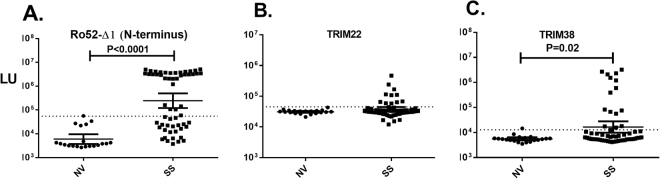
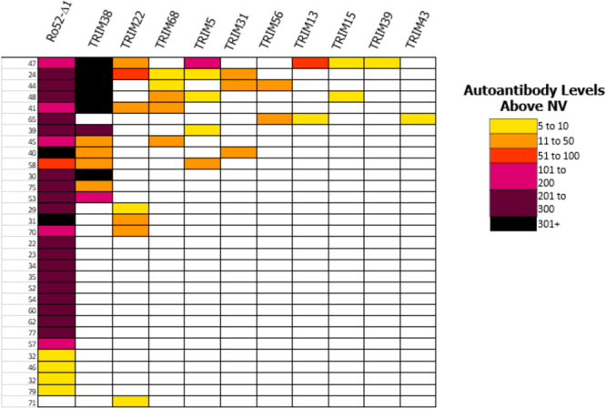
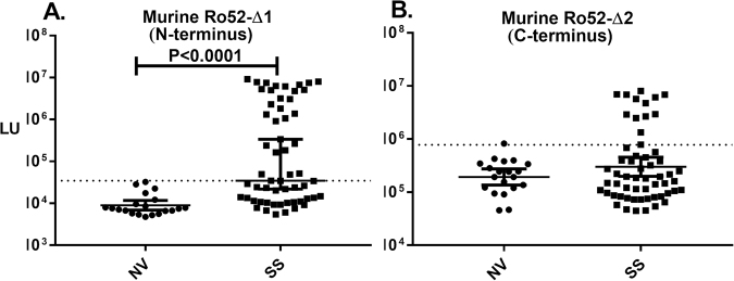
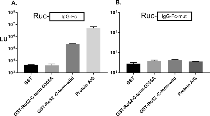
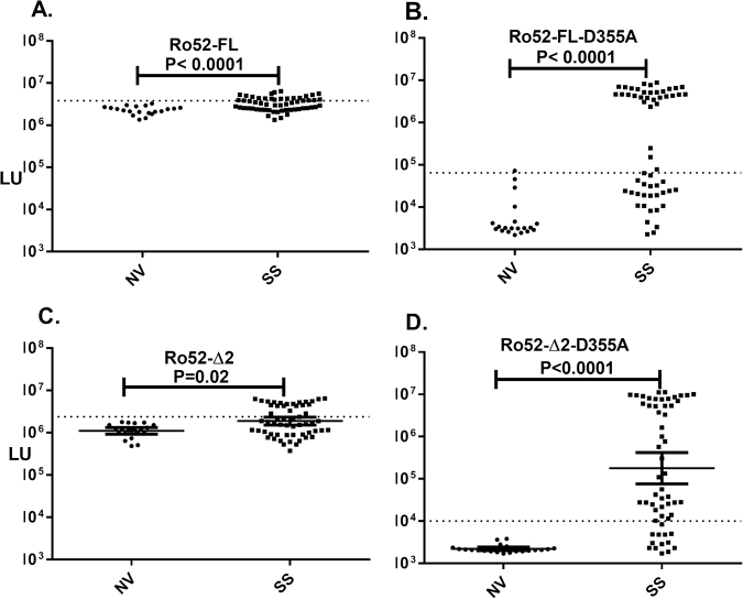
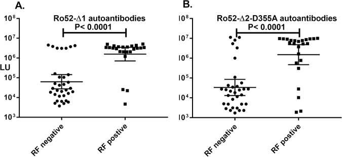
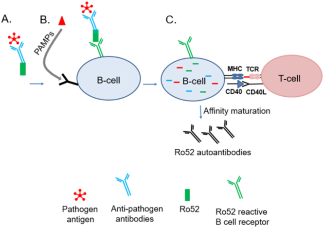
Ro52/TRIM21 plays a key role in antibody-dependent pathogen neutralization and is a major autoantigen in systemic lupus erythematosus, Sjögren's syndrome (SS), and other autoimmune diseases. Here we evaluated immunoreactivity against Ro52-related molecules in SS and healthy volunteers. Although most proteins examined were not antigenic, several TRIM paralogs, including TRIM22, and TRIM38, showed sporadic immunoreactivity in SS. In contrast, the murine Ro52 ortholog with limited linear homology demonstrated high levels of autoantibodies implicating the importance of shared conformational epitopes. To further explore the autoantigencity of Ro52, deletion and point mutant analyses were employed revealing previously hidden, robust autoantibodies directed against its C-terminal immunoglobulin-binding domain. Another autoantibody, rheumatoid factor, targeting the Fc region of IgG, strongly overlapped with Ro52 seropositivity (odds ratio 14; P < 0.0001). These convergent mechanistic findings support a model whereby intracellular Ro52-bound antibody-coated pathogen complexes, released or misprocessed from infected cells, drive autoantigenicity against Ro52 and the Fc region of IgG.


Conflict of interest statement

The authors declare no competing interests.

Figures

Figure 1
Figure 1
Detection of autoantibody against Ro52, TRIM22 and TRIM38 in SS. Scatter plot graphs represent autoantibody levels determined by LIPS in individual subjects from a cohort of 20 normal volunteers (NV) and 57 subjects with SS. The luminescence of autoantibody response (LU) for each subject are plotted on the Y-axis. (A) Autoantibodies against Ro52 were determined using a previously described N-terminal protein fragment (Ro52-Δ1). Autoantibodies against (B) TRIM22 and (C) TRIM38, two potential Ro52 co-regulated genes, are also shown. For each plot, the solid line represents the geometric mean plus the 95% CI. For each antigen, a cutoff value shown by the dotted line was based on the mean plus three SD of the autoantibody values observed in normal volunteers. P values were calculated using the Mann-Whitney U test and only P values < 0.05 are shown.
Figure 2
Figure 2
Heatmap analysis of autoantibodies against Ro52 and TRIM proteins in SS. Heatmap analysis of autoantibody responses against Ro52 and a panel of ten TRIM proteins in SS subjects are shown. Each row represents a single SS subject’s autoantibody profile. Clear boxes represent seronegative samples that matched the normal volunteer profile. Color coding shows antibody levels, expressed as integer multiples compared to the normal volunteer baseline. As denoted in the figure legend, these values in the SS subjects ranged from yellow (Z score from 5–10) to black (Z score < 301) reflecting extremely high autoantibody levels.
Figure 3
Figure 3
Detection of SS autoantibodies using murine Ro52 protein fragments. Autoantibodies were studied in the SS cohort using recombinant murine Ro52 proteins encompassing the (A) N-terminal and (B) C-terminal protein fragments. Cut-off values for each target were based on the antibody value obtained from the mean plus three SD for normal volunteers and is delineated by the dotted line. P values comparing the normal volunteers (NV) and SS subjects were calculated using the Mann-Whitney U test.
Figure 4
Figure 4
The C-terminus of Ro52 interacts with the Fc region of IgG1. Protein-protein interactions were detected using bead immobilized target proteins and a luciferase-tagged Fc-IgG recombinant protein. GST fusion proteins of wild type (Ro52-Δ2) and C-terminal mutant (Ro52-Δ2-D355A) Ro52 were tested for their ability to interact with (A) Renilla luciferase-IgG-Fc and (B) Renilla luciferase-IgG-Fc mutant. Following incubation, the beads were washed and then measured for LU. The mean from two experiments is shown along with the standard error. As a positive control, protein A/G beads were found to strongly bind IgG-Fc, but not the IgG-Fc mutant recombinant protein.
Figure 5
Figure 5
Robust autoantibodies are directed against the C-terminus of Ro52 in SS. Autoantibodies were examined in the cohort against (A) full-length wild type Ro52, (B) a full-length Ro52 mutant (Ro52-D355A), (C) the wild type C-terminus of Ro52 (Ro52-Δ2), and (D) a Ro52 C-terminal mutant (Ro52-Δ2-D355A). Each dot represents an individual subject from the cohort and the geometric mean of the antibody levels for normal volunteers (NV) and SS subjects is shown by the colored horizontal bar. The dotted lines for each antigen represent the cut-off values for determining seropositivity. P values were calculated using the Mann-Whitney U test.
Figure 6
Figure 6
C-terminal deletion mutants eliminate the detection of conformational autoantibodies directed against this region. Evaluation of autoantibodies are shown to different N-terminal and C-terminal Ro52 deletion mutants in cohort. Ro52 deletion mutants included: (A) Ro52-Δ5; an N-terminal Ro52 mutant of 140 amino acids, (B) Ro52-Δ1-zipless; an internal deletion mutant eliminating the leucine zipper in the N-terminus of Ro52 (C) Ro52-Δ6; a C-terminal encompassing the first half of the immunoglobulin-binding region and (D) Ro52-Δ7; a C-terminal deletion mutant encompassing the second half of the immunoglobulin-binding region.
Figure 7
Figure 7
Rheumatoid factor autoantibodies co-segregate with Ro52 autoantibodies. SS subjects were segregated by their RF status (positive or negative) and the autoantibody levels against the (A) Ro52-Δ1 and (B) Ro52-Δ2-D355A protein fragments were then plotted in the subjects from each group. Each dot represents an individual SS subject and the geometric mean of the antibody levels in each group is shown by the horizontal bar. The P value was calculated using the Mann-Whitney U test.
Figure 8
Figure 8
Model for the role of ADIN in anti-Ro52 autoantibody production. Ro52 is normally involved in antibody dependent intracellular neutralization (ADIN) of pathogens. (A) During infection, Ro52-bound antibody-coated pathogen complexes are released from dying cells. (B) In SS, autoreactive B-cell against Ro52 may bind and abnormally process the extracellular Ro52 along with the anti-pathogen antibody-antigen complex. The B-cell also receives co-stimulatory signals from pathogen–associated molecular pattern molecules (PAMPs). (C) Even though Ro52 was bound by the B-cell receptor, pathogen antigen is internalized, processed and presented by the B-cell. MHCII presentation of the pathogen antigen by the B-cell provides T-cell help fostered by co-stimulatory signals from B-T-cell interactions resulting in affinity maturation of the anti-Ro52 B-cells, which now produce high titer autoantibodies.

References

    1. Anderson JR, Gray KG, Beck JS, Kinnear WF. Precipitating autoantibodies in Sjogren’s disease. Lancet. 1961;2:456–460. doi: 10.1016/S0140-6736(61)92430-8. - DOI - PubMed
    1. Yoshimi R, Ueda A, Ozato K, Ishigatsubo Y. Clinical and pathological roles of Ro/SSA autoantibody system. Clin Dev Immunol. 2012;2012:606195. doi: 10.1155/2012/606195. - DOI - PMC - PubMed
    1. Chan EK, Hamel JC, Buyon JP, Tan EM. Molecular definition and sequence motifs of the 52-kD component of human SS-A/Ro autoantigen. J Clin Invest. 1991;87:68–76. doi: 10.1172/JCI115003. - DOI - PMC - PubMed
    1. Deutscher SL, Harley JB, Keene JD. Molecular analysis of the 60-kDa human Ro ribonucleoprotein. Proc Natl Acad Sci USA. 1988;85:9479–9483. doi: 10.1073/pnas.85.24.9479. - DOI - PMC - PubMed
    1. Ben-Chetrit E, Gandy BJ, Tan EM, Sullivan KF. Isolation and characterization of a cDNA clone encoding the 60-kD component of the human SS-A/Ro ribonucleoprotein autoantigen. J Clin Invest. 1989;83:1284–1292. doi: 10.1172/JCI114013. - DOI - PMC - PubMed

Publication types