. 2018 Feb 20;8(1):3337.
doi: 10.1038/s41598-018-21685-3.

Enhanced Biological Response of AVS-Functionalized Ti-6Al-4V Alloy through Covalent Immobilization of Collagen

Affiliations

Enhanced Biological Response of AVS-Functionalized Ti-6Al-4V Alloy through Covalent Immobilization of Collagen

Parsa Rezvanian et al. Sci Rep. .

Abstract

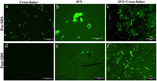
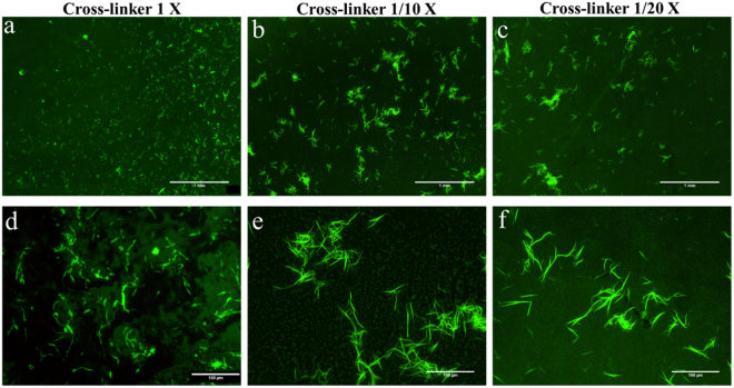
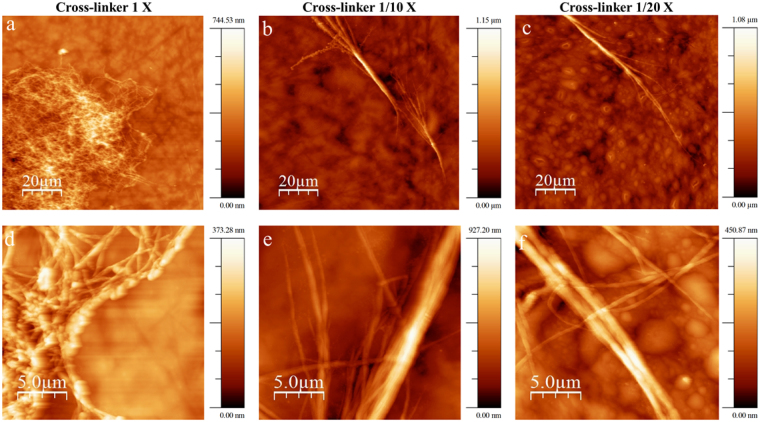
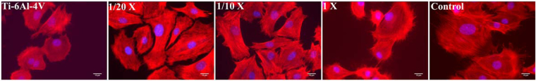
This study presents the development of an efficient procedure for covalently immobilizing collagen molecules on AVS-functionalized Ti-6Al-4V samples, and the assessment of the survival and proliferation of cells cultured on these substrates. Activated Vapor Silanization (AVS) is a versatile functionalization technique that allows obtaining a high density of active amine groups on the surface. A procedure is presented to covalently bind collagen to the functional layer using EDC/NHS as cross-linker. The covalently bound collagen proteins are characterized by fluorescence microscopy and atomic force microscopy and their stability is tested. The effect of the cross-linker concentration on the process is assessed. The concentration of the cross-linker is optimized and a reliable cleaning protocol is developed for the removal of the excess of carbodiimide from the samples. The results demonstrate that the covalent immobilization of collagen type I on Ti-6Al-4V substrates, using the optimized protocol, increases the number of viable cells present on the material. Consequently, AVS in combination with the carbodiimide chemistry appears as a robust method for the immobilization of proteins and, for the first time, it is shown that it can be used to enhance the biological response to the material.


Conflict of interest statement

The authors declare no competing interests.

Figures

Figure 1
Figure 1
Morphology of the MSC cells cultured on bare Ti-6Al-4V, adsorbed collagen on Ti-6Al-4V (Col-ads) and plastic control samples after 4 and 48 hour of seeding. Viable cells are stained green while the dead cells appear as red dots. Scale Bar: 100 μm.
Figure 2
Figure 2
Number of cells obtained by (a) cell counting from micrographs after 4 and 48 hours of seeding, and (b) XTT reaction after 48 hours of seeding for MSC cells on bare Ti-6Al-4V, collagen adsorbed on Ti-6Al-4V (Col-ads) and control samples. *Denotes p < 0.05.
Figure 3
Figure 3
Fluorescence microscopy images of samples incubated with FITC-tagged collagen on (a) bare Ti-6Al-4V incubated with EDC/NHS cross-linker before SDS treatment and (d) after SDS treatment, (b) AVS-functionalized Ti-6Al-4V without EDC/NHS cross-linker before SDS treatment and (e) after SDS treatment, and c) AVS-functionalized Ti-6Al-4V incubated with EDC/NHS cross-linker (Col-imm-f) before SDS treatment and (f) after SDS treatment. Scale Bar 100 μm.
Figure 4
Figure 4
AFM topography images of samples incubated with FITC-tagged collagen on (a) bare Ti-6Al-4V incubated with EDC/NHS cross-linker before SDS treatment and (d) after SDS treatment, (b) AVS-functionalized Ti-6Al-4V without EDC/NHS cross-linker before SDS treatment and (e) after SDS treatment, and (c) AVS-functionalized Ti-6Al-4V incubated with EDC/NHS cross-linker (Col-imm-f) before SDS treatment and (f) after SDS treatment. Scale Bar 5 µm.
Figure 5
Figure 5
Fluorescence microscopy images of functionalized Ti-6Al-4V samples incubated with collagen and different concentrations of EDC/NHS. (a,d) 1X concentration of EDC/NHS (b,e) 1/10X concentration of EDC/NHS (c,f) 1/20X concentration of EDC/NHS. (a,b,c) Scale Bar 1 mm and (d,e,f) Scale Bar: 100 μm.
Figure 6
Figure 6
AFM topography images of functionalized Ti-6Al-4V samples incubated with collagen and different concentration of EDC/NHS at different scan sizes (a,d) 1X concentration of EDC/NHS, (b,e) 1/10X concentration of EDC/NHS, and (c,f) 1/20X concentration of EDC/NHS.
Figure 7
Figure 7
MSC cells adhered on bare Ti-6Al-4V, functionalized Ti-6Al-4V with immobilized collagen containing 1X, 1/10X and 1/20X concentrations of EDC/NHS, and control sample at 4 and 48 hours after seeding. Viable cells are stained green, while the dead cells appear as red dots. Scale Bar 100 μm.
Figure 8
Figure 8
Number of MSC cells obtained by (a) cell counting from micrographs after 4 and 48 hours of seeding and (b) XTT reaction at 48 hours after seeding on bare Ti-6Al-4V, functionalized Ti-6Al-4V with immobilized collagen containing 1X, 1/10X and 1/20X concentrations of EDC/NHS, and control sample. *Denotes p < 0.05.
Figure 9
Figure 9
Fluorescence microscopy images of Phalloidin/Hoechst stained MSC cells adhered on bare Ti-6Al-4V, functionalized Ti-6Al-4V with immobilized collagen containing 1X, 1/10X and 1/20X concentrations of EDC/NHS, and control sample after 4 hours of seeding. Scale Bar 20 μm.
Figure 10
Figure 10
Surface area of MSCs on bare Ti-6Al-4V, functionalized Ti-6Al-4V with immobilized collagen containing 1X, 1/10X and 1/20X concentrations of EDC/NHS, and control sample after 4 hours of seeding. *Denotes p < 0.05 with respect to the control sample.

References

    1. Geetha M, Singh AK, Asokamani R, Gogia AK. Ti based biomaterials, the ultimate choice for orthopaedic implants - A review. Progress in Materials Science. 2009;54:397–425. doi: 10.1016/j.pmatsci.2008.06.004. - DOI
    1. Brunette, D. M., Tengvall, P., Textor, M. & Thomsen, P. In Titanium in Medicine: MaterialScience, Surface Science, Engineering, Biological Responses and Medical Applications (Springer Science & Business Media, 2012).
    1. Martin JY, et al. Effect of Titanium Surface-Roughness on Proliferation, Differentiation, and Protein-Synthesis of Human Osteoblast-Like Cells (Mg63) J. Biomed. Mater. Res. 1995;29:389–401. doi: 10.1002/jbm.820290314. - DOI - PubMed
    1. Davies JE. In vitro modeling of the bone/implant interface. Anat. Rec. 1996;245:426–445. doi: 10.1002/(SICI)1097-0185(199606)245:2<426::AID-AR21>3.0.CO;2-Q. - DOI - PubMed
    1. Le Guehennec L, Soueidan A, Layrolle P, Amouriq Y. Surface treatments of titanium dental implants for rapid osseointegration. Dental Materials. 2007;23:844–854. doi: 10.1016/j.dental.2006.06.025. - DOI - PubMed

Publication types