Review
. 2018 May;15(5):257-273.
doi: 10.1038/nrgastro.2018.5. Epub 2018 Feb 21.

Acid and the basis for cellular plasticity and reprogramming in gastric repair and cancer

Affiliations
Review

Acid and the basis for cellular plasticity and reprogramming in gastric repair and cancer

José B Sáenz et al. Nat Rev Gastroenterol Hepatol. 2018 May.

Abstract

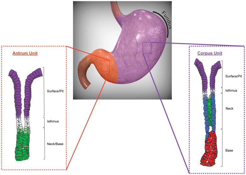
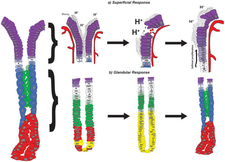
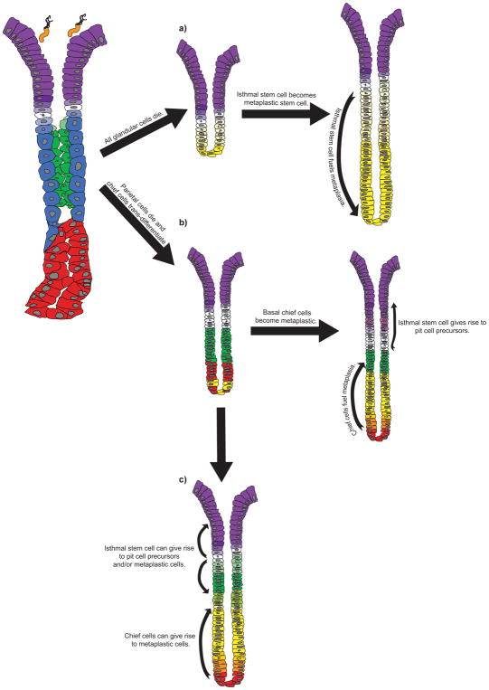
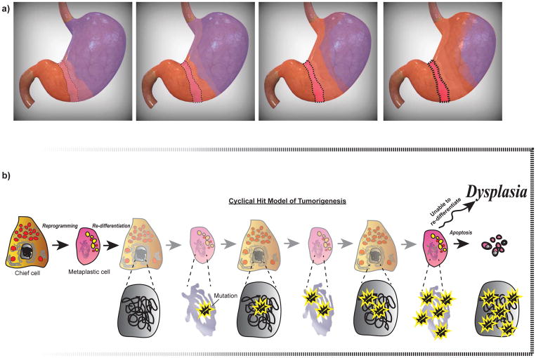
Subjected to countless daily injuries, the stomach still functions as a remarkably efficient digestive organ and microbial filter. In this Review, we follow the lead of the earliest gastroenterologists who were fascinated by the antiseptic and digestive powers of gastric secretions. We propose that it is easiest to understand how the stomach responds to injury by stressing the central role of the most important gastric secretion, acid. The stomach follows two basic patterns of adaptation. The superficial response is a pattern whereby the surface epithelial cells migrate and rapidly proliferate to repair erosions induced by acid or other irritants. The stomach can also adapt through a glandular response when the source of acid is lost or compromised (that is, the process of oxyntic atrophy). We primarily review the mechanisms governing the glandular response, which is characterized by a metaplastic change in cellular differentiation known as spasmolytic polypeptide-expressing metaplasia (SPEM). We propose that the stomach, like other organs, exhibits marked cellular plasticity: the glandular response involves reprogramming mature cells to serve as auxiliary stem cells that replace lost cells. Unfortunately, such plasticity might mean that the gastric epithelium undergoes cycles of differentiation and de-differentiation that increase the risk of accumulating cancer-predisposing mutations.


Conflict of interest statement

Competing interests

The authors declare no competing interests.

Figures

Figure 1
Figure 1. The anatomic and glandular organization of the human stomach
The human stomach can be divided into two anatomic regions, the corpus (purple) and the antrum (orange). These regions are characterized by the cellular composition of their glands, with corpus units (purple inset) defined by acid-producing parietal cells (blue) and zymogenic chief cells (red). The antrum unit (orange inset) is largely devoid of parietal cells and chief cells and instead comprises mucous cells (green) that extend down to the gland base. Both corpus and antrum units contain an isthmus region, made up of proliferative stem cells (white), and a surface/pit region, with pit cells (purple) extending up from the isthmus to the luminal surface. Pre-parietal (light blue), pre-pit (light purple), and pre-mucous (light green) cells are also shown. Note that endocrine and tuft cells in both the corpus and antrum have been omitted. The gastric fundus has been outlined. Corpus and antrum units have been adapted from Willet and Mills. The human stomach was modified from an original image obtained from turbosquid.com.
Figure 2
Figure 2. The superficial and glandular responses in the gastric corpus
The corpus unit (left) responds to gastric injury through two main mechanisms, the superficial response (a) and the glandular response (b). (a) The surface epithelium (left) consists of pit cells (purple) that produce a viscous mucus barrier that protects against endogenous (e.g., acid, denoted by H+) and exogenous (not shown) injury. Breaches in the surface epithelium resulting from acid oversecretion, decreased mucus production, and/or ischemia can lead to ulcers and/or bleeding (middle). The superficial injury response restores the protective barrier of the surface epithelium by increasing mucus production, restoring local blood flow, and re-establishing epithelial integrity through restitution and cellular proliferation (right). (b) The glandular injury response correlates with the loss of acid-producing parietal cells (blue) and a replacement of the deeper glandular epithelium with metaplastic cells (yellow) that co-express markers of mucous neck cells (green) and chief cells (red). This metaplastic response (middle) is often termed Spasmolytic Polypeptide-Expressing Metaplasia (SPEM) and represents a transient response to re-establish homeostasis (right). Pre-pit (light purple), pre-parietal (light blue), and pre-mucous neck (light green) cells are also shown. The superficial and glandular responses are not mutually exclusive and can occur simultaneously within the same corpus unit.
Figure 3
Figure 3. Distinguishing the possible cellular origins of SPEM
Three possible mechanisms for the initiation of SPEM and the replacement of depleted glandular epithelium are presented. For the isthmal stem cell (white) to serve as the unique cell of origin for SPEM (a), all of the glandular cells - parietal cells (blue), chief cells (red), and mucous neck cells (green) - must die and be replaced with cells derived from a proliferative, isthmal stem cell (yellow) that has trans-differentiated to become a stem cell for metaplastic cells. (b) Others, have hypothesized that SPEM arises from the trans-differentiation of chief cells into metaplastic cells following the loss of parietal cells. These basal metaplastic cells (yellow and orange) repopulate the corpus unit from the base up, while isthmal proliferation results only in an expansion of pit cells (purple), a process known as foveolar hyperplasia. Another hybrid model can also be envisioned wherein both the isthmal stem cells and chief cells can contribute to SPEM (c). In this scenario, two foci of proliferation may be dedicated to generating different cells along the gland axis, with the isthmal stem cell giving rise to pit cell precursors (light purple) and/or mucous neck cell precursors (light green), and the chief cells giving rise to metaplastic cells (yellow and orange). Bacteria represent Helicobacter pylori.
Figure 4
Figure 4. The expansion of SPEM and the Cyclical Hit model: a possible mechanism for dysplasia
(a) The topographic expansion of SPEM during sustained glandular injury results in the “antralization” of the corpus, such that corpus units become metaplastic and morphologically resemble antral units. The antralization of the corpus likely emerges from an initial focus of metaplasia (boxed red area) at the transition between corpus (purple) and antrum (orange) and expands proximally along the lesser curvature before spreading to the greater curvature. The transition zone (light purple) represents a dynamic, hybrid region that progresses along the leading edge of antralization. The earliest sites of antralization will have the longest history of metaplasia with associated dedifferentiation-redifferentiation cycles, increased risk of accumulation of mutations, and an increased likelihood that those mutations will seed dysplasia/neoplasia: this increased risk is denoted by an increasing color saturation of the boxed region at the corpus-antrum border. (b) The Cyclical Hit Model for the development of gastric dysplasia is presented. As post-mitotic chief cells become metaplastic and re-enter the cell cycle to proliferate and fuel metaplasia, they can accumulate genetic mutations (shown with yellow outlined symbol) through replicative stress. Chief cells within the initial focus of metaplasia (boxed area in a), a region that would have sustained the longest duration of glandular injury, harbor genetic mutations that can become unmasked and prevent re-differentiation, either leading to apoptosis or potentially serving as a cell of origin for dysplasia.

References

    1. De Fourcroy AF. Élémens d’Histoire Naturelle et de Chimie. Cuchet; Paris: 1791. pp. 357–62.
    1. Spallanzani L. Dissertations Relative to the Natural History of Animals and Vegetables. Vol. 1. J. Murray; London: 1789. This was one of the first treatises focusing on the stomach and its role in digestion.
    1. Ferlay J, et al. Cancer incidence and mortality worldwide: sources, methods and major patterns in GLOBOCAN 2012. Int J Cancer. 2015;136:E359–386. - PubMed
    1. Colquhoun A, et al. Global patterns of cardia and non-cardia gastric cancer incidence in 2012. Gut. 2015;64:1881–1888. - PubMed
    1. Carter AJ, Nguyen CN. A comparison of cancer burden and research spending reveals discrepancies in the distribution of research funding. BMC Public Health. 2012;12:526. - PMC - PubMed

Publication types