. 2017 Dec 26;9(5):6062-6074.
doi: 10.18632/oncotarget.23681. eCollection 2018 Jan 19.

[Neratinib + Valproate] exposure permanently reduces ERBB1 and RAS expression in 4T1 mammary tumors and enhances M1 macrophage infiltration

Affiliations

[Neratinib + Valproate] exposure permanently reduces ERBB1 and RAS expression in 4T1 mammary tumors and enhances M1 macrophage infiltration

Laurence Booth et al. Oncotarget. .

Abstract

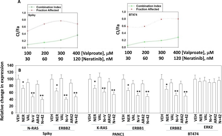
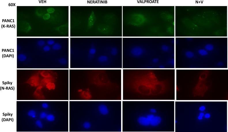
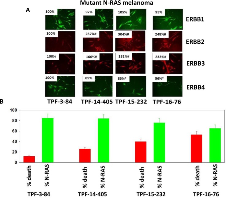
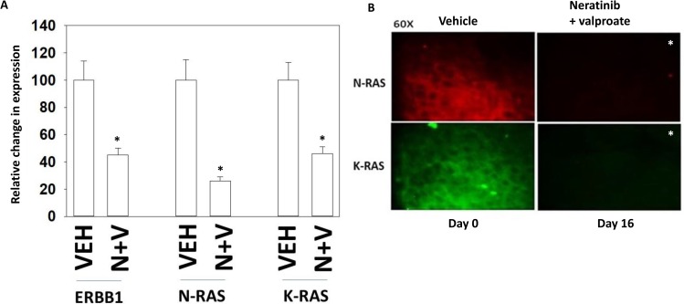
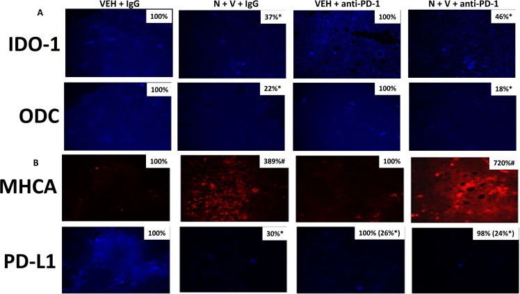
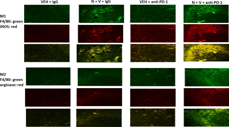
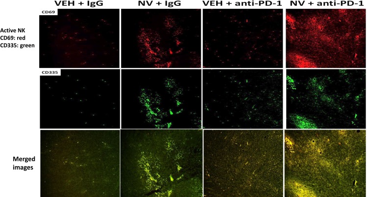
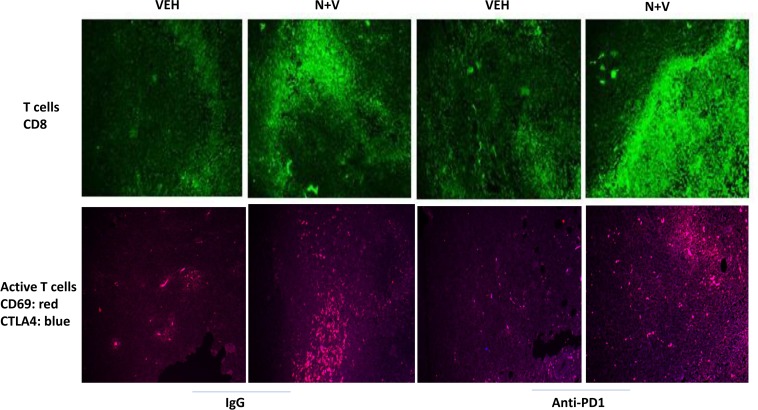
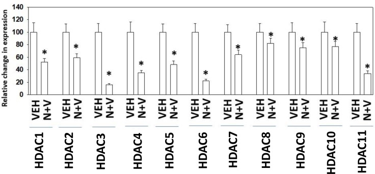
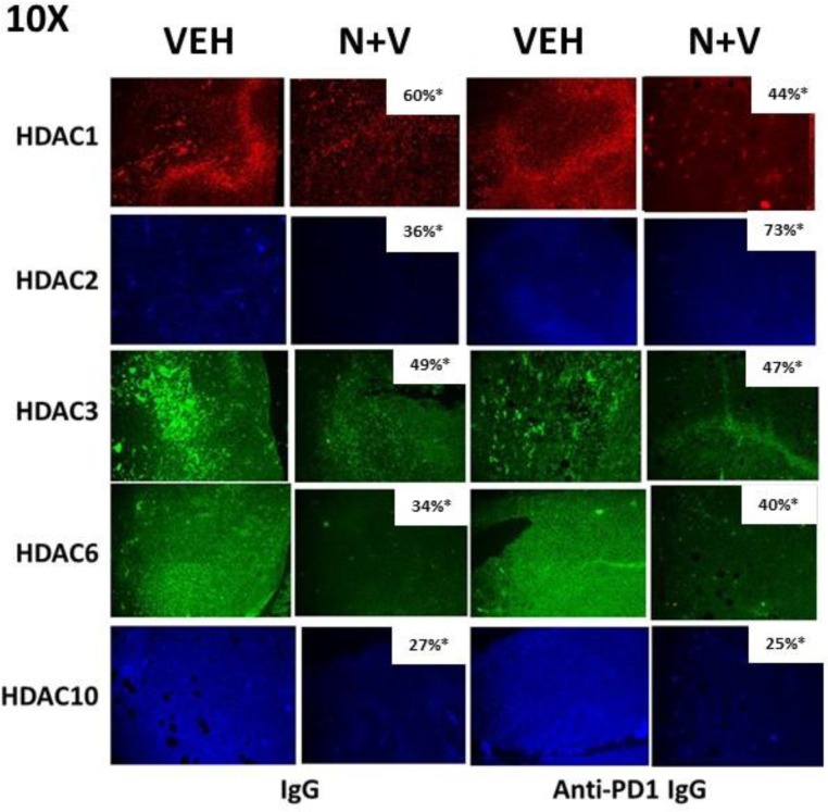
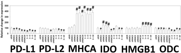
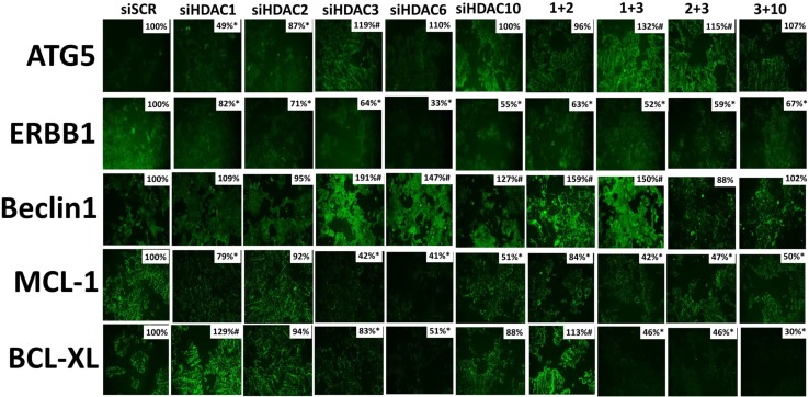
The irreversible ERBB1/2/4 inhibitor neratinib has been shown in vitro to rapidly reduce the expression of ERBB1/2/4 and RAS proteins via autophagic/lysosomal degradation. We have recently demonstrated that neratinib and valproate interact to suppress the growth of 4T1 mammary tumors but had not defined whether the [neratinib + valproate] drug combination, in a mouse, had altered the biology of the 4T1 cells. Exposure of 4T1 mammary tumors to [neratinib + valproate] for three days resulted, two weeks later, in tumors that expressed less ERBB1, K-RAS, N-RAS, indoleamine-pyrrole 2,3-dioxygenase (IDO-1), ornithine decarboxylase (ODC) and had increased Class I MHCA expression. Tumors previously exposed to [neratinib + valproate] grew more slowly than those exposed to vehicle control and contained more CD8+ cells and activated NK cells. M1 but not M2 macrophage infiltration was significantly enhanced by the drug combination. In vitro exposure of 4T1 tumor cells to [neratinib + valproate] variably reduced the expression of histone deacetylases 1-11. In vivo, prior exposure of tumors to [neratinib + valproate] permanently reduced the expression of HDACs 1-3, 6 and 10. Combined knock down of HDACs 1/2/3 or of 3/10 rapidly reduced the expression IDO-1, and ODC and increased the expression of MHCA. H&E staining of normal tissues at animal nadir revealed no obvious cyto-architectural differences between control and drug-treated animals. We conclude that [neratinib + valproate] evolves 4T1 tumors to grow more slowly and to be more sensitive to checkpoint immunotherapy antibodies.

Keywords: autophagy; neratinib; receptor tyrosine kinase; valproate.


Conflict of interest statement

CONFLICTS OF INTEREST None.

Figures

Figure 1
Figure 1. Neratinib and HDAC inhibitors synergize to kill cancer cells
(A) Spiky cells and BT474 cells were plated in 6-well plates in sextuplicate as individual cells (500 cells per well). After 12 h the cells were treated with vehicle control, neratinib, niraparib or the drugs combined, at the indicated concentrations in the figure, at a fixed ratio. After 24 h, the media is removed, the cells washed with warm drug-free media, and fresh drug-free media placed on the cells. After 10 days, colonies of > 50 cells have formed and the cells are fixed in place and stained with crystal violet. The plating efficiency under each treatment condition is determined and the fraction affected determined. Synergy was determined using the Calcusyn for Windows program using the method of Cho and Talalay (n = 2 independent studies in sextuplicate). A combination index of less than 1.00 indicates a synergy of drug interaction. The colony formation/fraction affected are plotted on the same graph. (B) Spiky, PANC1 and BT474 cells were treated with vehicle control, neratinib (50 nM), sodium valproate (250 µM), AR42 (600 nM) or the drugs in combination for 6 h as indicated. The cells were fixed in place and immunostaining performed to determine the expression of the indicated proteins at 10× magnification (data from multiple separate images & treatments ±SEM) *p < 0.05 less than vehicle control; **p < 0.05 less than neratinib alone value.
Figure 2
Figure 2. Neratinib and HDAC inhibitors interact to reduce the expression of mutant N-RAS and mutant K-RAS
Spiky and PANC1 cells that express a mutant N-RAS and a mutant K-RAS, respectively, were treated with vehicle control, neratinib (50 nM), sodium valproate (250 µM), or the drugs in combination for 6h as indicated. The cells were fixed in place and immunostaining performed with DAPI counter-stain to determine the expression levels and cellular localization of N-RAS and K-RAS at 60× magnification.
Figure 3
Figure 3. Neratinib and valproate reduce N-RAS expression in PDX melanoma isolates and variably enhance tumor cell killing
(A) Mutant N-RAS expressing PDX isolates of melanoma from the University of Pittsburgh cell bank were grown for 24 h, fixed, and stained to determine the protein expression of ERBB1, ERBB2, ERBB3 and ERBB4. (n = 3 replicates of 40 cells each assessed for their fluorescence intensity ± SEM). *p < 0.05 less than value in TPF-3-84; #p < 0.05 greater than value in TPF-3-84. (B) RED: Melanoma isolates were treated with vehicle control or with [neratinib (50 nM) + valproate (250 µM)] for 24 h. Cell viability after 24 h exposure was determined by live/dead assay. (n = 3 ± SEM) #p < 0.05 greater than vehicle control. GREEN: Melanoma isolates were treated with vehicle control or with [neratinib (50 nM) + valproate (250 µM)] for 6h. Cells were fixed in place and immunostaining performed to determine the expression level of N-RAS. (n = 3 replicates of 40 cells each assessed for their fluorescence intensity ± SEM). *p < 0.05 less than value in vehicle control.
Figure 4
Figure 4. Tumors previously exposed to [neratinib + valproate] have a sustained reduction in the expression of ERBB1, N-RAS and K-RAS
(A) 4T1 tumors, removed on Day 16 were fixed, paraffin embedded and sectioned (5 μm). Sections were renatured, blocked and immunostaining performed to determine the expression of ERBB1, N-RAS and of K-RAS. Images were taken at 10× magnification. (data from multiple separate images & treatments ± SEM) *p < 0.05 less than vehicle control. (B) 4T1 tumors, removed on Day 16 were fixed, paraffin embedded and sectioned (5 μm). Sections were renatured, blocked and immunostaining performed to determine the expression of N-RAS and K-RAS. Images were taken at 60× magnification. (data from multiple separate images & treatments ± SEM) *p < 0.05 less than vehicle control.
Figure 5
Figure 5. Prior [neratinib + valproate] exposure reduces IDO-1, ODC and PD-L1 expression and enhances MHCA expression in 4T1 tumors
(A) 4T1 tumors, removed on Day 16 were fixed, paraffin embedded and sectioned (5 μm). Sections were renatured, blocked and immunostaining performed to determine the expression of IDO-1 and of ODC. Images were taken at 10× magnification. (data from multiple separate images & treatments ± SEM) *p < 0.05 less than vehicle control. (B) 4T1 tumors, removed on Day 16 were fixed, paraffin embedded and sectioned (5 μm). Sections were renatured, blocked and immunostaining performed to determine the expression of MHCA and of PD-L1. Images were taken at 10× magnification. (data from multiple separate images & treatments ± SEM) *p < 0.05 less than vehicle control.
Figure 6
Figure 6. Prior [neratinib + valproate] exposure enhances the invasion of active NK cells, activated T cells and M1 polarized macrophages into 4T1 tumors
4T1 tumors, removed on Day 16 were fixed, paraffin embedded and sectioned (5 μm). Sections were renatured, blocked and immunostaining performed to determine the expression of F4/80, iNOS and arginase within the tumor. Images were taken at 10× magnification. Fluorescence intensity staining levels for each antibody in multiple sections were obtained to determine the relative staining intensities under each condition of F4/80, iNOS and arginase (n = 3 ± SEM, p < 0.05 staining intensity of iNOS greater than intensity of arginase).
Figure 7
Figure 7. [Neratinib + valproate] promotes activated NK cell tumor infiltration, that is enhanced by an anti-PD-1 antibody
4T1 tumors, removed on Day 16 were fixed, paraffin embedded and sectioned (5 μm). Sections were renatured, blocked and immunostaining performed to determine the expression of CD69 and of CD335 within the tumor. Images were taken at 10× magnification and presented with elevated contrast to remove low-level background staining.
Figure 8
Figure 8. [Neratinib + valproate] increases the levels of CD8+ and CD69+CTLA4+ cells in 4T1 tumors
4T1 tumors, removed on Day 16 were fixed, paraffin embedded and sectioned (5 μm). Sections were renatured, blocked and immunostaining performed to determine the expression of CD8 or of [CD69 + CTLA4] within the tumor. Images were taken at 10× magnification. Using Adobe Photoshop 6.0 the staining/co-staining images for CD8+ and [CD69+CTLA4+] were obtained. Red + Green staining = yellow; Red + Blue staining = mauve.
Figure 9
Figure 9. Exposure of 4T1 cells to [neratinib + valproate] rapidly reduces HDAC expression in vitro
4T1 cells were treated with vehicle control, neratinib (50 nM), sodium valproate (250 µM) or the drugs combined for 6 h. Cells were fixed in place and immunostaining performed to determine the expression levels of HDACs 1-11. (n = 3 replicates of 40 cells each assessed for their fluorescence intensity ± SEM). *p < 0.05 less than value in vehicle control.
Figure 10
Figure 10. Prior exposure of 4T1 tumors in vivo to [neratinib + valproate] permanently reduces HDAC expression in the surviving 4T1 cells
4T1 tumors, removed on Day 16 were fixed, paraffin embedded and sectioned (5 μm). Sections were renatured, blocked and immunostaining performed to determine the expression of HDAC1, HDAC2, HDAC3, HDAC6 and HDAC10 within the tumor. Images were taken at 10× magnification and presented with elevated contrast to remove low-level background staining. Fluorescence intensity staining levels for each antibody in multiple sections were obtained to determine the relative staining intensities the HDACs (n = 3 ± SEM). *p < 0.05 less than corresponding value in vehicle control tumors.
Figure 11
Figure 11. Knock down of HDACs reduces PD-L1, PD-L2, IDO-1 and ODC expression and enhances MHCA expression in 4T1 cells
4T1 cells were transfected with an siRNA control (siSCR) or with siRNA molecules to knock down the expression of HDAC1, HDAC2, HDAC3, HDAC6, HDAC10, or with combinations of HDAC knock down as indicated in the Figure. Twenty-four h after transfection cells were fixed in place and immunostaining performed to determine the staining intensity under each knock down condition of PD-L1, PD-L2, MHCA, IDO-1, HMGB1 and ODC. (data from multiple separate images & treatments ± SEM) *p < 0.05 less than siSCR control; #p < 0.05 greater than siSCR control.
Figure 12
Figure 12. Knock down of HDAC expression reduces the levels of ERBB1, BCL-XL and MCL-1 expression in 4T1 cells
4T1 cells were transfected with an siRNA control (siSCR) or with siRNA molecules to knock down the expression of HDAC1, HDAC2, HDAC3, HDAC6, HDAC10, or with combinations of HDAC knock down as indicated in the Figure. Twenty-four h after transfection cells were fixed in place and immunostaining performed to determine the staining intensity under each knock down condition of ATG5, ERBB1, Beclin1, MCL-1 and BCL-XL. (data from multiple separate images & treatments ± SEM) *p < 0.05 less than siSCR control; #p < 0.05 greater than siSCR control.

References

    1. Booth L, Roberts JL, Poklepovic A, Avogadri-Connors F, Cutler RE, Lalani AS, Dent P. HDAC inhibitors enhance neratinib activity and when combined enhance the actions of an anti-PD-1 immunomodulatory antibody in vivo. Oncotarget. 2017;8:90262–90277. https://doi.org/10.18632/oncotarget.21660. - DOI - PMC - PubMed
    1. Booth L, Roberts JL, Poklepovic A, Kirkwood J, Sander C, Avogadri-Connors F, Cutler RE, Lalani AS, Dent P. The levels of mutant K-RAS and mutant N-RAS are rapidly reduced by the irreversible ERBB1/2/4 inhibitor neratinib. Cancer Biol Ther. 2017 Dec 8 https://doi.org/10.1080/15384047.2017.1394556. [Epub ahead of print] - DOI - PMC - PubMed
    1. Zhang Y, Zhang J, Liu C, Du S, Feng L, Luan X, Zhang Y, Shi Y, Wang T, Wu Y, Cheng W, Meng S, Li M, et al. Neratinib induces ErbB2 ubiquitylation and endocytic degradation via HSP90 dissociation in breast cancer cells. Cancer Lett. 2016;382:176–185. - PubMed
    1. Booth L, Roberts JL, Sander C, Lee J, Kirkwood JM, Poklepovic A, Dent P. The HDAC inhibitor AR42 interacts with pazopanib to kill trametinib/dabrafenib-resistant melanoma cells in vitro and in vivo. Oncotarget. 2017;8:16367–16386. https://doi.org/10.18632/oncotarget.14829. - DOI - PMC - PubMed
    1. Booth L, Roberts JL, Poklepovic A, Gordon S, Dent P. PDE5 inhibitors enhance the lethality of pemetrexed through inhibition of multiple chaperone proteins and via the actions of cyclic GMP and nitric oxide. Oncotarget. 2017;8:1449–1468. https://doi.org/10.18632/oncotarget.13640. - DOI - PMC - PubMed