. 2018 Feb 19;5(1):ENEURO.0042-17.2018.
doi: 10.1523/ENEURO.0042-17.2018. eCollection 2018 Jan-Feb.

Sequences Flanking the Gephyrin-Binding Site of GlyRβ Tune Receptor Stabilization at Synapses

Affiliations

Sequences Flanking the Gephyrin-Binding Site of GlyRβ Tune Receptor Stabilization at Synapses

Nora Grünewald et al. eNeuro. .

Abstract

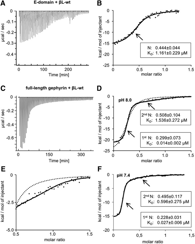
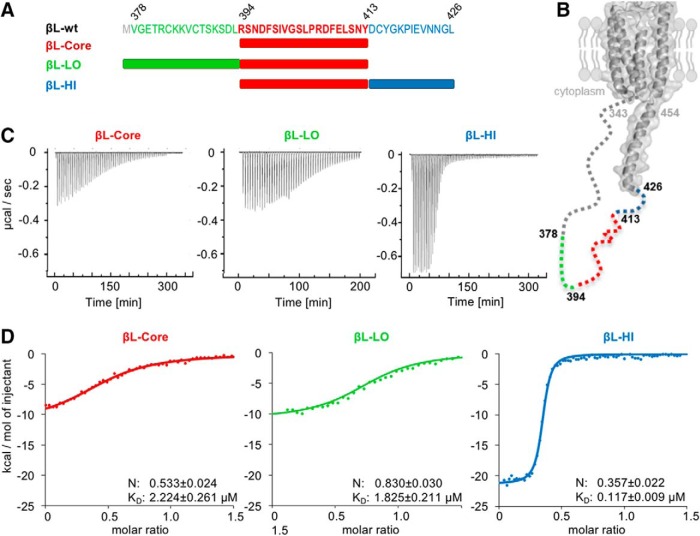
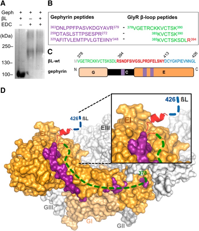
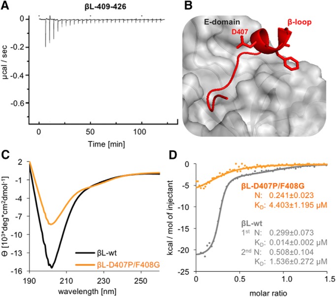
The efficacy of synaptic transmission is determined by the number of neurotransmitter receptors at synapses. Their recruitment depends upon the availability of postsynaptic scaffolding molecules that interact with specific binding sequences of the receptor. At inhibitory synapses, gephyrin is the major scaffold protein that mediates the accumulation of heteromeric glycine receptors (GlyRs) via the cytoplasmic loop in the β-subunit (β-loop). This binding involves high- and low-affinity interactions, but the molecular mechanism of this bimodal binding and its implication in GlyR stabilization at synapses remain unknown. We have approached this question using a combination of quantitative biochemical tools and high-density single molecule tracking in cultured rat spinal cord neurons. The high-affinity binding site could be identified and was shown to rely on the formation of a 310-helix C-terminal to the β-loop core gephyrin-binding motif. This site plays a structural role in shaping the core motif and represents the major contributor to the synaptic confinement of GlyRs by gephyrin. The N-terminal flanking sequence promotes lower affinity interactions by occupying newly identified binding sites on gephyrin. Despite its low affinity, this binding site plays a modulatory role in tuning the mobility of the receptor. Together, the GlyR β-loop sequences flanking the core-binding site differentially regulate the affinity of the receptor for gephyrin and its trapping at synapses. Our experimental approach thus bridges the gap between thermodynamic aspects of receptor-scaffold interactions and functional receptor stabilization at synapses in living cells.

Keywords: bimodal binding; binding site; gephyrin; glycine receptor; receptor clustering.


Figures

Figure 1.
Figure 1.
Binding properties of GlyR β-loop to full-length gephyrin or the isolated E-domain. A, Representative ITC titration profile of βL-wt (378–426; 281 µM) into GephE (31 µM) at pH 8.0. The recorded peaks were corrected by baseline-corrected injection heats. B, Binding isotherms (dots) of integrated binding heats were fitted to a one-site model (black line). The average dissociation constant (KD) and binding stoichiometry with GephE (N) of five independent experiments are given. C, Representative ITC titration profile of βL-wt (327 µM) into gephyrin (29 µM). D, Binding isotherm (dots) of integrated binding heats were fitted to a two-site model (black line) or a one-site model (dotted line). An individual measurement of βL-wt binding to gephyrin is shown alongside with averaged thermodynamic parameters of both sites (binding stoichiometry N and dissociation constant KD). Binding enthalpies (ΔH in kcal/mol) for βL-wt high and low affinity were compared using an unpaired two-tailed t test: p = 0.0005 βL-wt high-affinity site n = 3 versus βL-wt low-affinity site n = 3. E, Magnification of the graph represented in D, showing the fitted curves of the binding isotherm of βL-wt and gephyrin (dots) derived from the two-site (black line) or the one-site (dotted line) binding model. F, ITC data showing the bimodal binding between βL-wt and gephyrin at pH 7.4. Binding isotherms of βL-wt (378–426; 248 µM) into gephyrin (28.6 µM) at pH 7.4. Binding isotherms (dots) of integrated binding heats were fitted to a two-site model (black line). An individual measurement of βL-wt binding to gephyrin is shown alongside with averaged thermodynamic parameters of both sites (binding stoichiometry N and dissociation constant KD). Binding affinities and enthalpies for βL-wt high- and low-affinity binding sites at pH 8.0 and 7.4 were compared using an unpaired two-tailed t test: p = 0.2143 KD βL-wt high-affinity sites n = 3; p = 0.0958 KD βL-wt low-affinity sites n = 3; p = 0.1889 ΔH βL-wt high-affinity sites n = 3; p = 0.0849 ΔH βL-wt low-affinity sites n = 3.
Figure 2.
Figure 2.
Dissection of the bimodal binding between GlyRβ and gephyrin. A, GlyR β-loop peptides include full-length βL378-426 (βL-wt), C-terminal (βL-LO, green), and N-terminal (βL-HI, blue) truncations as well as a core region (βL-Core, red). B, Structural model of the GlyR β-subunit based on the crystal structure of the nicotinic acetylcholine receptor (Unwin, 2005). Structural information corresponding to GlyRβ residues 343–426 of the ICD is lacking and therefore depicted with a dashed line. The position of the analyzed GlyR β-loop peptides is depicted. C, Representative ITC titration profiles of βL-Core, βL-LO, and βL-HI (250–300 µM each) to 20–30 µM gephyrin under similar conditions. D, Fitting of the ITC binding isotherms (dots) of βL-Core, βL-LO, and βL-HI to a one-site binding model (colored traces). Representative recordings are shown together with averaged KD values and binding stoichiometry with gephyrin (N). Data were compared using an unpaired two-tailed t test: p = 0.0008 KD of βL-wt high-affinity site n = 3 versus βL-Core n = 9; p = 0.3234 KD of βL-Core n = 9 versus βL-LO n = 5; p < 0.0001 N of βL-Core n = 9 versus βL-LO n = 5; p = 0.0003 KD of βL-Core n = 9 versus βL-HI n = 4; p = 0.001 N of βL-Core n = 9 versus βL-HI n = 4.
Figure 3.
Figure 3.
Extension of the GlyRβ binding site on gephyrin. A, EDC-based crosslinking of βL-wt (378–426) and gephyrin. Gephyrin (Geph) and GlyR β-loop (βL) were treated (lanes 2 and 3) or not treated (lane 1) with EDC and separated by 6% SDS-PAGE (Coomassie staining). B, Identification of crosslinked peptides. Bands corresponding to the protein complexes shown in A were extracted and analyzed by peptide mass fingerprinting. The crosslinked peptides of gephyrin (purple) and the β-loop (green, red) were identified several times. C, Amino acid sequence of βL-wt with the localization of N- (green, 378–393) and C-terminal (blue, 414–426) flanking sequences of the core gephyrin-binding site (red, 394–426). Schematic representation of gephyrin domains with highlighted positions of identified peptides in the C- and E-domain (purple). D, Surface representation of a modeled trimeric full-length gephyrin (modified from (Belaidi and Schwarz, 2013) with highlighted peptides identified in the crosslinked gephyrin-GlyR β-loop complex. Dashed lines indicate regions in the βL for which structural information is lacking. Gephyrin protomer II and III: light gray; protomer I: E-domain, orange and G-domain, light orange; βL core sequence: red; βL N-terminal flanking sequence: green; βL C-terminal flanking sequence: blue; and crosslinked peptides of the gephyrin E-domain: purple.
Figure 4.
Figure 4.
Impact of the GlyR β-loop conformation on gephyrin binding. A, ITC titration profile of βL409–426 (445 µM) into gephyrin (25 µM). B, The GlyR β-loop in association with GephE adopts a short 310-helix formed by residues 406–410 (Kim et al., 2006). Two residues, Asp407 and Phe408, were mutated to proline and glycine (βL-D407P/F408G) to block the formation of the 310-helix. C, βL-wt and βL-D407P/F408G (both 0.21 mg/ml) folding was compared by CD spectroscopy. The mean residue ellipticity (θ) was plotted against the respective wavelength. D, Comparison of the binding isotherms of representative measurements using 327 µM βL-wt peptide (gray dots, same data as in Fig. 1D,E) and 315 µM βL-D407P/F408G (orange dots) with 29 or 32 µM gephyrin, respectively. Curve calculation was performed based on a two-site model for βL-wt (gray line) and a one-site model for βL-D407P/F408G (orange line). Data were compared using an unpaired two-tailed t test: p = 0.0267 KD of βL-wt high-affinity site n = 3 versus βL-D407P/F408G n = 4.
Figure 5.
Figure 5.
Membrane diffusion of TMD-βL variants in spinal cord neurons. A, B, sptPALM was done using Dendra2-tagged TMD-βL variants in cultured neurons as described in Materials and Methods. Single molecule trajectories were recorded in 10,000 frames at an acquisition rate of 15 ms (red traces). Active synapses were identified using FM 4-64 labeling (binarized fluorescence images shown in white). Left, High-density sptPALM of dendritic segments expressing TMD-βL-WT (A) or the gephyrin binding-deficient construct TMD-βL-geph- (B). Right, Zoomed recordings showing confinement of TMD-βL-WT at synapses (A) as opposed to the high mobility of TMD-βL-geph- (B). Scale bar: 5 µm (left panels); pixel size of FM-labeled synapses: 160 nm (right panels). C, D, Comparison of the areas explored by the TMD-βL variants at synapses (C) and in the extrasynaptic compartment (D), represented by the mean value (colored dots), the median, 25% and 75% quartiles of the trajectories (boxes). Explored areas were normalized by the number of detections for each trajectory. Data were compared via one-way ANOVA (Kruskal–Wallis test) followed by a post hoc Dunn’s multiple comparison test: all pairs were significantly different from one another with p < 0.001. E, F, Cumulative histogram of diffusion coefficients of TMD-βL variants in spinal cord neurons. Diffusion coefficients at synapses vary according to the strength of βL-gephyrin binding (E). The variants display comparable diffusion behaviors at extrasynaptic locations (F). Data were compared with one-way ANOVA (Kruskal–Wallis test) followed by a post hoc Dunn’s multiple comparison test: for all pairs, Deff was significantly different with p < 0.001, except for TMD-βL-ΔCore versus TMD-βL-D407P/F408G at synapses with p > 0.05 (median values and quartiles with statistical comparison are given in Tables 1, 4).
Figure 6.
Figure 6.
Single molecule diffusion of full-length GlyR complexes in spinal cord neurons. A, Deff (left panel) of mEos4b-tagged GlyRs at synapses were determined by sptPALM, using wild-type GlyRβ subunits (black trace) and the variants GlyRβ-HI (blue), GlyRβ-LO (green), GlyRβ-D407P/F408G (yellow), and GlyRβ-geph- (red). The dotted line indicates the median Deff value of mEos4b-GlyRβ-wt trajectories in a fixed sample (0.02 µm2/s) that is the limit of resolution in our recordings. The explored areas (normalized by the number of detections, right panel) are represented by their mean (colored dots), median, 25% and 75% quartiles (boxes) of the trajectory population. B, Distribution of synaptic trajectories (data shown in A) with Deff values >0.02 µm2/s. Differences in the diffusion coefficients (left) and the corresponding areas (right) of the GlyRβ variants are evident for the thresholded data. Data were compared with one-way ANOVA (Kruskal–Wallis test) followed by a post hoc Dunn’s multiple comparison test: explored areas and Deff values were significantly different for all pairs (p < 0.0001), except for GlyRβ-LO versus GlyRβ-D407P/F408G with p > 0.05 (median values and quartiles are given in Table 4). C, Deff (left panel) and explored areas (right) of GlyRβ variants outside of synapses (significantly different between all conditions, p < 0.001).
Figure 7.
Figure 7.
Correlation of in vitro binding affinities and diffusion coefficients. The binding affinities of βL-WT (high-affinity site), βL-HI and βL-D407P/F408G determined by ITC correlate with their Deff obtained by sptPALM recordings in living spinal cord neurons (TMD-βL variants).

References

    1. Belaidi AA, Schwarz G (2013) Metal insertion into the molybdenum cofactor: product-substrate channelling demonstrates the functional origin of domain fusion in gephyrin. Biochem J 450:149–157. 10.1042/BJ20121078 - DOI - PubMed
    1. Biron Z, Khare S, Samson AO, Hayek Y, Naider F, Anglister J (2002) A monomeric 3(10)-helix is formed in water by a 13-residue peptide representing the neutralizing determinant of HIV-1 on gp41. Biochemistry 41:12687–12696. 10.1021/bi026261y - DOI - PubMed
    1. Burgos CF, Castro PA, Mariqueo T, Bunster M, Guzmán L, Aguayo LG (2015) Evidence for α-helices in the large intracellular domain mediating modulation of the α1-glycine receptor by ethanol and Gβγ. J Pharmacol Exp Ther 352:148–155. 10.1124/jpet.114.217976 - DOI - PMC - PubMed
    1. Choquet D, Triller A (2003) The role of receptor diffusion in the organization of the postsynaptic membrane. Nat Rev Neurosci 4:251–265. 10.1038/nrn1077 - DOI - PubMed
    1. Choquet D, Triller A (2013) The dynamic synapse. Neuron 80:691–703. 10.1016/j.neuron.2013.10.013 - DOI - PubMed

Publication types