. 2018 Feb;70(2):103-114.
doi: 10.1007/s10858-017-0162-1. Epub 2018 Jan 18.

Segmental isotopic labeling of HIV-1 capsid protein assemblies for solid state NMR

Affiliations

Segmental isotopic labeling of HIV-1 capsid protein assemblies for solid state NMR

Sebanti Gupta et al. J Biomol NMR. 2018 Feb.

Abstract

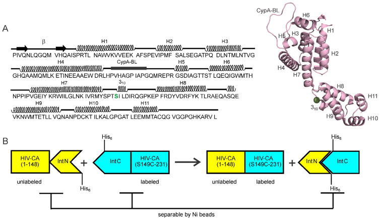
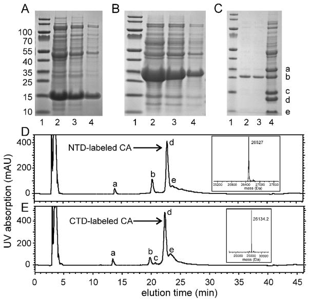
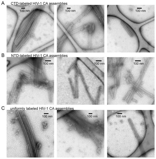
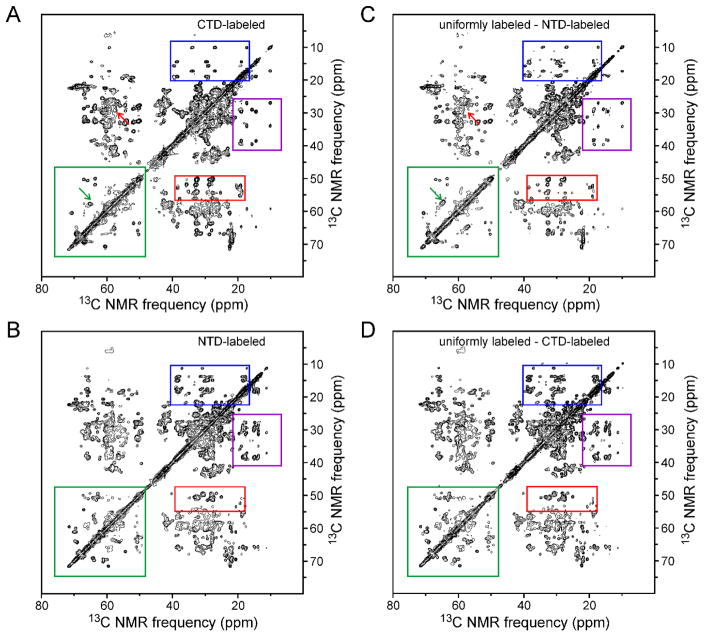
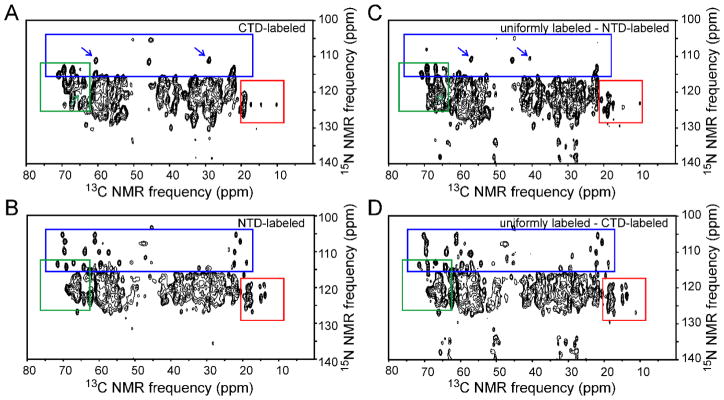
Recent studies of noncrystalline HIV-1 capsid protein (CA) assemblies by our laboratory and by Polenova and coworkers (Protein Sci 19:716-730, 2010; J Mol Biol 426:1109-1127, 2014; J Biol Chem 291:13098-13112, 2016; J Am Chem Soc 138:8538-8546, 2016; J Am Chem Soc 138:12029-12032, 2016; J Am Chem Soc 134:6455-6466, 2012; J Am Chem Soc 132:1976-1987, 2010; J Am Chem Soc 135:17793-17803, 2013; Proc Natl Acad Sci USA 112:14617-14622, 2015; J Am Chem Soc 138:14066-14075, 2016) have established the capability of solid state nuclear magnetic resonance (NMR) measurements to provide site-specific structural and dynamical information that is not available from other types of measurements. Nonetheless, the relatively high molecular weight of HIV-1 CA leads to congestion of solid state NMR spectra of fully isotopically labeled assemblies that has been an impediment to further progress. Here we describe an efficient protocol for production of segmentally labeled HIV-1 CA samples in which either the N-terminal domain (NTD) or the C-terminal domain (CTD) is uniformly 15N,13C-labeled. Segmental labeling is achieved by trans-splicing, using the DnaE split intein. Comparisons of two-dimensional solid state NMR spectra of fully labeled and segmentally labeled tubular CA assemblies show substantial improvements in spectral resolution. The molecular structure of HIV-1 assemblies is not significantly perturbed by the single Ser-to-Cys substitution that we introduce between NTD and CTD segments, as required for trans-splicing.

Keywords: HIV-1 capsid; Intein; Protein assembly; Protein structure; Segmental labeling; Solid state nmr.


Figures

Figure 1
Figure 1
(A) HIV-1 CA sequence, with secondary structure elements indicated above the sequence. The splicing site is at the NTD-CTD linker region, highlighted in green. A cartoon representation of the CA monomer structure (Protein Data Bank file 4XFX) shows the eleven helical segments (H1–H7 of NTD, H8–H11 of CTD), the cyclophilin A binding loop (CypA-BL), and a 310 helix immediately after the linker segment between NTD and CTD. (B) Schematic representation of the plasmid design and the purification strategy for the preparation of segmentally labeled HIV-1 CA. In this case, the CTD segment would be labeled.
Figure 2
Figure 2
(A) SDS-PAGE analysis of the cell lysate from overexpresssion of the CTD-intein fusion protein. Lane 1 contains molecular weight markers. Lanes 2–4 contain decreasing quantities of the lysate. The band near 15 kDa is the CTD-intein fusion protein. Gels were stained using Imperial protein stain from ThermoFisher Scientific. (B) Same as panel A, but for the NTD-intein fusion protein, which is the band near 30 kDa. (C) SDS-PAGE analysis of the ligation mixture after 40 h of incubation (lane 4) and the purified ligation product (lanes 2 and 3). Bands marked a–e are the NTD-intein fusion protein, ligated HIV-1 CA, the CTD-intein fusion protein, the N-terminal intein segment of Npu DnaE, and the C-terminal intein segment, respectively. (D) Reverse phase HPLC chromatogram of the ligation mixture after 40 h of incubation to produce full-length CA with a uniformly 15N,13C-labeled NTD segment. UV absorption peaks (280 nm) marked a–e are the C-terminal intein segment, the CTD-intein fusion protein, the N-terminal intein segment, the ligated CA product, and the NTD-intein fusion protein, respectively. The inset shows the mass spectrum of peak d, after deconvolution of multi-charged mass data from electrospray ionization. (E) Same as panel D, but for production of full-length CA with a uniformly 15N,13C-labeled CTD segment.
Figure 3
Figure 3
Characterization of the HIV-1 CA assemblies by TEM. Rows A–C are images of negatively stained tubular assemblies formed by CTD-labeled CA, NTD-labeled CA, and uniformly labeled CA, respectively.
Figure 4
Figure 4
(A,B) Aliphatic regions of 2D solid state 13C-13C NMR spectra of CTD-labeled CA assemblies and NTD-labeled CA assemblies, respectively. Color-coded rectangles indicate regions where clear differences in crosspeak patterns are observed. (C) 2D spectrum obtained by subtracting the spectrum of NTD-labeled CA assemblies from that of fully-labeled CA assemblies. (D) 2D spectrum obtained by subtracting the spectrum of CTD-labeled CA assemblies from that of fully-labeled CA assemblies. Green arrows in panels A and C indicate a crosspeak from S149 that is present in panel C but absent from panel A, due to the S149C substitution in the segmentally labeled samples. Red arrows indicate a crosspeak from C149 or C198 that is present in panel A but absent from panel C. Contour levels increase by successive factors of 1.7.
Figure 5
Figure 5
Same as Fig. 4, but for 2D NCACX spectra of segmentally and fully labeled HIV-1 CA assemblies. Arrows in panels A and C indicate crosspeaks from C218 that are shifted due to the difference in C198-C218 disulfide formation in the segmentally labeled and fully labeled samples, resulting from reducing conditions maintained during CA self-assembly. Contour levels increase by successive factors of 1.5.
Figure 6
Figure 6
Same as Fig. 4, but for 2D NCOCX spectra of segmentally and fully labeled HIV-1 CA assemblies. Contour levels increase by successive factors of 1.5.

References

    1. Ganser-Pornillos BK, Yeager M, Sundquist WI. The structural biology of HIV assembly. Curr Opin Struct Biol. 2008;18:203–217. - PMC - PubMed
    1. Briggs JAG, Kräusslich HG. The molecular architecture of HIV. J Mol Biol. 2011;410:491–500. - PubMed
    1. Engelman A, Cherepanov P. The structural biology of HIV-1: Mechanistic and therapeutic insights. Nature Rev Microbiol. 2012;10:279–290. - PMC - PubMed
    1. Gamble TR, Yoo SH, Vajdos FF, vonSchwedler UK, Worthylake DK, Wang H, McCutcheon JP, Sundquist WI, Hill CP. Structure of the carboxyl-terminal dimerization domain of the HIV-1 capsid protein. Science. 1997;278:849–853. - PubMed
    1. Gitti RK, Lee BM, Walker J, Summers MF, Yoo S, Sundquist WI. Structure of the amino-terminal core domain of the HIV-1 capsid protein. Science. 1996;273:231–235. - PubMed

LinkOut - more resources