

Difference in regional neural fluctuations and functional connectivity in Crohn's disease: a resting-state functional MRI study
- PMID: 29464530
- PMCID: PMC6218319
- DOI: 10.1007/s11682-018-9850-z
Difference in regional neural fluctuations and functional connectivity in Crohn's disease: a resting-state functional MRI study
Abstract
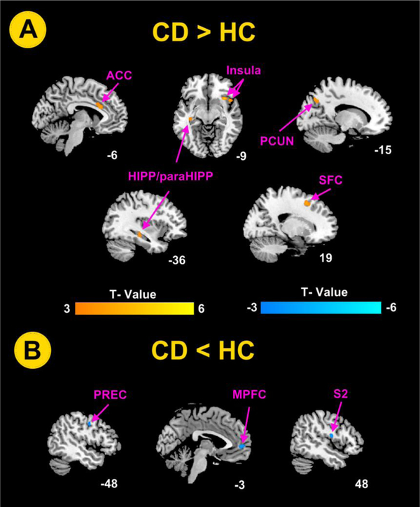
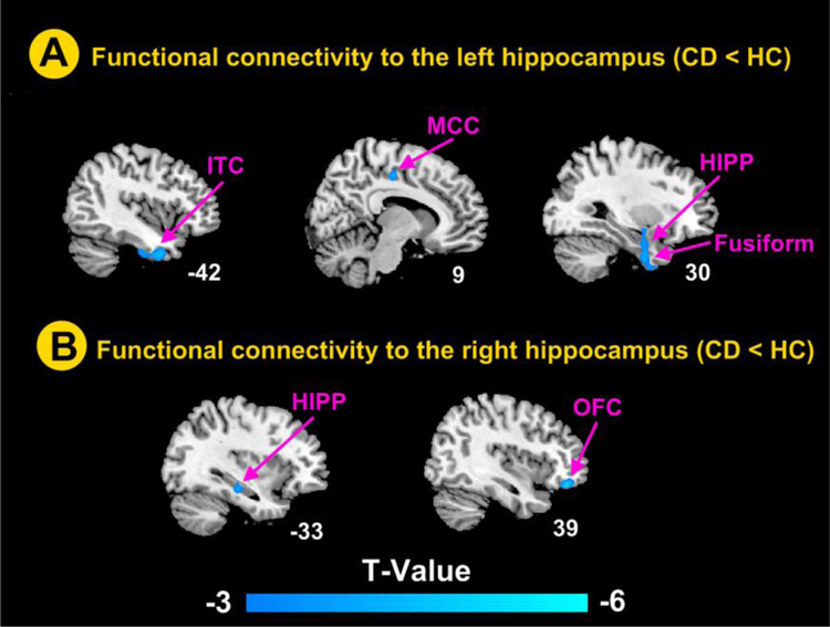
Patients with Crohn's disease (CD) are shown to have abnormal changes in brain structures. This study aimed to further investigate whether these patients have abnormal brain activities and network connectivity. Sixty patients with CD and 40 healthy controls (HCs) underwent resting-state functional magnetic resonance imaging (fMRI) scans. Amplitude of low-frequency fluctuation (ALFF) and seed-based functional connectivity (FC) were used to assess differences in spontaneous regional brain activity and functional connectivity. Compared to the HCs, patients with CD showed significantly higher ALFF values in hippocampus and parahippocampus (HIPP/paraHIPP), anterior cingulate cortex, insula, superior frontal cortex and precuneus. The ALFF values were significantly lower in secondary somatosensory cortex (S2), precentral gyrus, and medial prefrontal cortex. Functional connectivities between left HIPP and left inferior temporal cortex, and right middle cingulate cortex, HIPP, and fusiform area were significantly lower. The functional connectivities between right HIPP and right inferior orbitofrontal cortex and left HIPP were also significantly lower. Patients with CD showed higher or lower spontaneous activity in multiple brain regions. Altered activities in these brain regions may collectively reflect abnormal function and regulation of visceral pain and sensation, external environmental monitoring, and cognitive processing in these patients. Lower functional connectivity of the hippocampus-limbic system was observed in these patients. These findings may provide more information to elucidate the neurobiological mechanisms of the disease.
Keywords: Amplitude of low-frequency fluctuation; Crohn’s disease; Functional connectivity; Resting-state functional MRI.
Conflict of interest statement
Figures
References
-
- Agostini A, Benuzzi F, Filippini N, Bertani A, Scarcelli A, Farinelli V et al. (2013). New insights into the brain involvement in patients with Crohn′s disease: a voxel-based morphometry study. Neurogastroenterology & Motility, 25(2), 147–e82. - PubMed
-
- Al OY & Aziz Q (2014). The brain-gut axis in health and disease. Advances in Experimental Medicine & Biology, 817, 135–153. - PubMed
-
- Bao CH, Liu P, Liu HR, Wu LY, Shi Y, Chen WF et al. (2015). Alterations in Brain Grey Matter Structures in Patients With Crohn’s Disease and Their Correlation With Psychological Distress. Journal of Crohn S & Colitis, 9(7), 532–540. - PubMed
MeSH terms
Grants and funding
LinkOut - more resources
Full Text Sources
Other Literature Sources
Medical
