. 2018 May;12(5):611-629.
doi: 10.1002/1878-0261.12184. Epub 2018 Mar 14.

CK1ε and p120-catenin control Ror2 function in noncanonical Wnt signaling

Affiliations

CK1ε and p120-catenin control Ror2 function in noncanonical Wnt signaling

Josué Curto et al. Mol Oncol. 2018 May.

Abstract

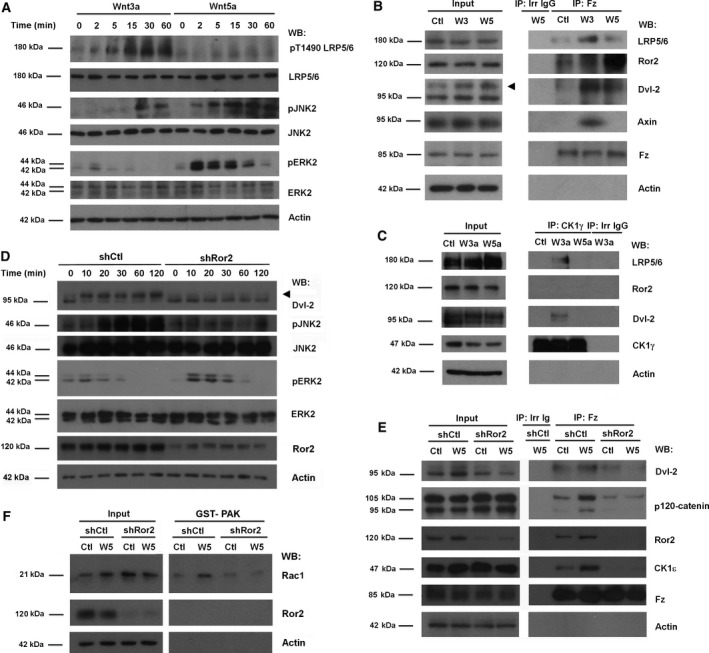
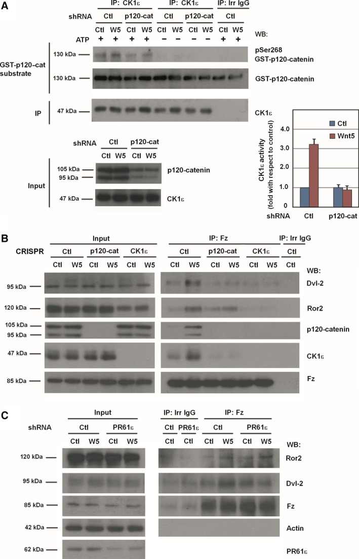
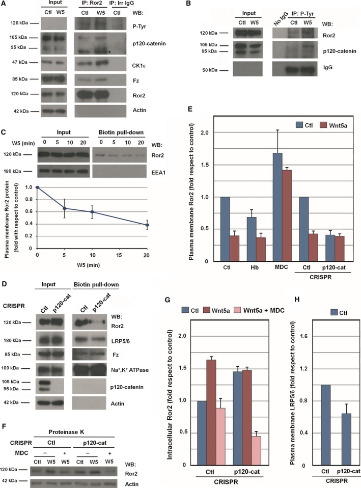
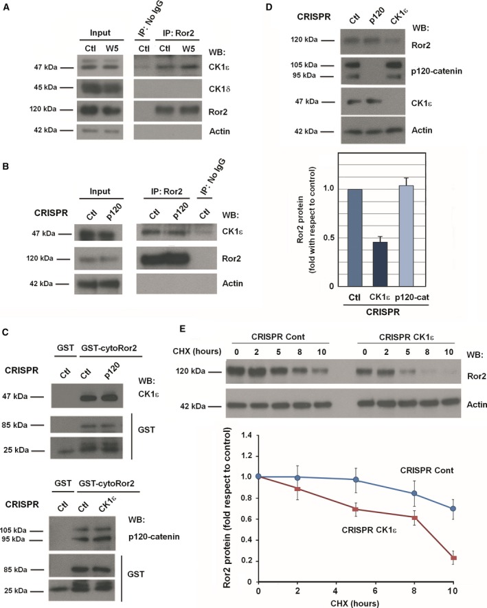
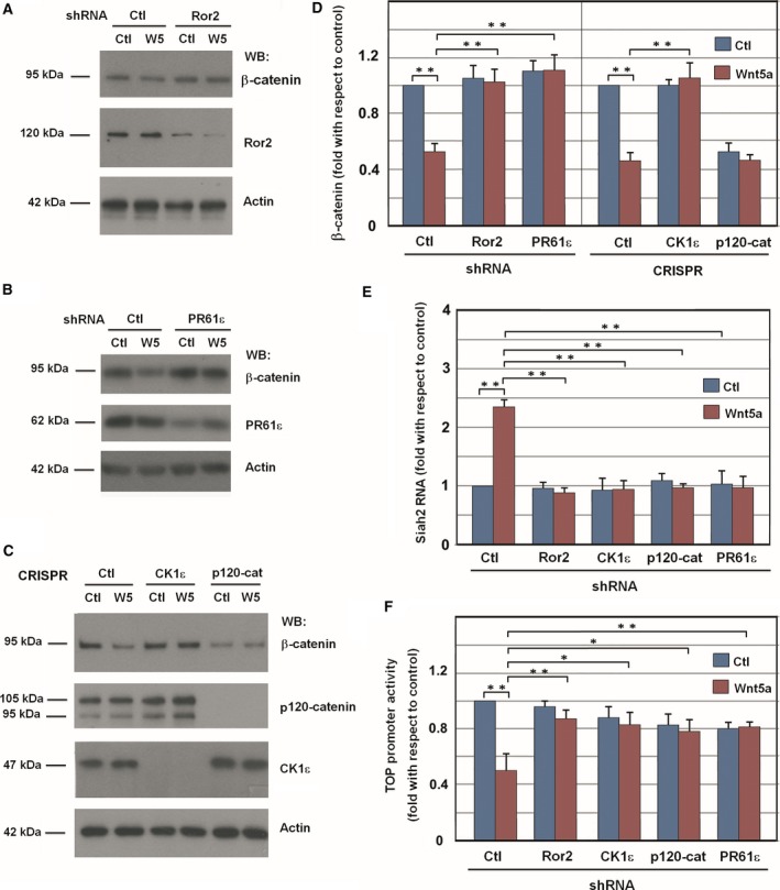
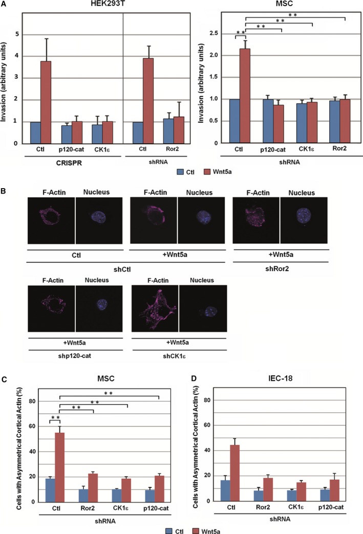
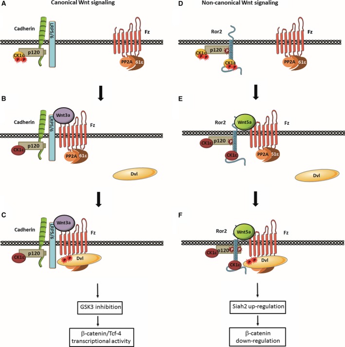
Canonical and noncanonical Wnt pathways share some common elements but differ in the responses they evoke. Similar to Wnt ligands acting through the canonical pathway, Wnts that activate the noncanonical signaling, such as Wnt5a, promote Disheveled (Dvl) phosphorylation and its binding to the Frizzled (Fz) Wnt receptor complex. The protein kinase CK1ε is required for Dvl/Fz association in both canonical and noncanonical signaling. Here we show that differently to its binding to canonical Wnt receptor complex, CK1ε does not require p120-catenin for the association with the Wnt5a co-receptor Ror2. Wnt5a promotes the formation of the Ror2-Fz complex and enables the activation of Ror2-bound CK1ε by Fz-associated protein phosphatase 2A. Moreover, CK1ε also regulates Ror2 protein levels; CK1ε association stabilizes Ror2, which undergoes lysosomal-dependent degradation in the absence of this kinase. Although p120-catenin is not required for CK1ε association with Ror2, it also participates in this signaling pathway as p120-catenin binds and maintains Ror2 at the plasma membrane; in p120-depleted cells, Ror2 is rapidly internalized through a clathrin-dependent mechanism. Accordingly, downregulation of p120-catenin or CK1ε affects late responses to Wnt5a that are also sensitive to Ror2, such as SIAH2 transcription, cell invasion, or cortical actin polarization. Our results explain how CK1ε is activated by noncanonical Wnt and identify p120-catenin and CK1ε as two critical factors controlling Ror2 function.

Keywords: CK1ε; Ror2; noncanonical Wnt; p120-catenin.


Figures

Figure 1
Figure 1
Noncanonical Wnt promotes Ror2‐dependent Dvl2 binding to Fz. (A) HEK293T cells were stimulated with Wnt3a‐ or Wnt5a‐conditioned medium for the indicated times. Cells were lysed, and proteins were analyzed by WB with specific antibodies. JNK2, ERK2, and LRP5/6 phosphorylation were determined with anti‐phospho antibodies against JNK (Thr183/Tyr185, Thr221/Tyr223), ERK (Thr202/Tyr2014), or LRP5/6 (Thr1490). (B, C) HEK293T cells treated with control, Wnt3a‐ or Wnt5a‐conditioned medium for 15 min were lysed, and total Fz (B) or CK1γ (C) were immunoprecipitated with specific antibodies. Associated proteins were analyzed by WB. (D) HEK293T cells depleted of Ror2 with specific shRNA were stimulated with control or Wnt5a‐conditioned medium for the indicated times. Cells were lysed, and Dvl2 phosphorylation was determined by the shift in the molecular weight of Dvl2, and JNK2 and ERK2 phosphorylation as above. The arrow indicates phosphorylated Dvl2. (E, F) HEK293T cells depleted of Ror2 were treated with control or Wnt5a‐conditioned medium for 5 min (E) or 1 h (F). In (F), a GSTPAK pull‐down assay was performed, and active Rac1 was determined by WB. In (E), Fz was immunoprecipitated from total extracts, and associated proteins were analyzed by WB.
Figure 2
Figure 2
p120‐catenin, CK1ε, and PR61ε are necessary for the activation of noncanonical Wnt pathway. (A) Control or p120‐catenin‐depleted HEK293T cells were treated with control or Wnt5a‐conditioned medium for 15 min. CK1ε was immunoprecipitated from total cell extracts, and the immunocomplex was incubated with 2 pmol of recombinant GST–p120‐catenin in CK1 phosphorylation conditions. Phosphorylation of Ser268 in GST–p120‐catenin was analyzed by WB with a specific PSer268 p120‐catenin antibody. Signal was densitometered, normalized with respect to the GST–p120‐catenin, and represented. The quantification of three different experiments is shown (mean ± SD). The extent of p120‐catenin downregulation by the shRNA is shown in the bottom panel. (B) Fz2 was immunoprecipitated from control, p120‐catenin, or CKHEK293T CRISPR whole‐cell extracts treated with control or Wnt5a‐conditioned medium for 5 min. Protein complexes were analyzed by WB with the indicated antibodies. (C) HEK293T cells depleted of PR61ε using specific shRNA, or a scrambled shRNA as control, were treated with control or Wnt5a‐conditioned medium for 5 min. Fz2 was immunoprecipitated from total cell extracts, and the protein complex was analyzed by WB.
Figure 3
Figure 3
Ror2 internalization is controlled by p120‐catenin. HEK293T cells were treated with control or Wnt5a‐conditioned medium for 5 min. Cells were lysed, and Ror2 (A) or tyrosine‐phosphorylated proteins (B) were immunoprecipitated with specific antibodies. Associated proteins were analyzed by WB. The asterisk in (A) indicates an unspecific band. (C, D) Surface proteins were biotinylated in HEK293T cells treated with control or Wnt5a‐conditioned medium for the indicated times (C), or in control and p120‐catenin HEK293T CRISPR cells (D). A pull‐down assay was performed with NeutrAvidin Agarose, and biotinylated membrane proteins were analyzed by WB. In (C, bottom), Ror2 protein levels at plasma membrane were quantified by analyzing three independent experiments (mean ± SD). (E) Control or p120‐catenin HEK293T CRISPR cells were pretreated with 20 ng·mL −1 herbimycin (Hb) for 1 h, or with 50 μm monodansylcadaverine (MDC) for 30 min, as indicated. Cells were then stimulated with control or Wnt5a‐conditioned medium for an additional 20 min. After cell surface biotinylation, lysates were precipitated with NeutrAvidin Agarose. The amount of cell surface Ror2 was quantified and represented. (F) Control or p120‐catenin HEK293T CRISPR cells were pretreated with 50 μm MDC for 30 min. Cells were then stimulated with control or Wnt5a‐conditioned medium for an additional 20 min with MDC, as indicated. Intact cells were treated with proteinase K for 10 min. Total cell extracts were prepared, and total Ror2 levels were analyzed by WB. (G, H) Autoradiograms from the three different experiments performed in (D) and (F) were quantified and represented. In (G), intracellular Ror2 levels were quantified, and in (H), LRP5/6 plasma membrane levels were quantified.
Figure 4
Figure 4
CK1ε binds and stabilizes Ror2. (A, B) Control or p120‐catenin HEK293T CRISPR cells were treated with control or Wnt5a‐conditioned medium for 5 min, and Ror2 was immunoprecipitated from total cell extracts. Associated proteins were analyzed by WB. (C) Pull‐down assays were performed by incubating 700 μg total cell extracts from control, p120‐catenin, or CKHEK293T CRISPR cells with 10 pmol of GST–cyto‐Ror2. Protein complexes were affinity purified and analyzed by WB. (D) Control, p120‐catenin, and CKHEK293T CRISPR cells were lysed and analyzed by WB (top). Autoradiograms from three different experiments were quantified, and total Ror2 protein levels are shown (mean ± SD) (bottom). (E) Control or CKHEK293T CRISPR were treated with 50 μg·mL −1 cycloheximide for the indicated times. Cells were lysed, and Ror2 protein levels were analyzed by WB (top). Autoradiograms from four different experiments performed were quantified using quantity one software (Bio‐Rad, Hercules, CA, USA) and represented for each time point with respect to the control (mean ± SD) (bottom).
Figure 5
Figure 5
Downregulation of Ror2, p120‐catenin, CK1ε, or PR61ε prevents the β‐catenin down‐modulation induced by Wnt5a. HEK293T cells were depleted of Ror2 (A) or PR61ε (B) using specific shRNA; a scrambled shRNA was used as a control. After 48 h, cells were stimulated with control or Wnt5a‐conditioned medium overnight, and β‐catenin levels were analyzed by WB from total cell extracts. (C) Control, p120‐catenin, and CKCRISPR HEK293T cells were treated with control or Wnt5a‐conditioned medium overnight, and β‐catenin was analyzed by WB. (D) β‐catenin levels were quantified by analyzing three independent experiments performed in (A–C) (mean ± SD). **< 0.01. (E) RNA was isolated from control, Ror2, CK1ε, p120‐catenin, and PR61ε‐depleted HEK293T cells stimulated overnight with control or Wnt5a‐conditioned medium. Expression of SIAH2 was assessed by semi‐quantitative RTPCR. Results are presented as mean ± SD from three independent experiments. *< 0.05; **< 0.01. (F) β‐catenin transcriptional activity was determined using the TOP‐Flash reporter plasmid in SW‐480 cells transfected with the indicated shRNA. pTK‐Renilla plasmid was transfected to normalize the efficiency of transfection. Relative luciferase activity was determined 48 h after transfection. Cells were treated with Wnt5a for 16 h before cell lysis. FOP‐Flash plasmid was also transfected as a control; the activity of this promoter was always lower than 1% of the value of obtained with TOP‐Flash. The mean ± SD of four experiments is shown. **< 0.01.
Figure 6
Figure 6
The co‐receptor Ror2, p120‐catenin, and CK1ε are necessary for cell invasion and cortical actin polarization induced by noncanonical Wnt. (A) HEK293T (left) or MSC cells (right) were seeded in Transwell chambers containing 1 mg·mL −1 collagen type I. CRISPR cells or cells transfected with the indicated shRNA were used with control or Wnt5a‐conditioned medium added to the lower chamber. After 16 h (MSC) or 36 h (HEK293) of incubation, cells were fixed and stained with crystal violet, and optical density was quantified at 590 nm. Results are presented as mean ± range from two independent experiments (left), or as mean ± SD from three independent experiments (right). **P <0.01. (B) Control MSCs show a polarized cell shape at the single‐cell level in a Wnt5a‐dependent manner. Cells were transfected with the indicated shRNA and a GFP expression vector and then plated on Matrigel for 2 h with control or Wnt5a‐conditioned medium, fixed, and stained for F‐actin and nucleus (with Dapi). (C) At least 100 GFP‐positive cells were counted for each condition, and cells with polarized actin were represented as percentage of total cells. Results are presented as mean ± SD from three independent experiments. **P <0.01. (D) IEC‐18 cells were transfected with the indicated shRNA and a GFP expression vector and stained for F‐actin. The percentage of GFP‐positive cells showing cortical actin was represented as above. Results are presented as mean ± range from two independent experiments.
Figure 7
Figure 7
Model for the initiation of Ror2‐dependent noncanonical Wnt signaling. In the canonical Wnt pathway, Ser‐phosphorylated and inactive CK1ε (orange) binds to the LRP5/6 co‐receptor through cadherin and p120‐catenin (A); upon Wnt3a stimulation, PP2A, associated with Fz through the PR61ε regulatory subunit, becomes closer to LRP5/6‐bound CK1ε, allowing it to dephosphorylate and thus activate this kinase (B). Active CK1ε (dark red) phosphorylates either Dvl2, Fz, or both, thereby facilitating the interaction of Dvl2 with the receptor complex (C) and enabling the further reactions of this pathway, which lead to GSK3 inactivation and stimulation of β‐catenin/TCF‐4 transcriptional activity. Wnt5a activates noncanonical signaling using the same receptor but a different co‐receptor – namely, Ror2, which interacts with CK1ε both directly and indirectly through p120‐catenin (D). Association with p120‐catenin requires tyrosine phosphorylation of this protein and protects Ror2 from clathrin‐mediated internalization. Similar to the canonical Wnt pathway, Wnt5a‐induced assembly of the Fz–Ror2 complex enables CK1ε dephosphorylation and activation by PP2A (E), recruitment of Dvl2 to the complex (F) and an increase of p120‐catenin Tyr phosphorylation, which enhances its interaction with Ror2. Dvl2 binding to the complex is required for downstream signaling events, such as Rac1 and JNK activation, Siah2 expression, and β‐catenin downregulation.

Similar articles

Cited by

References

    1. Alba‐Castellón L, Olivera‐Salguero R, Mestre‐Farrera A, Peña R, Herrera M, Bonilla F, Casal JI, Baulida J, Peña C and García de Herreros A (2016) Snail1‐dependent activation of cancer‐associated fibroblast controls epithelial tumor cell invasion and metastasis. Cancer Res 76, 6205–6217. - PubMed
    1. Bernatík O, Ganji RS, Dijksterhuis JP, Konik P, Cervenka I, Polonio T, Krejci P, Schulte G and Bryja V (2011) Sequential activation and inactivation of Dishevelled in the Wnt/beta‐catenin pathway by casein kinases. J Biol Chem 286, 10396–10410. - PMC - PubMed
    1. Bernatík O, Šedová K, Schille C, Ganji RS, Červenka I, Trantírek L, Schambony A, Zdráhal Z and Bryja V (2014) Functional analysis of dishevelled‐3 phosphorylation identifies distinct mechanisms driven by casein kinase 1ϵ and frizzled5. J Biol Chem 289, 23520–23533. - PMC - PubMed
    1. Bilic J, Huang YL, Davidson G, Zimmermann T, Cruciat CM, Bienz M and Niehrs C (2007) Wnt induces LRP6 signalosomes and promotes dishevelled‐dependent LRP6 phosphorylation. Science 316, 1619–1622. - PubMed
    1. Borcherding N, Kusner D, Kolb R, Xie Q, Li W, Yuan F, Velez G, Askeland R, Weigel RJ and Zhang W (2015) Paracrine WNT5A Signaling Inhibits Expansion of Tumor‐Initiating Cells. Cancer Res 75, 1972–1982. - PMC - PubMed

Publication types

LinkOut - more resources