. 2018 Feb 21;8(2):119.
doi: 10.3390/nano8020119.

Visualization of MMP-2 Activity Using Dual-Probe Nanoparticles to Detect Potential Metastatic Cancer Cells

Affiliations

Visualization of MMP-2 Activity Using Dual-Probe Nanoparticles to Detect Potential Metastatic Cancer Cells

Aeju Lee et al. Nanomaterials (Basel). .

Abstract

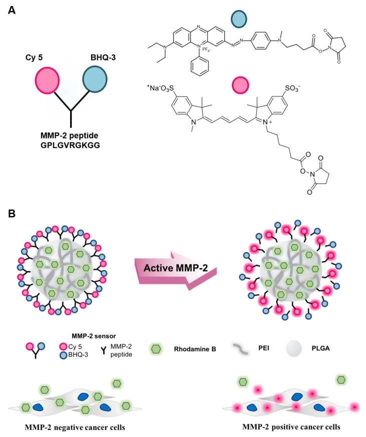
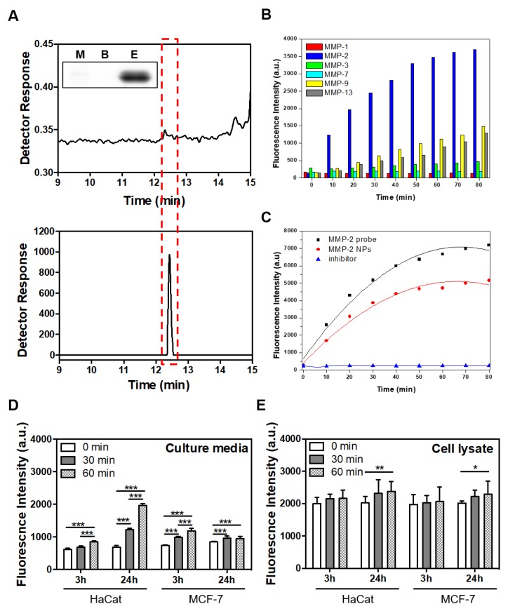
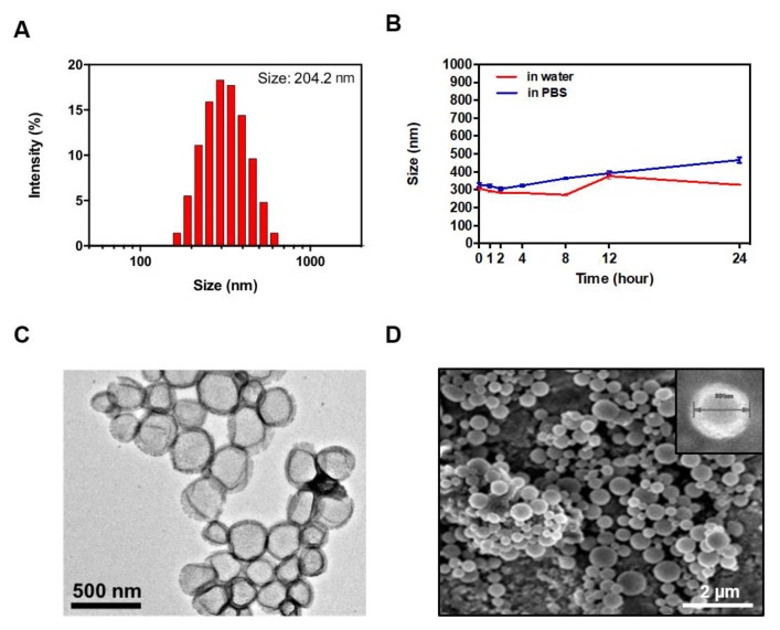
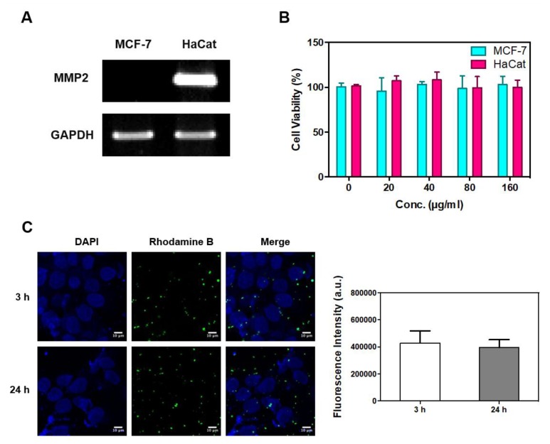
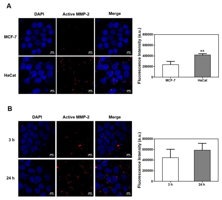
Matrix metalloproteinases (MMPs) are a family of zinc-dependent enzymes capable of degrading extracellular matrix components. Previous studies have shown that the upregulation of MMP-2 is closely related to metastatic cancers. While Western blotting, zymography, and Enzyme-Linked Immunosorbent Assays (ELISA) can be used to measure the amount of MMP-2 activity, it is not possible to visualize the dynamic MMP-2 activities of cancer cells using these techniques. In this study, MMP-2-activated poly(lactic-co-glycolic acid) with polyethylenimine (MMP-2-PLGA-PEI) nanoparticles were developed to visualize time-dependent MMP-2 activities. The MMP-2-PLGA-PEI nanoparticles contain MMP-2-activated probes that were detectable via fluorescence microscopy only in the presence of MMP-2 activity, while the Rhodamine-based probes in the nanoparticles were used to continuously visualize the location of the nanoparticles. This approach allowed us to visualize MMP-2 activities in cancer cells and their microenvironment. Our results showed that the MMP-2-PLGA-PEI nanoparticles were able to distinguish between MMP-2-positive (HaCat) and MMP-2-negative (MCF-7) cells. While the MMP-2-PLGA-PEI nanoparticles gave fluorescent signals recovered by active recombinant MMP-2, there was no signal recovery in the presence of an MMP-2 inhibitor. In conclusion, MMP-2-PLGA-PEI nanoparticles are an effective tool to visualize dynamic MMP-2 activities of potential metastatic cancer cells.

Keywords: PLGA-PEI nanoparticles; active matrix metalloproteinase-2; imaging sensor; metastasis.


Conflict of interest statement

The authors declare no conflict of interest.

Figures

Figure 1
Figure 1
Schematic illustration of the matrix metalloproteinase-2 (MMP-2)-activated peptide sensor and MMP-2-activated poly(lactic-co-glycolic acid) with polyethylenimine (MMP-2-PLGA-PEI) nanoparticles. (A) Chemical structure of the MMP-2-activated peptide sensor. The sensor consisted of a near-infrared fluorescence dye (Cy5), MMP-2 substrate peptide, and a dark quencher (BHQ-3); (B) Schematic diagram of the MMP-2-PLGA-PEI nanoparticles. The fluorescence signal was recovered only in the presence of MMP-2-positive cancer cells.
Figure 2
Figure 2
In vitro characterization of the MMP-2-activated peptide sensor and MMP-2-PLGA-PEI nanoparticles. (A) Analysis of MMP-2-activated peptide sensor fluorescence recovery by SDS-PAGE (upper panel; inset box: M-marker, B-TCNB buffer, E-Enzyme) and HPLC analysis results with MMP-2 enzyme (lower panel) and without MMP-2 enzyme (upper panel) indicated by the red dotted box. The MMP-2-activated peptide sensor was incubated with active recombinant MMP-2 for 30 min; (B) Signal recovery after incubation of the MMP-2-activated peptide sensor in the presence of various recombinant MMPs (MMP-1, -2, -3, -7, -9, -13) for 80 min at 37 °C; (C) MMP-2 fluorescence signal recovery from the MMP-2-activated peptide sensor, MMP-2-PLGA-PEI nanoparticles, and inhibitor-treated MMP-2-activated peptide sensor during incubation with recombinant MMP-2 for 80 min at 37 °C; (D) Analysis of MMP-2-PLGA-PEI fluorescence recovery after incubation with cell culture media; (E) MMP-2-PLGA-PEI fluorescence recovery of the cell lysate from MCF-7 and HaCat cells. Cell culture media and cell lysates were collected after culturing in fresh media for 3 h and 24 h. MMP-2 fluorescence signal recovery was confirmed at 0, 30, and 60 min. (* indicates p < 0.05, ** indicates p < 0.01, *** indicates p < 0.005).
Figure 3
Figure 3
Characterization of MMP-2-PLGA-PEI nanoparticles. (A) Size distribution in aqueous solutions; (B) Stability tests at 37 °C in water and physiological solution (PBS, pH 7.4); (C) Transmission emission microscopy (TEM) images of the nanoparticles; (D) Scanning electron microscopy (SEM) images of the nanoparticles.
Figure 4
Figure 4
Confirmation of MMP-2 expression, cellular cytotoxicity, and MMP-2-PLGA-PEI nanoparticle uptake. (A) MMP-2 mRNA level in MCF-7 and HaCat cells was analyzed by RT-PCR. Glyceraldehyde 3-phosphate dehydrogenase (GAPDH) was used as an internal control; (B) In vitro cell cytotoxicity analysis for MCF-7 (cyan) and HaCat (pink) cells after a 30 min incubation with PLGA-PEI nanoparticles; (C) Confocal microscopy analysis of cellular uptake of Rhodamine B lipid PLGA-PEI nanoparticles. Results are presented as mean ± SD (n = 3) (Scale bar: 10 µm).
Figure 5
Figure 5
MMP-2 fluorescence signal recovery according to cell line and culture time. (A) Activity of MMP-2 in MCF-7 and HaCat cells by confocal imaging using MMP-2-PLGA-PEI nanoparticles. After culturing in fresh media for 24 h, the cells were incubated with MMP-3-PLGA-PEI nanoparticles for 30 min. Mean signal intensities were calculated by the imaging software (n = 3); (B) Cell culture time–dependent activity of MMP-2 in HaCat cells. Cells were precultured in serum-free media for 3 h and 24 h, respectively, and then incubated with MMP-2-PLGA-PEI nanoparticles for 30 min. Mean signal intensities were calculated by the imaging software (n = 3). (** indicates p < 0.01.) (Scale bar: 10 µm).

References

    1. Kessenbrock K., Plaks V., Werb Z. Matrix metalloproteinases: Regulators of the tumor microenvironment. Cell. 2010;141:52–67. doi: 10.1016/j.cell.2010.03.015. - DOI - PMC - PubMed
    1. Egeblad M., Werb Z. New functions for the matrix metalloproteinases in cancer progression. Nat. Rev. Cancer. 2002;2:161–174. doi: 10.1038/nrc745. - DOI - PubMed
    1. Huppertz B., Kertschanska S., Demir A.Y., Frank H.G., Kaufmann P. Immunohistochemistry of matrix metalloproteinases (MMP), their substrates, and their inhibitors (TIMP) during trophoblast invasion in the human placenta. Cell. Tissue Res. 1998;291:133–148. doi: 10.1007/s004410050987. - DOI - PubMed
    1. Wysocki A.B., Staiano-Coico L., Grinnell F. Wound fluid from chronic leg ulcers contains elevated levels of metalloproteinases MMP-2 and MMP-9. J. Invest. Dermatol. 1993;101:64–68. doi: 10.1111/1523-1747.ep12359590. - DOI - PubMed
    1. Jackson H.W., Defamie V., Waterhouse P., Khokha R. Timps: Versatile extracellular regulators in cancer. Nat. Rev. Cancer. 2017;17:38–53. doi: 10.1038/nrc.2016.115. - DOI - PubMed