. 2018 Mar 1;555(7694):117-120.
doi: 10.1038/nature25502. Epub 2018 Feb 21.

The cryo-electron microscopy structure of huntingtin

Affiliations

The cryo-electron microscopy structure of huntingtin

Qiang Guo et al. Nature. .

Abstract

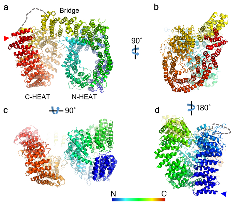
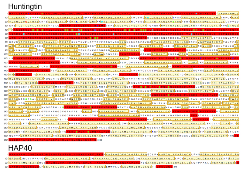
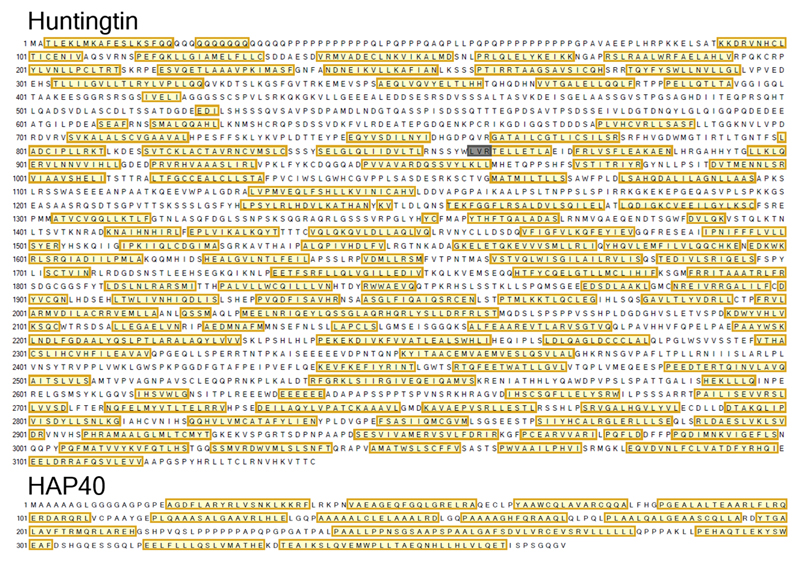
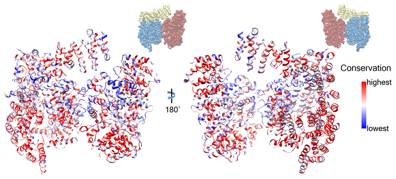
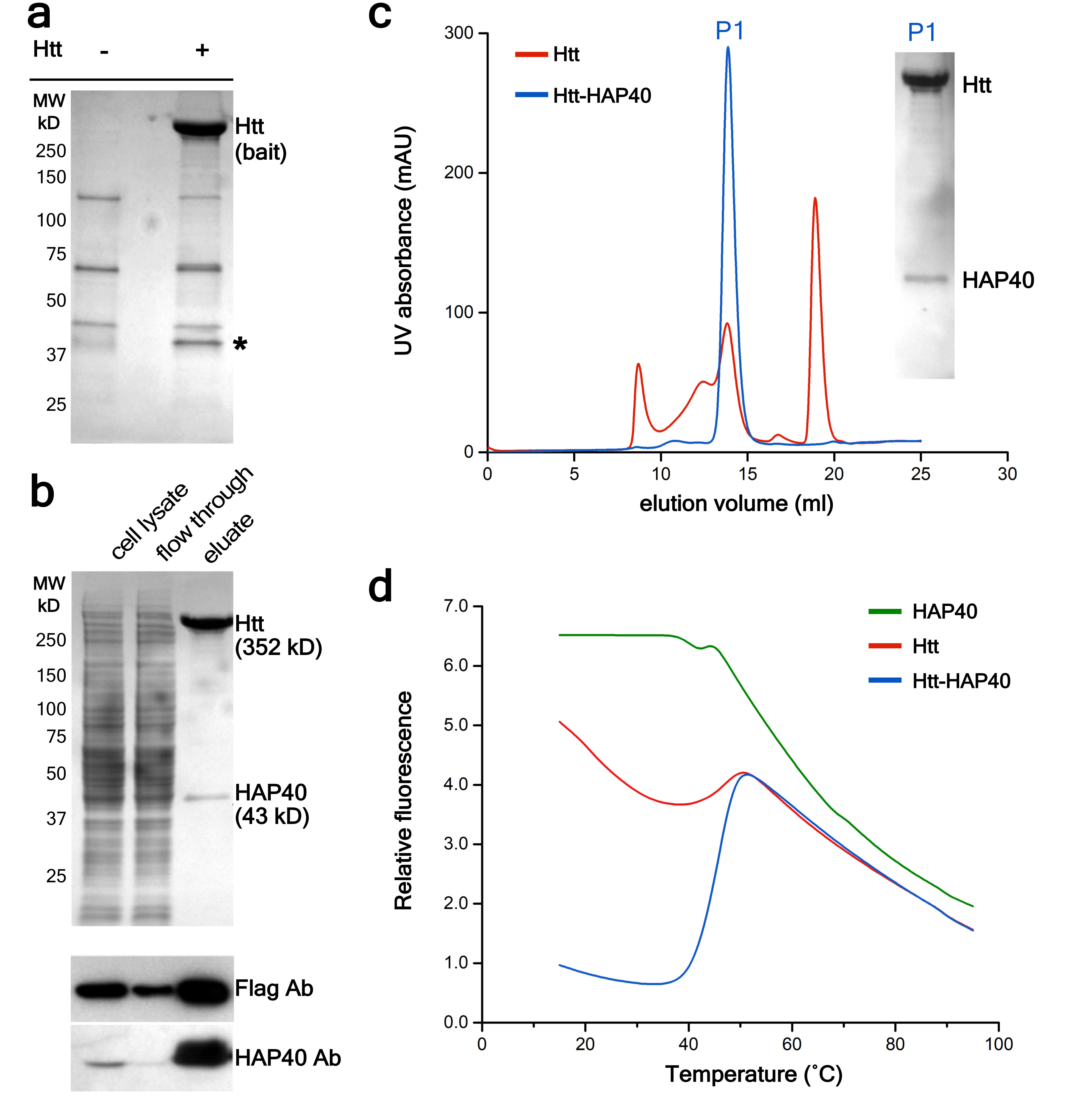
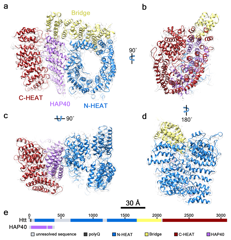
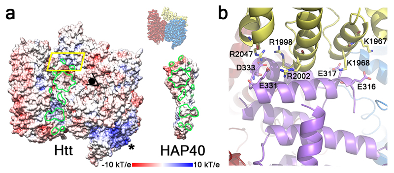
Huntingtin (HTT) is a large (348 kDa) protein that is essential for embryonic development and is involved in diverse cellular activities such as vesicular transport, endocytosis, autophagy and the regulation of transcription. Although an integrative understanding of the biological functions of HTT is lacking, the large number of identified HTT interactors suggests that it serves as a protein-protein interaction hub. Furthermore, Huntington's disease is caused by a mutation in the HTT gene, resulting in a pathogenic expansion of a polyglutamine repeat at the amino terminus of HTT. However, only limited structural information regarding HTT is currently available. Here we use cryo-electron microscopy to determine the structure of full-length human HTT in a complex with HTT-associated protein 40 (HAP40; encoded by three F8A genes in humans) to an overall resolution of 4 Å. HTT is largely α-helical and consists of three major domains. The amino- and carboxy-terminal domains contain multiple HEAT (huntingtin, elongation factor 3, protein phosphatase 2A and lipid kinase TOR) repeats arranged in a solenoid fashion. These domains are connected by a smaller bridge domain containing different types of tandem repeats. HAP40 is also largely α-helical and has a tetratricopeptide repeat-like organization. HAP40 binds in a cleft and contacts the three HTT domains by hydrophobic and electrostatic interactions, thereby stabilizing the conformation of HTT. These data rationalize previous biochemical results and pave the way for improved understanding of the diverse cellular functions of HTT.


Conflict of interest statement

The authors declare no competing financial interests.

Figures

Extended Data Fig. 1
Extended Data Fig. 1. Sedimentation analysis by rate-zonal ultracentrifugation.
a, FLAG-tag purified Htt (top) and Strep-tag purified Htt-HAP40 complex (bottom) analysed by rate-zonal ultracentrifugation followed by SDS-PAGE and Coomassie staining. 25 fractions from 5-20 % sucrose gradients were collected from the bottom of the tube, here showing fractions 1-18. While Htt alone was present in fractions 1-18, the Htt-HAP40 complex was found mainly in fractions 15-17, indicating lower conformational heterogeneity. b, Western blot analysis of fractions 10-18 of the Htt-HAP40 complex. Independent experiments with similar results (n): n=3. For gel source data, see Supplementary Figure 1.
Extended Data Fig. 2
Extended Data Fig. 2. Cryo-EM analysis of the Htt-HAP40 complex.
a, Representative micrograph of Htt-HAP40 complex. b, 2D class averages. c, Fourier shell correlation (FSC) plots. Cyan, gold-standard FSC curve; orange, FSC curve calculated between the cryo-EM map and refined atomic model. 0.143 and 0.5 FCS cut-off values, respectively, were used as indicated. The (initial/final) numbers of micrographs and particles were 707/635 and 418,627/98,310, respectively. d, Final density map of the Htt-HAP40 complex colored according to local resolution. The map was low-pass filtered to 4.0 Å and sharpened with a B-factor of -174 Å2. e, Detail of the electron density maps (mesh) for parts of Htt and HAP40.
Extended Data Fig. 3
Extended Data Fig. 3. Atomic model of Htt within the Htt-HAP40 complex.
The atomic model is shown in ribbon representation with the indicated rainbow color code from N-terminus (blue arrowhead in d) to C- terminus (red arrowhead in a). a, b, c, d show different views of the complex as indicated. Dashed lines mark unresolved regions.
Extended Data Fig. 4
Extended Data Fig. 4. Amino acid sequences of 17QHtt and HAP40.
Structural elements of the atomic models are indicated as follows: not visible in the model (red box), unstructured region (no box) and α-helix (yellow box). The sites of previously reported proteases cleavage and post-translational modifications of Htt,,, are indicated as follows: acetylation (dark blue), palmitoylation (red), phosphorylation (green) and proteolytic cleavage (cyan).
Extended Data Fig. 5
Extended Data Fig. 5. PSIPRED secondary structure predictions for Htt and HAP40.
Structural elements are indicated as follows: unstructured region (no box), α-helix (yellow box) and β-sheet (grey box).
Extended Data Fig. 6
Extended Data Fig. 6. Truncation analysis of HAP40 binding to Htt.
a, Schematic representation of the HAP40 constructs studied (all C-terminally Strep-tagged). b, HAP40 constructs were co-expressed with C-terminally FLAG-tagged 17QHtt, immunoprecipitated using Strep-Tactin beads and analyzed by Western blot. Lanes are indicated as follows: 1, cell lysates; 2, cell lysates upon incubation with Strep beads; 3, Strep beads eluates. Note that full-length HAP40 and a construct lacking the central domain immunoprecipitate Htt, but not deletions of the N- and C-terminal regions of HAP40. Independent experiments with similar results (n): n=2. For gel source data, see Supplementary Figure 1.
Extended Data Fig. 7
Extended Data Fig. 7. Evolutionary analysis of Htt.
The human Htt model is shown in ribbon representation, colored according to sequence conservation across 16 metazoan species (Homo sapiens, Rattus norvegicus, Mus musculus, Sus scrofa, Bos Taurus, Canis familiaris, Monodelphis domestica, Gallus gallus, Danio rerio, Tetraodon nigroviridis, Fugu rubripes, Ciona savignyi, Ciona intestinalis, Strongylocentrotus purpuratus, Tribolium castaneum, Apis mellifera), using a previously reported sequence alignment. At the top, the orientation of the Htt-HAP40 complex is indicated with respect to Fig. 2.
Extended Data Fig. 8
Extended Data Fig. 8. Workflow for initial model validation for 3D reconstruction of the Htt-HAP40 complex.
A subset of particles with well-resolved 2D average were used for initial model generation using RELION or SPHIRE. The resulting models were used as reference for 3D classification of all the good particles. A featureless sphere was also used as classification reference. Most of the particles were classified to identical structures with sufficient detail, indicating no reference bias in the reconstruction.
Fig. 1
Fig. 1. Purification of the Htt-HAP40 complex.
a, Identification of HAP40 as a major Htt interactor in HEK293 cells expressing low levels of FLAG-tagged 46QHtt (+) or not expressing FLAG-tagged Htt (-). Coomassie-stained gel after PAGE following FLAG-affinity purification. The band indicated by * was identified as HAP40 by mass spectrometry and Western blot. b, Purification of the Htt-HAP40 complex from HEK293-based cells expressing FLAG-tagged 17QHtt and Strep-tagged HAP40. Cleared lysates were incubated with Strep-Tactin beads and washed with desthiobiotin to elute bound proteins. Top, Coomassie-staining. Bottom, Western blot. c, elution profile of Htt alone (red) versus the Htt-HAP40 complex (blue). Inset, Coomassie-staining of the P1 peak of the elution profile of the Htt-HAP40 complex. d, Thermal unfolding and complex stabilization. Melting curves of Htt, HAP40 and the Htt-HAP40 complex obtained by differential scanning fluorimetry. Independent experiments with similar results (n): a: n=2; b: n=3; c: n=3; d: n=2. For source data, see Supplementary Figure 1.
Fig. 2
Fig. 2. Architecture of the Htt-HAP40 complex.
The reconstructed density map filtered according to local resolution is shown as a translucent surface. The atomic model is superimposed in ribbon representation, with domains color-coded as follows: Htt N-HEAT domain, blue; Htt bridge domain, yellow; Htt C-HEAT domain, maroon; HAP40, purple. a, b, c, d show different views of the complex as indicated. e, Schematic domain organization of Htt and HAP40.
Fig. 3
Fig. 3. Structure of Htt domains.
a, N-HEAT domain. The insertion between N-HEAT repeat 6 and 7 is shown in green, with the unresolved sequence as a dashed line. b, C-HEAT domain, with the insertions between HEAT repeats shown in green (between C-HEAT repeats 1 and 2) and teal (between C-HEAT repeats 2 and 3). c, Bridge domain. In a-c helices forming part of tandem repeats are shown as rods in similar colors, and other helices as ribbons. At the top right of a-c, the part of Htt shown in each panel is highlighted in the Htt-HAP40 map using the color code of Fig. 2. d, Back view of Htt highlighting the interaction region (inset) between loops of N- and C-HEAT. The unresolved sequence at the C-terminus of the bridge domain is shown as a yellow dashed line.
Fig. 4
Fig. 4. Structural basis of the Htt-HAP40 interaction.
a, “Open book” view of the complex in surface representation displaying electrostatic potential. Htt-HAP40 contact areas are circled in green. * marks the positively charged surface formed by N-HEAT repeats 2-4. At the top right, the orientation of the Htt-HAP40 complex in this panel with respect to Fig. 2 is indicated. b, Detailed view of the region boxed in yellow using the color code of Fig. 2. Residues involved in electrostatic interactions are displayed as sticks.

References

    1. Saudou F, Humbert S. The Biology of Huntingtin. Neuron. 2016;89:910–926. - PubMed
    1. Zuccato C, Cattaneo E. Huntington’s Disease. Oxford University Press; 2014.
    1. Kaltenbach LS, et al. Huntingtin interacting proteins are genetic modifiers of neurodegeneration. PLoS genetics. 2007;3:e82. - PMC - PubMed
    1. Shirasaki DI, et al. Network organization of the huntingtin proteomic interactome in mammalian brain. Neuron. 2012;75:41–57. - PMC - PubMed
    1. MacDonald ME, et al. A novel gene containing a trinucleotide repeat that is expanded and unstable on Huntington's disease chromosomes. Cell. 1993;72:971–983. - PubMed

Publication types