. 2018 Feb 21;13(2):e0192098.
doi: 10.1371/journal.pone.0192098. eCollection 2018.

Cross-reactive microbial peptides can modulate HIV-specific CD8+ T cell responses

Affiliations

Cross-reactive microbial peptides can modulate HIV-specific CD8+ T cell responses

Christopher W Pohlmeyer et al. PLoS One. .

Abstract

Heterologous immunity is an important aspect of the adaptive immune response. We hypothesized that this process could modulate the HIV-1-specific CD8+ T cell response, which has been shown to play an important role in HIV-1 immunity and control. We found that stimulation of peripheral blood mononuclear cells (PBMCs) from HIV-1-positive subjects with microbial peptides that were cross-reactive with immunodominant HIV-1 epitopes resulted in dramatic expansion of HIV-1-specific CD8+ T cells. Interestingly, the TCR repertoire of HIV-1-specific CD8+ T cells generated by ex vivo stimulation of PBMCs using HIV-1 peptide was different from that of cells stimulated with cross-reactive microbial peptides in some HIV-1-positive subjects. Despite these differences, CD8+ T cells stimulated with either HIV-1 or cross-reactive peptides effectively suppressed HIV-1 replication in autologous CD4+ T cells. These data suggest that exposure to cross-reactive microbial antigens can modulate HIV-1-specific immunity.


Conflict of interest statement

Competing Interests: The authors have declared that no competing interests exist

Figures

Fig 1
Fig 1. Cross-reactive microbial peptides stimulate HIV-1-specific CD8+ T cells.
(A) Flow cytometric plots of outgrowth of KK10-specific CD8+ T cells from PBMCs (subject ES9) after six-day stimulation with no peptide, CEF pooled peptides, KK10 peptide, or KK10CR peptides. (B) Summarized results for outgrowth of KK10-specific CD8+ T cells from seven HIV-1-positive subject samples stimulated with CEF pooled peptides, KK10 peptide, or individual KK10CR peptides. (C) Flow cytometric plots of outgrowth of SL9-specific CD8+ T cells from PBMCs (subject CP2A) after six-day stimulation with no peptide, CEF pooled peptides, SL9 peptide, or SL9CR peptides. (D) Summarized results for outgrowth of SL9-specific CD8+ T cells from eight HIV-positive subject samples stimulated with CEF pooled peptides, SL9 peptide, or individual SL9CR peptides.
Fig 2
Fig 2. KK10 and KK10-cross-reactive microbial peptides differentially expand KK10-specific CD8+ T cells.
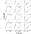
(A) IFN-y release by ELISpot from HIV-positive HLA-B*27+ subject PBMCs stimulated with dilutions of KK10 or KK10CR peptides. (B) Characterization of KK10-specific TCR repertoires after PBMC stimulation with KK10 or KK10CR peptides. PBMCs from HIV-1-positive HLA-B*27+ subjects were stimulated with peptides for six days, and KK10-specific CD8+ T cells were sorted from fresh (unstimulated), KK10-stimulated, or KK10CR-stimulated PBMCs. Frequencies of unique TCR clones as measured by diversity in the TCR-β CDR3 region were quantified by ImmunoSEQ deep sequencing. Each dot shows the frequency of a single TCR clone in the specified KK10-specific CD8+ T cell populations. TCR clones that have the same frequency in both populations compared on a plot fall on the diagonal line. Magenta dots indicate TCR clones that are present at significantly different frequencies (minimum 20 reads in either condition, p<0.005, Fisher’s Exact test with Benjamini-Hochberg correction. S1 Table shows all associated p-values).
Fig 3
Fig 3. SL9 and SL9-cross-reactive microbial peptides differentially expand SL9-specific CD8+ T cells.
Characterization of SL9-specific TCR repertoires after PBMC stimulation with SL9 or SL9CR peptides. PBMCs from HIV-1-positive HLA-A2+ subjects were stimulated with peptides for six days, and SL9-specific CD8+ T cells were sorted from fresh (unstimulated), SL9-stimulated, or SL9CR-stimulated PBMCs. Frequencies of unique TCR clones as measured by diversity in the TCR-β CDR3 region were quantified by ImmunoSEQ deep sequencing. Each dot shows the frequency of a single TCR clone in the specified SL9-specific CD8+ T cell populations. TCR clones that have the same frequency in both populations compared on a plot fall on the diagonal line. Magenta dots indicate TCR clones that are present at significantly different frequencies (minimum 20 reads in either condition, p<0.005, Fisher’s Exact test with Benjamini-Hochberg correction. S2 Table shows all associated p-values).
Fig 4
Fig 4. PBMCs stimulated with cross-reactive microbial peptides can suppress HIV-1 infection.
(A-B) Results of PBMC suppression assay. PBMCs from HIV-1-positive HLA-B*27+ subjects were stimulated for five days with no peptide, CEF pooled peptides, KK10 peptide, or KK10CR peptide and then infected with highly concentrated IIIB virus. After 40 hours, infection of CD3+/CD8- cells was measured by staining for intracellular HIV-1 Gag. Values below 20% (in grey) fall below the limit of quantification. Part A shows flow cytometric results for subject ES31; part B shows summarized results for five subjects with CEF pooled peptides, KK10 peptide, and individual KK10CR peptides (C-D) PBMCs from HIV-1-positive HLA-A2+ subjects were stimulated for five days with no peptide, CEF pooled peptides, SL9 peptide, or SL9CR peptide and then infected with highly concentrated IIIB virus. After 40 hours, infection of CD3+/CD8- cells was measured by staining for intracellular HIV-1 Gag. Part C shows flow cytometric results for subject CP37; part D shows summarized results for seven subjects with CEF pooled peptides, SL9 peptide, and individual SL9CR peptides.
Fig 5
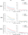
Fig 5. CD8+ T cells stimulated with cross-reactive microbial peptides can suppress HIV-1 infection.
Purified CD4+ T cells from three HIV-1-positive HLA-B*27+ subjects were spinoculated with single-round NL4-3 GFP reporter virus and then co-cultured with peptide-stimulated or unstimulated (fresh ex vivo) autologous CD8+ T cells. Viral suppression was measured at four different effector-to-target cell ratios. Left panels show suppression of wild-type (WT) NL4-3 reporter virus, and right panels show suppression of NL4-3 harboring the R264K and L268 mutations in KK10, which prevent KK10 presentation on HLA-B*27 MHC molecules.
Fig 6
Fig 6. Cross-reactive microbial peptides do not expand HIV-1-specific CD8+ T cells in HIV-negative donors.
25 HIV-negative HLA-B*27+ donors were screened for the presence of KK10-specific CD8+ T cells. Donor PBMCs were stimulated with KK10 peptide, KK10CR peptides or negative control CEF pooled peptides for six days, and the percentage of (A) KK10-specific and (B) IFN-γ+Perforin+ CD8+ T cells was quantified by flow cytometry.

References

    1. Koup RA, Safrit JT, Cao Y, Andrews CA, McLeod G, Borkowsky W, et al. Temporal association of cellular immune responses with the initial control of viremia in primary human immunodeficiency virus type 1 syndrome. J Virol. 1994;68: 4650–4655. - PMC - PubMed
    1. Borrow P, Lewicki H, Hahn BH, Shaw GM, Oldstone MB. Virus-specific CD8+ cytotoxic T-lymphocyte activity associated with control of viremia in primary human immunodeficiency virus type 1 infection. J Virol. 1994; 68: 6103–6110. - PMC - PubMed
    1. Schmitz JE, Kuroda MJ, Santra S, Sasseville VG, Simon MA, Lifton MA, et al. Control of viremia in simian immunodeficiency virus infection by CD8+ lymphocytes. Science. 1999;283: 857–860. - PubMed
    1. Jin X, Bauer DE, Tuttleton SE, Lewin S, Gettie A, Blanchard J, et al. Dramatic rise in plasma viremia after CD8(+) T cell depletion in simian immunodeficiency virus-infected macaques. J Exp Med. 1999;189: 991–998. - PMC - PubMed
    1. Migueles SA, Laborico AC, Shupert WL, Sabbaghian MS, Rabin R, Hallahan CW, et al. HIV-specific CD8+ T cell proliferation is coupled to perforin expression and is maintained in nonprogressors. Nat Immunol. 2002;3: 1061–1068. doi: 10.1038/ni845 - DOI - PubMed

Publication types