. 2018 Feb 20;22(8):2190-2205.
doi: 10.1016/j.celrep.2018.01.087.

A Roadmap for Human Liver Differentiation from Pluripotent Stem Cells

Affiliations

A Roadmap for Human Liver Differentiation from Pluripotent Stem Cells

Lay Teng Ang et al. Cell Rep. .

Abstract

How are closely related lineages, including liver, pancreas, and intestines, diversified from a common endodermal origin? Here, we apply principles learned from developmental biology to rapidly reconstitute liver progenitors from human pluripotent stem cells (hPSCs). Mapping the formation of multiple endodermal lineages revealed how alternate endodermal fates (e.g., pancreas and intestines) are restricted during liver commitment. Human liver fate was encoded by combinations of inductive and repressive extracellular signals at different doses. However, these signaling combinations were temporally re-interpreted: cellular competence to respond to retinoid, WNT, TGF-β, and other signals sharply changed within 24 hr. Consequently, temporally dynamic manipulation of extracellular signals was imperative to suppress the production of unwanted cell fates across six consecutive developmental junctures. This efficiently generated 94.1% ± 7.35% TBX3+HNF4A+ human liver bud progenitors and 81.5% ± 3.2% FAH+ hepatocyte-like cells by days 6 and 18 of hPSC differentiation, respectively; the latter improved short-term survival in the Fah-/-Rag2-/-Il2rg-/- mouse model of liver failure.

Keywords: efficient differentiation; human liver development; pluripotent stem cells; progenitor; signaling.


Figures

Figure 1
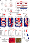
Figure 1. Differentiation of Human Definitive Endoderm into Posterior Foregut
(A) Liver development in the mouse embryo between E5.5 to E9.5 depicting Mixl1+ primitive streak, Sox17+ definitive endoderm, Hnf4a+ foregut, and Tbx3+ liver bud progenitors. (B) Overview of human PSC differentiation strategy in this study. (C) Percentage of MIXL1-GFP+ cells using MIXL1-GFP knockin hESC reporter line (Loh et al., 2014). (D) Percentage of SOX17-mCherry+ cells using SOX17-mCherry knockin hESC reporter line (Loh et al., 2014). (E) Markers expressed in E9.5 mouse liver bud progenitors. (F) Strategy to treat definitive endoderm (DE) with RA or TGF-β modulators on the day-2 to day-3 interval to produce day-3 posterior foregut (PFG) and assaying subsequent effects on liver bud gene expression by day 6, as shown in (H)–(J). (G) Transient treatment on the day-2 to day-3 interval with ATRA or TTNPB markedly improves AFP expression in day-6 hPSC-derived liver bud progenitors on top of base media condition A83 + B + F (A83 + B + F: A8301, 1 μM; BMP4, 30 ng/mL; FGF2, 10 ng/mL), as shown by immunostaining with a DAPI nuclear counterstain. Scale bar, 1 mm. (H) qPCR gene expression of day-5 liver bud cells generated from endoderm cells briefly treated on the day-2 to day-3 interval with a retinoid inhibitor (BMS: BMS493, 10 μM) or ATRA of varying doses (0.1 mM, 0.5 μM, 1 μM, or 2 μM) on top of base media condition A83 (A83: A8301, 1 μM). (I) qPCR gene expression of day-6 liver bud cells generated from endoderm cells briefly treated on the day-2 to day-3 interval with a TGF-β inhibitor A83 (A83: A8301, 1 μM) or a TGF-β agonist (A10: ACTIVIN, 10 ng/mL) on top of base media condition ATRA (ATRA: 2 μM). (J) qPCR gene expression of day-5 liver bud cells generated from endoderm cells briefly treated on the day-2 to day-3 interval with a BMP inhibitor DM (DM: DM3189, 250 nM) or a BMP agonist (B3: BMP4, 3 ng/mL) on top of base media condition RA + A83 (RA: ATRA, 2 μM; A83: A8301, 1 μM).
Figure 2
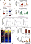
Figure 2. Accelerated Generation of Human TBX3+HNF4A+AFP+ Liver Bud Progenitors by Day 6 of PSC Differentiation
(A) Development of liver bud in mouse embryos. Strategy to treat posterior foregut with RA, TGF-β, PKA, BMP, and WNT modulators on the day-4 to day-6 interval to produce day-6 liver bud progenitors and assaying effects on liver bud gene expression by day 6, as shown in (C)–(F). (B) Temporally dynamic signals govern liver differentiation. (C) qPCR gene expression of day-6 liver bud cells generated from endoderm treated on the day-4 to day-6 interval with a retinoid inhibitor (BMS: BMS493, 10 μM) or varying doses of a retinoid agonist (ATRA, 0.1 μM, 0.5 μM, 1 μM, and 2 μM) on top of base media condition A83B10 (A83B10: A8301, 1 μM; BMP4, 10 ng/mL) and qPCR gene expression of day-6 hPSC-derived midgut/hindgut (MHG) or pancreatic endoderm (PAN) cells. (D) qPCR gene expression of day-6 liver bud cells generated from endoderm treated on the day-4 to day-6 interval with a TGF-β inhibitor (A83: A8301, 1 μM) or ACTIVIN (10 ng/mL) on top of base media condition B10 (B10: BMP4, 10 ng/mL). Day-6 hPSC-derived pancreatic endoderm (PAN) or midgut/hindgut cells were included as controls. (E) qPCR gene expression of day-5 liver bud cells generated from endoderm treated on the day-4 to day-5 interval with a BMP inhibitor (DM: DM3189, 250 nM) or varying doses of BMP4 (B, 10–100 ng/mL) in the presence of base media condition A10 (A10: ACTIVIN at 10 ng/mL). Midgut/hindgut denotes hPSC-derived midgut/hindgut cells. (F) Gene expression of day-6 liver bud cells after 2-day treatment of PKA inhibitor (RpCAMP, 100 μM) or PKA agonist (BrCAMP, 1 mM) in the presence of base media condition A83B10 (A83B10: A8301, 1 μM; BMP4, 10 ng/mL) during a day-4 to day-5 interval and day-6 hPSC-derived midgut/hindgut, as shown by qPCR. (G) Percentage of day-6 liver bud progenitors positive for AFP from the differentiation of 3 hPSC lines (H1, H7, and H9) as shown by intracellular FACS. (H) Day-6H1hPSC-derived liver progenitors generated using SR2 were immunostained for AFP, HNF4A, and TBX3 with a DAPI nuclear counterstain. Scalebar, 500 μm. (I) Gene expression of day-6 liver progenitors, pancreatic endoderm, and midgut/hindgut progenitors derived from hPSCs for HNF1A, PDX1, and CDX2.Error bars represent ± SE.
Figure 3
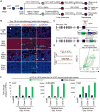
Figure 3. High-Throughput Screening Identifies Liver-Specific Surface Markers during PSC Differentiation
(A) Schematic diagram of liver differentiation approaches including SR2 and other methods (Zhao et al., 2013; Si-Tayeb et al., 2010; Avior et al., 2015; Carpentier et al., 2016). Primitive-streak-inducing conditions: ACP, ACTIVIN + CHIR99201 + PI103 (Loh et al., 2014); AWH, ACTIVIN + Wnt3a + HGF (Avior et al., 2015); StemDiff, STEMdiff definitive endoderm differentiation kit. Endoderm-inducing condition: ADP, ACTIVIN + DM3189 + PI103 (Loh et al., 2014). Liver progenitors were analyzed at day 6 (Zhao et al., 2013; Si Tayeb et al., 2010) or day 7 (Avior et al., 2015; Carpentier et al., 2016), respectively, as described in the respective studies. (B) qPCR gene expression of day-6H1 hPSC-derived liver progenitors generated using SR2 or other methods (Si-Tayeb et al., 2010; Zhao et al., 2013; Avior et al., 2015; Carpentier et al., 2016). (C) Strategy to conduct high-throughput FACS screening of surface markers expressed on H7 hPSCs, day-2 H7 hPSC-derived endoderm, and day-6 H7 hPSC-derived liver bud progenitors. (D) Venn diagram of surface markers expressed on hPSCs, day-2 hPSC-derived definitive endoderm, and day-6 hPSC-derived liver bud progenitors. (E) hPSCs, hPSC-derived definitive endoderm, and liver bud progenitors stained for CD10, CD184, and CD99, as shown by live-cell FACS. (F) Surface markers expressed on hPSC and day-2 hPSC-derived definitive endoderm and day-6 liver progenitors; summary of present work.
Figure 4
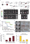
Figure 4. Generation of Enriched Populations of ALBUMIN+ and FAH+ Immature Hepatocytes by Day 18 of PSC Differentiation
(A) Diagram depicting liver development in mouse embryos between E9.5 and birth and the expression of various liver genes. dpc, days post-coitum. (B) A schematic of the present work showing the differentiation of human liver bud progenitors into bile duct cells or hepatocytes. A8301, TGF-β inhibitor; DAPT, Notch inhibitor; FSK, forskolin; AAP, ascorbic acid-2-phosphate. (C and D) qPCR gene expression of day-6 hPSC-derived liver bud (LB) progenitors before and after 2-day treatment of 10 μM DAPT in the absence or presence of (C) 10 μM dexamethasone (Dex) or (D) a TGF-β inhibitor (A83: A83-01, 1 μM) or ACTIVIN (10, 30, or 50 ng/mL) on top of base media condition. (E) ALBUMIN intracellular FACS analysis of hPSCs or day-18 hepatocytes derived from hPSC lines H1, H7, and H9. (F) Global microarray gene expression of hPSCs, day-2 definitive endoderm, day-6 liver bud (LB) progenitors, and day-12 hPSC-derived hepatic progenitors that were either induced using the full combination (base) or with the individual omission of either DAPT, forskolin, ascorbic acid-2-phosphate, or insulin during the liver bud → hepatic progenitor differentiation step to reveal genes whose expression is regulated by DAPT, FSK, AAP, and insulin (INS). 4 biological replicates were used for the microarray analyses. (G) ALBUMIN levels detected in culture medium grown with hPSC-derived or primary adult human hepatocytes as measured by ELISA with a human-specific ALBUMIN antibody; “Base” denotes culture medium alone. (H) CYP3A4 activity of hPSCs, HepG2, and hPSC-derived hepatocytes (Day-18 Hep), as measured by P450-Glo luciferase assays and abolished by CYP3A4 inhibitor ketoconazole; signal is further normalized to cell number baseline, as measured by CellTiter-Glo. Error bars represent ± SE.
Figure 5
Figure 5. Generation of a FAH-2A-Clover Knockin H1 hPSC Reporter Line Using CRISPR/Cas9-Mediated Gene Editing
(A) Schematic diagram of liver differentiation approaches, including SR2 or other methods (Zhao et al., 2013; Si-Tayeb et al., 2010). Primitive-streak-inducing conditions: ACP, ACTIVIN + CHIR99201 + PI103 (Loh et al., 2014). Endoderm-inducing condition: ADP, ACTIVIN + DM3189 + PI103 (Loh et al., 2014). Hepatocyte-inducing condition: BODFsRAI, BMP4 + OSM + dexamethasone (Dex) + forskolin + Ro4929097 + AA2P + insulin. For detailed methods, see Supplemental Information. (B) Day-18 hepatocyte-like cells generated by 3 methods (SR2 [the method described in the present work] or previously reported methods [Si-Tayeb et al., 2010; Zhao et al., 2013]) were immunostained for carbamoyl phosphate synthetase 1 (CPS1), ALBUMIN (ALB), and Alpha-1 anti-trypsin (AAT) with a DAPI nuclear counterstain, and multiple fields were stitched into 1 image. (C) Schematic illustration of the strategy to insert a P2A-Clover reporter cassette into the FAH locus. A Gly-Ser-Gly (gsg) sequence was added to the start of the P2A sequence; eSpCas9, enhanced specificity Cas9; PGK1, Phosphoglycerate kinase 1 promoter; Blast, Blasticidin drug resistance gene. (D) Wild-type and knockin alleles of the FAH locus sequenced by PCR. (E) FACs analysis of the day-18 hPSC-derived hepatocyte-like population revealed that it was 83.7% FAH-Clover+. (F) FACS-sorted FAH-Clover+ day-18 H1-derived hepatocyte-like cells (green bars) were significantly enriched for liver genes FAH, ALBUMIN, and HGD mRNAs when compared with FAH-Clover− cells (black bars) as shown by qPCR. Consistent results were obtained across multiple independent FAH-2A-Clover knockin H1 hPSC reporter clones (#1-3). Error bars represent ± SE.
Figure 6
Figure 6. Human PSC-Derived Hepatocytes Engraft Neonatal and Adult Fah–/– Rag2–/– Ilr2g–/– Mice and Improve Short-Term Survival
(A) Strategy to inject hPSC-derived hepatocytes into the livers of neonatal FRG mice (0–72 hr old) or adult Fah–/–Rag2–/–Ilr2g–/– (FRG) mice (4–6 weeks old) and to subsequently induce liver injury; H9 EF1A-BCL2-2A-GFP; UBC-tdTomato-2A-Luciferase hPSCs (Loh et al., 2016) were used to generate hepatocytes for injections. (B) Bioluminescent imaging of adult FRG mice that were intrahepatically injected with Luciferase+day-18 hPSC-derived hepatocytes while they were neonates. Liver injury was induced at weeks 4 to 6 by withdrawing NTBC; the remaining mice at 3 or 10 weeks post-injury are shown. (C) Adult FRG mice were injected with Luciferase+ day-18 hPSC-derived hepatocytes. Kaplan-Meier's survival curves depicting the percent survival (after NTBC withdrawal) of adult FRG mice that had either been injected with day-18 hPSC-derived hepatocytes (n = 11), primary adult human hepatocytes (red line; n = 5), or media-only control (blue line; n = 12); Mantel-Cox log-rank test, *p < 0.05, from 3 independent experiments. (D) Adult FRG mice were intrasplenically injected with day-18 hPSC-derived or primary adult human hepatocytes and NTBC was withheld. One month later, engrafted livers stained positive for human ALBUMIN (brown; at 5× or 20× magnifications), as shown by immunostaining. Scale bars, 200 μm. Image is representative of 5 livers. (E) In vivo secretion of human serum ALBUMIN secretion into the bloodstream in vivo by hPSC-derived or primary adult human hepatocytes, as measured by ELISA, 1 month post-transplant. (F) Reduction of bilirubin levels in the serum of mice transplanted with hPSC-derived or primary adult human hepatocytes versus negative or no-cell media-only control, 1 month post-transplant. Error bars represent ± SE.
Figure 7
Figure 7. A Signaling Roadmap to Efficiently Generate Human Liver Cells from PSCs
Summary of the present work. Inh., inhibitor; hi., high; lo., low; Ascorbic, ascorbic acid-2-phosphate. Optionally, Wnt inhibitor was added on days 4–5, and TGF-β inhibitor was added on days 7–8. Surface markers expressed on hPSC, day-2 hPSC-derived definitive endoderm, and day-6 liver bud progenitors are detailed in Results and Figure 3.

References

    1. Agarwal S, Holton KL, Lanza R. Efficient differentiation of functional hepatocytes from human embryonic stem cells. Stem Cells. 2008;26:1117–1127. - PubMed
    1. Avior Y, Levy G, Zimerman M, Kitsberg D, Schwartz R, Sadeh R, Moussaieff A, Cohen M, Itskovitz-Eldor J, Nahmias Y. Microbial-derived lithocholic acid and vitamin K2 drive the metabolic maturation of pluripotent stem cells-derived and fetal hepatocytes. Hepatology. 2015;62:265–278. - PubMed
    1. Azuma H, Paulk N, Ranade A, Dorrell C, Al-Dhalimy M, Ellis E, Strom S, Kay MA, Finegold M, Grompe M. Robust expansion of human hepatocytes in Fah-/-/Rag2-/-/Il2rg-/- mice. Nat Biotechnol. 2007;25:903–910. - PMC - PubMed
    1. Basma H, Soto-Gutiérrez A, Yannam GR, Liu L, Ito R, Yamamoto T, Ellis E, Carson SD, Sato S, Chen Y, et al. Differentiation and transplantation of human embryonic stem cell-derived hepatocytes. Gastroenterology. 2009;136:990–999. - PMC - PubMed
    1. Bayha E, Jørgensen MC, Serup P, Grapin-Botton A. Retinoic acid signaling organizes endodermal organ specification along the entire antero-posterior axis. PLoS One. 2009;4:e5845. - PMC - PubMed

Publication types

MeSH terms