. 2018 Feb 20;48(2):327-338.e5.
doi: 10.1016/j.immuni.2018.01.015.

T Cells in Nonlymphoid Tissues Give Rise to Lymph-Node-Resident Memory T Cells

Affiliations

T Cells in Nonlymphoid Tissues Give Rise to Lymph-Node-Resident Memory T Cells

Lalit K Beura et al. Immunity. .

Abstract

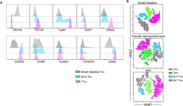
Immunosurveillance of secondary lymphoid organs (SLO) is performed by central memory T cells that recirculate through blood. Resident memory T (Trm) cells remain parked in nonlymphoid tissues and often stably express CD69. We recently identified Trm cells within SLO, but the origin and phenotype of these cells remains unclear. Using parabiosis of "dirty" mice, we found that CD69 expression is insufficient to infer stable residence of SLO Trm cells. Restimulation of nonlymphoid memory CD8+ T cells within the skin or mucosa resulted in a substantial increase in bona fide Trm cells specifically within draining lymph nodes. SLO Trm cells derived from emigrants from nonlymphoid tissues and shared some transcriptional and phenotypic signatures associated with nonlymphoid Trm cells. These data indicate that nonlymphoid cells can give rise to SLO Trm cells and suggest vaccination strategies by which memory CD8+ T cell immunosurveillance can be regionalized to specific lymph nodes.

Keywords: local recall immunization; non-lymphoid tissue; regionalized immunosurveillance; resident memory T cells; secondary lymphoid organs.


Figures

Figure 1
Figure 1. CD69 expression is insufficient to infer residence
(A) LN CD8+ T cell phenotypes were compared between specific-pathogen free (SPF) mice, mice obtained from pet stores (Pet store) and SPF mice cohoused for at least 60 days with pet store partners (Cohoused). Representative flow plots from three independent experiments (n=3 mice per group/repeat) are shown. Plots are gated on CD8+ T lymphocytes isolated from pooled inguinal and mesenteric LNs. (B) The frequency of CD69+ cells among the indicated CD8+ T cell subsets isolated from LNs of SPF, Pet store and Cohoused mice. Data are representative of three independent experiments with 3 mice/group per experiment. (C) Experimental scheme for testing residence of LN CD8+ T cells in Cohoused mice. (D) Blood and LNs in each parabiont were analyzed by flow cytometry 15-23 days post-parabiosis for the presence of partner and host-derived cells within each indicated CD8+ T cell subset. (E) The phenotype of host and partner LN CD44lo and CD44hi CD8+ T cells. (F) The frequency of CD69+ cells in each indicated CD8+ T cell subset in LNs of parabiotic pairs. Data are representative of three independent experiments with 3 parabiotic pairs per experiment (for D, E & F). ns= not significant, ** p<0.01, **** p<0.0001, Two-way ANOVA with Sidak's multiple comparison test (for B & F). Bars indicate mean ± S.E.M.
Figure 2
Figure 2. LN Trm cells are broadly distributed after LCMV infection
(A&B) The phenotype of memory P14 CD8+ T cells isolated from the indicated location 45 days after LCMV infection. Representative data shown, 4 mice/group per experiment. (C&D) CD45.1+ P14 immune chimeric mice (host) were surgically attached to naïve CD45.2+ mice (partner). Four weeks post-parabiosis, the phenotype of P14 CD8+ T cells were measured in both host and partner parabionts. Representative data shown, two experiments with 4 parabiotic pairs per experiment (for C & D). * p<0.05, **** p<0.0001, Two-way ANOVA with Sidak's multiple comparison test (for D). Bars indicate mean ± S.E.M. See also figure S1.
Figure 3
Figure 3. Nonlymphoid tissue restimulation amplifies regionalized LN Trm cells
(A-C) LCMV immune chimeric mice were challenged with gp33 peptide transcervically (t.c.). 45 days later, the phenotype of P14 memory CD8+ T cells was compared between FRT draining (iliac) and non-draining (cervical) LNs. Data are representative of two independent experiments with 5 mice/group per experiment. (D-F) LCMV immune chimeras that were rechallenged with gp33 (or SIINFEKL control peptide) t.c. (as in A) were conjoined to naïve mice. 30 days later, FRT draining (iliac) and non-draining (cervical) LNs were interrogated for the presence and phenotype of P14 CD8+ T cells. Data are representative of two separate experiments with 4 parabiosis pairs/per experiment. (G-I) VSV-ova OT-I immune chimeric mice were challenged with SIINFEKL on the left skin flank. 30-45 days later, contralateral (non-draining) and ipsilateral (draining) LNs were interrogated for the presence and phenotype of OT-I CD8+ T cells. ns= not significant, * p<0.05, **** p<0.0001, Two-way ANOVA with Sidak's multiple comparison test (for C & F). Wilcoxon matched-pairs signed rank test (for I). Bars indicate mean ± S.E.M. See also figure S2 and S3.
Figure 4
Figure 4. Local restimulation increases memory CD8+ T cells within all regions of draining LN
VSV-ova OT-I immune chimeric mice were challenged with SIINFEKL peptide in the left flank (as in figure 3G). 30 days later, contralateral (right inguinal) and ipsilateral (left inguinal) LNs were examined. (A) Representative Immunofluorescence staining of frozen LN sections (scale bars, 200 μm; CD169, yellow; B220, cyan; OT-I, red). (B) Enlarged view of LNs shown in A (scale bars, 50 μm; follicle border marked with white dotted lines). (C) OT-I CD8+ T cell density in various areas of contralateral and ipsilateral inguinal LNs was analyzed by quantitative immunofluorescence microscopy. The fold increase in the abundance of OT-I CD8+ T cells in the ipsilateral LN compared to its contralateral counterpart is indicated on the top of each graph. Data are representative of two separate experiments with four mice per experiment. * p<0.05. Wilcoxon matched-pairs signed rank test.
Figure 5
Figure 5. SLO Trm cells share some phenotypic signatures with nonlymphoid Trm cells
Sixty days after infection, P14 CD8+ T cells were isolated from LCMV immune chimeras and examined by flow cytometry. (A) Histograms comparing Tcm cells and CD69+ CD62Llo (SLO Trm) cells isolated from mesenteric LN to Trm cells isolated from the small intestine epithelium. (B) tSNE maps of the overlay of P14 Trm cells isolated from each indicated NLT (small intestine, female reproductive tract, or skin) on the Tcm, Tem, and SLO Trm cells isolated from each respective draining LN (mesenteric, iliac, or inguinal). Data are representative of two separate experiments with four mice per experiment.
Figure 6
Figure 6. SLO Trm cells expressed some gene sets in common with nonlymphoid Trm cells and others that were shared with Tcm cells
The indicated P14 memory CD8+ T cell subsets were sorted from spleen or FRT 70 days after LCMV infection, and microarray analysis was performed. (A) A heatmap showing the expression of 25 of the 37 genes previously reported to be differentially expressed between Tcm and Trm isolated from various NLT (Mackay et al., 2013). (B) From our dataset, a Venn diagram of all genes differentially expressed by spleen and/or FRT Trm cells as compared to Tcm cells. (C) A pairwise comparison of the fold change in expression of selected genes in spleen Trm cells and FRT Trm cells relative to Tcm cells.
Figure 7
Figure 7. SLO Trm cells can arise from nonlymphoid T cells
(A) LCMV immune chimeras were challenged with gp33 peptide t.c. Two days later, uterus was examined for the localization of CD45.1+ P14 and Lyve-1+ lymphatic vessels by immunofluorescence microscopy (P14 = cyan, Lyve-1 = red, CD8β = yellow). Scale bars= 200μm (upper), 40 μm (lower). Number of P14 cells within Lyve-1+ lymphatic vessels in the FRT between gp33 and mock challenged (Ctrl) animals are compared in the right. (B-D) VSVova immune chimeric mice were made with photoconvertible OT-I-Kaede CD8+ T cells. 40 days later, mice were challenged with SIINFEKL peptide on the left flank, then exposed to violet light. 12h later, the indicated tissues were examined for the presence of photoconverted OT-I (Kaede-red+). Data are representative of three separate experiments with 3 mice per group per experiment. (E&F) CD90.1+ P14 immune chimeric mice were treated with CD90.1 depleting antibody and then challenged with gp33 t.c. 30 days later, iliac LN was examined for the presence of P14 (in F, top row gated on CD8 T cells, bottom row gated on CD90.1+ CD8+ T cells). Data are representative of two independent experiments with 5 mice per group/experiment. (G-H) Flank skins from CD90.1+ VSVova OT-I immune chimeras were engrafted onto the upper flank of CD45.1+ VSVova OT-I immune chimeras. 30 days later, graft site and surrounding skin was tattooed with SIINFEKL peptide. 20 days later, ipsilateral and contralateral LNs were examined for the presence of skin-graft derived OT-I CD8+ T cells. Data are representative of two experiments with 4 mice/experiment. * p<0.05. Two-way ANOVA with Sidak's multiple comparison test (for C). Bars indicate mean ± S.E.M. See also figure S4 and S5.

References

    1. Anderson KG, Mayer-Barber K, Sung H, Beura L, James BR, Taylor JJ, Qunaj L, Griffith TS, Vezys V, Barber DL, Masopust D. Intravascular staining for discrimination of vascular and tissue leukocytes. Nat Protoc. 2014;9:209–22. - PMC - PubMed
    1. Beura LK, Anderson KG, Schenkel JM, Locquiao JJ, Fraser KA, Vezys V, Pepper M, Masopust D. Lymphocytic choriomeningitis virus persistence promotes effector-like memory differentiation and enhances mucosal T cell distribution. J Leukoc Biol. 2015;97:217–25. - PMC - PubMed
    1. Beura LK, Hamilton SE, Bi K, Schenkel JM, Odumade OA, Casey KA, Thompson EA, Fraser KA, Rosato PC, Filali-Mouhim A, Sekaly RP, Jenkins MK, Vezys V, Haining WN, Jameson SC, Masopust D. Normalizing the environment recapitulates adult human immune traits in laboratory mice. Nature. 2016;532:512–6. - PMC - PubMed
    1. Beura LK, Mitchell JS, Thompson EA, Schenkel JM, Mohammed J, Wijeyesinghe S, Fonseca R, Burbach BJ, Hickman HD, Vezys V, Fife BT, Masopust D. Intravital mucosal imaging of CD8+ resident memory T cells shows tissue-autonomous recall responses that amplify secondary memory. Nat Immunol. 2018;19:173–182. - PMC - PubMed
    1. Casey KA, Fraser KA, Schenkel JM, Moran A, Abt MC, Beura LK, Lucas PJ, Artis D, Wherry EJ, Hogquist K, Vezys V, Masopust D. Antigen-independent differentiation and maintenance of effector-like resident memory T cells in tissues. J Immunol. 2012;188:4866–75. - PMC - PubMed

Publication types