. 2018 Jun;24(3):303-308.
doi: 10.1177/1591019917754039. Epub 2018 Feb 21.

Effect of metal artifact reduction software on image quality of C-arm cone-beam computed tomography during intracranial aneurysm treatment

Affiliations

Effect of metal artifact reduction software on image quality of C-arm cone-beam computed tomography during intracranial aneurysm treatment

Yukiko Enomoto et al. Interv Neuroradiol. 2018 Jun.

Abstract

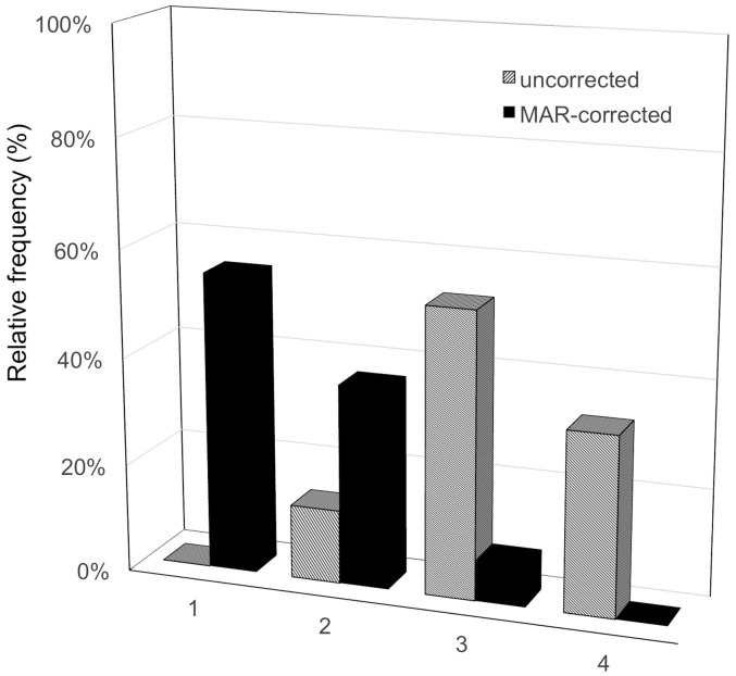
Background and purpose C-arm cone-beam computed tomography (CBCT) has the drawback that image quality is degraded by artifacts caused by implanted metal objects. We evaluated whether metal artifact reduction (MAR) prototype software can improve the subjective image quality of CBCT images of patients with intracranial aneurysms treated with coils or clips. Materials and methods Forty-four patients with intracranial aneurysms implanted with coils (40 patients) or clips (four patients) underwent one CBCT scan from which uncorrected and MAR-corrected CBCT image datasets were reconstructed. Three blinded readers evaluated the image quality of the image sets using a four-point scale (1: Excellent, 2: Good, 3: Poor, 4: Bad). The median scores of the three readers of uncorrected and MAR-corrected images were compared with the paired Wilcoxon signed-rank and inter-reader agreement of change scores was assessed by weighted kappa statistics. The readers also recorded new clinical findings, such as intracranial hemorrhage, air, or surrounding anatomical structures on MAR-corrected images. Results The image quality of MAR-corrected CBCT images was significantly improved compared with the uncorrected CBCT image ( p < 0.001). Additional clinical findings were seen on CBCT images of 70.4% of patients after MAR correction. Conclusion MAR software improved image quality of CBCT images degraded by metal artifacts.

Keywords: Cone-beam computed tomography; cerebral aneurysm; endovascular treatment.


Figures

Figure 1.
Figure 1.
Comparison of the distribution of four-point metal artifact reduction (MAR) scale of the 44 patients between uncorrected and MAR-corrected cone-beam computed tomography images.
Figure 2.
Figure 2.
Case 1. An 80-year-old woman with unruptured left IC-Pcom aneurysm was incidentally detected when she was referred to the hospital after a traffic accident. The aneurysm became larger year by year from 10 to 14 mm and finally stent-assisted coil embolization was planned (a). When a vascular reconstruction stent (Enterprise, Johnson & Johnson CODMAN, Miami, FL, USA) was deployed at the neck of the aneurysm, she suddenly went into cardiac arrest (b). She was promptly revived by cardiopulmonary resuscitation. Uncorrected-CBCT image could not reveal whether hemorrhagic complication had occurred (c), but the MAR-corrected image showed that hemorrhagic complication had not occurred (d). The cardiologist diagnosed the cause of the cardiac arrest as ventricular fibrillation after an overdose of anesthetic agent, and administered medical treatments. The aneurysm was filled with additional coils (e) and successfully obliterated (f). IC-Pcom: internal carotid-posterior communicating artery; CBCT: cone-beam computed tomography; MAR: metal artifact reduction.
Figure 3.
Figure 3.
Case 2. A 55-year-old woman with a history of subarachnoid hemorrhage previously underwent clipping for ruptured aneurysms (left ICA and MCA aneurysm), and again presented with subarachnoid hemorrhage due to a ruptured right IC-Pcom aneurysm that was treated with an emergency coil embolization. CTA (a), DSA (b), and conventional CT (c) at onset time. Uncorrected-CBCT image (d) could not reveal the presence of subarachnoid hemorrhage (arrows). The MAR-corrected image provides better image quality (e). CBCT: cone-beam computed tomography; CT: computed tomography; CTA: computed tomography angiography; DSA: digital subtraction angiography; ICA: internal carotid artery; IC-Pcom: internal carotid-posterior communicating artery; MAR: metal artifact reduction; MCA: middle cerebral artery.
Figure 4.
Figure 4.
Case 3. A 73-year-old woman with partial thrombosed left IC cavernous aneurysm was treated with stent-assisted coil embolization. The postoperative CBCT images were degraded by massive metal artifacts (a), but the MAR-corrected CBCT images clearly demonstrated the aneurysm wall, intra-aneurysmal thrombus, and the positional relationship between the coil mass and the aneurysm itself (arrows; (b)). IC: internal carotid; CBCT: cone-beam computed tomography; MAR: metal artifact reduction.

References

    1. Levy E, Koebbe CJ, Horowitz MB, et al. Rupture of intracranial aneurysms during endovascular coiling: Management and outcomes. Neurosurgery 2001; 49: 807–813. - PubMed
    1. Stapleton CJ, Walcott BP, Butler WE, et al. Neurological outcomes following intraprocedural rerupture during coil embolization of ruptured intracranial aneurysm. J Neurosurg 2015; 122: 128–135. - PubMed
    1. Elijovich L, Higashida RT, Lawton MT, et al. Predictors and outcomes of intraprocedural rupture in patients treated for ruptured intracranial aneurysms: The CARAT study. Stroke 2008; 39: 1501–1506. - PubMed
    1. Tummala RP, Chu RM, Madison MT, et al. Outcomes after aneurysm ruptured during endovascular coil embolization. Neurosurgery 2001; 49: 1059–1066; discussion 1066–1067. - PubMed
    1. Psychogios MN, Buhk JH, Schramm P, et al. Feasibility of angiographic CT in peri-interventional diagnostic imaging: A comparative study with multidetector CT. AJNR Am J Neuroradiol 2010; 31: 1226–1231. - PMC - PubMed