

Hearing impairment in MELAS: new prospective in clinical use of microRNA, a systematic review
- PMID: 29466997
- PMCID: PMC5822652
- DOI: 10.1186/s13023-018-0770-1
Hearing impairment in MELAS: new prospective in clinical use of microRNA, a systematic review
Abstract
Aim: To evaluate the feasibility of microRNAs (miR) in clinical use to fill in the gap of current methodology commonly used to test hearing impairment in MELAS patients.
Material and method: A literature review was performed using the following keywords, i.e., MELAS, Hearing Loss, Hearing Impairment, Temporal Bone, Otoacustic Emission (OTOAE), Auditory Brain Response (ABR), and microRNA. We reviewed the literature and focused on the aspect of the temporal bone, the results of electrophysiological tests in human clinical studies, and the use of miR for detecting lesions in the cochlea in patients with MELAS.
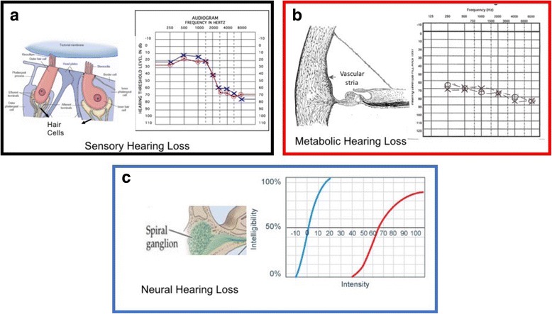
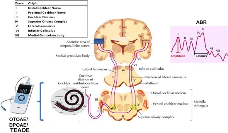
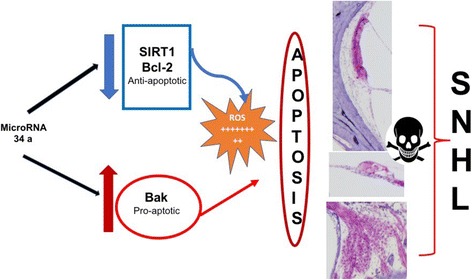
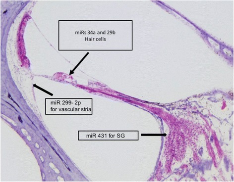
Results: In patients with MELAS, Spiral Ganglions (SG), stria vascularis (SV), and hair cells are damaged, and these damages affect in different ways various structures of the temporal bone. The function of these cells is typically investigated using OTOAE and ABR, but in patients with MELAS these tests provide inconsistent results, since OTOAE response is absent and ABR is normal. The normal ABR responses are unexpected given the SG loss in the temporal bone. Recent studies in humans and animals have shown that miRs, and in particular miRs 34a, 29b, 76, 96, and 431, can detect damage in the cells of the cochlea with high sensitivity. Studies that focus on the temporal bone aspects have reported that miRs increase is correlated with the death of specific cells of the inner ear. MiR - 9/9* was identified as a biomarker of human brain damage, miRs levels increase might be related to damage in the central auditory pathways and these increased levels could identify the damage with higher sensitivity and several months before than electrophysiological testing.
Conclusion: We suggest that due to their accuracy and sensitivity, miRs might help monitor the progression of SNHL in patients with MELAS.
Keywords: Auditory brain response; Diagnosis; Hearing impairment; Hearing loss; MELAS; Otoacustic emission; microRNA.
Conflict of interest statement
Ethics approval and consent to participate
n/a
Consent for publication
All authors are agree with publication policy and approved this version of the paper.
Competing interests
The authors declare that they have no competing interests.
Publisher’s Note
Springer Nature remains neutral with regard to jurisdictional claims in published maps and institutional affiliations.
Figures
References
-
- Angelini C. Genetic Neuromuscular Disorders. Cham: Springer; 2014. MELAS (myopathy, encephalopathy, lactic acidosis, stroke-like episodes)
-
- Kullar PJ, Quail J, Lindsey P, Ja W, Horvath R, Yu-Wai-Man P, Gorman GS, Taylor RW, Ng Y, McFarland R, Moore BCJ, Chinnery PF. Both mitochondrial DNA and mitonuclear gene mutations can cause hearing loss through cochlear dysfunction. Brain. 2016;139:1–5. doi: 10.1093/brain/aww051. - DOI - PMC - PubMed
Publication types
MeSH terms
Substances
Grants and funding
LinkOut - more resources
Full Text Sources
Other Literature Sources
Medical
Miscellaneous
