. 2018 Feb 21;15(1):53.
doi: 10.1186/s12974-018-1093-9.

Neuregulin-1 elicits a regulatory immune response following traumatic spinal cord injury

Affiliations

Neuregulin-1 elicits a regulatory immune response following traumatic spinal cord injury

Arsalan Alizadeh et al. J Neuroinflammation. .

Abstract

Background: Spinal cord injury (SCI) triggers a robust neuroinflammatory response that governs secondary injury mechanisms with both degenerative and pro-regenerative effects. Identifying new immunomodulatory therapies to promote the supportive aspect of immune response is critically needed for the treatment of SCI. We previously demonstrated that SCI results in acute and permanent depletion of the neuronally derived Neuregulin-1 (Nrg-1) in the spinal cord. Increasing the dysregulated level of Nrg-1 through acute intrathecal Nrg-1 treatment enhanced endogenous cell replacement and promoted white matter preservation and functional recovery in rat SCI. Moreover, we identified a neuroprotective role for Nrg-1 in moderating the activity of resident astrocytes and microglia following injury. To date, the impact of Nrg-1 on immune response in SCI has not yet been investigated. In this study, we elucidated the effect of systemic Nrg-1 therapy on the recruitment and function of macrophages, T cells, and B cells, three major leukocyte populations involved in neuroinflammatory processes following SCI.

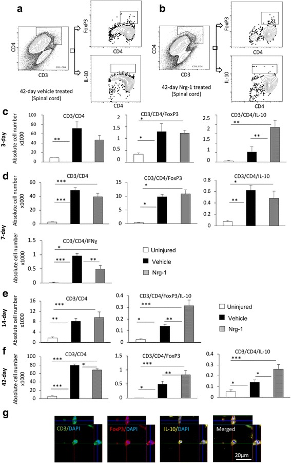
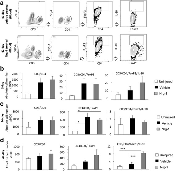
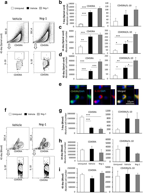
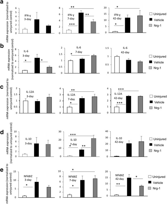
Methods: We utilized a clinically relevant model of moderately severe compressive SCI in female Sprague-Dawley rats. Nrg-1 (2 μg/day) or saline was delivered subcutaneously through osmotic mini-pumps starting 30 min after SCI. We conducted flow cytometry, quantitative real-time PCR, and immunohistochemistry at acute, subacute, and chronic stages of SCI to investigate the effects of Nrg-1 treatment on systemic and spinal cord immune response as well as cytokine, chemokine, and antibody production.

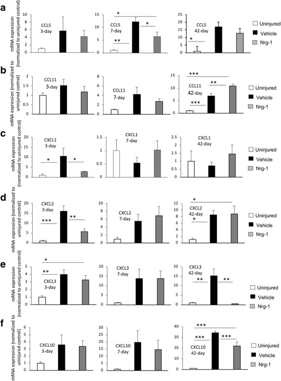
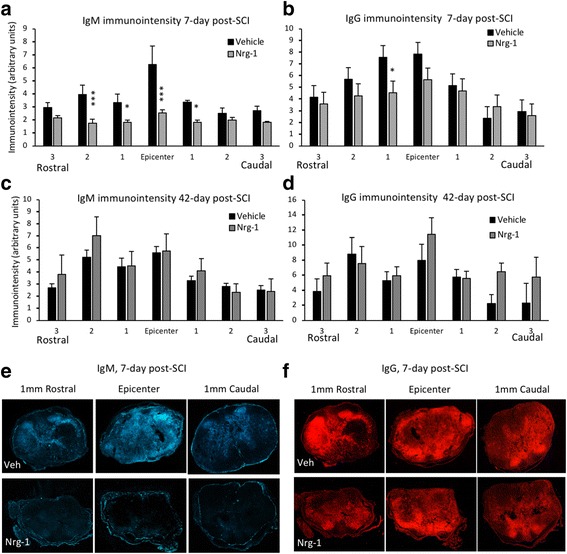
Results: We provide novel evidence that Nrg-1 promotes a pro-regenerative immune response after SCI. Bioavailability of Nrg-1 stimulated a regulatory phenotype in T and B cells and augmented the population of M2 macrophages in the spinal cord and blood during the acute and chronic stages of SCI. Importantly, Nrg-1 fostered a more balanced microenvironment in the injured spinal cord by attenuating antibody deposition and expression of pro-inflammatory cytokines and chemokines while upregulating pro-regenerative mediators.

Conclusion: We provide the first evidence of a significant regulatory role for Nrg-1 in neuroinflammation after SCI. Importantly, the present study establishes the promise of systemic Nrg-1 treatment as a candidate immunotherapy for traumatic SCI and other CNS neuroinflammatory conditions.

Keywords: Autoantibodies; B cells; Cytokines and chemokines; Macrophages; Neuregulin-1; Neuroinflammation; Rat; Spinal cord injury; T cells.


Conflict of interest statement

Ethics approval

All experimental protocols in this study involving animals were approved by the Animal Care Committee of University of Manitoba in accordance with the guidelines and policies established by the Canadian Council on Animal Care, protocol number: 15030 (AC11067).

Consent for publication

Not applicable.

Competing interests

The authors declare that they have no competing interests.

Publisher’s Note

Springer Nature remains neutral with regard to jurisdictional claims in published maps and institutional affiliations.

Figures

Fig. 1
Fig. 1
Nrg-1 treatment alters M1 and M2 macrophage populations after SCI. a, b Representative images of the gating strategy for flow cytometry are provided for infiltrated spinal cord macrophages at 42-day post-injury under each treatment group. c Flow cytometric analysis 3 days post-SCI showed a robust increase in the number of macrophages (CD45+CD68+) in the injured spinal cord. Nrg-1-treated animals demonstrated a significantly higher number of macrophages compared to the vehicle-treated group. Phenotypical analysis of macrophages at 3-day time-point showed no significant difference in the number of infiltrated M1 macrophages (CD45+CD68+CD86+) between vehicle- and Nrg-1-treated groups. However, Nrg-1-treated SCI animals had a significantly higher population of M2 macrophages (CD45+CD68+CD163+ and CD45+CD68+CD163+IL-10+) in the spinal cord. d, e At 7 and 14 days post-SCI, the total number of tissue macrophages and their M1 subpopulation was significantly higher in the injured animals compared to the uninjured group, while M2 macrophage population remained unaltered. There was also no significant difference in the total number of macrophages and their M1 or M2 phenotype between the vehicle- and Nrg-1-treated groups. f At 42 days post-SCI, the number of infiltrated macrophages was 30 times less than the acute 3-day time-point in SCI baseline condition but still significantly higher than uninjured animals. Compared to both vehicle and uninjured rats, Nrg-1-treated animals showed a significantly higher number of M1 macrophages. There was no significant difference in the number of M2 macrophages between the vehicle and Nrg-1 treatment groups at this time-point, although Nrg-1-treated rats had a significantly higher number of M2 macrophages as compared to uninjured animals. g Immunohistochemical analysis verified the presence of M1 (CD68+CD86+) and M2 (CD68+CD163+) macrophages in the perilesional area (N = 5/group/time-point, *p < 0.05, **p < 0.01, ***p < 0.001, one-way ANOVA followed by Holm-Sidak post hoc test)
Fig. 2
Fig. 2
Nrg-1 promotes Treg cell response in the injured spinal cord. a, b Representative images of the gating strategy for flow cytometry after singlet selection are provided for vehicle- and Nrg-1-treated groups. c At 3-day post-injury, the number of CD3+CD4+ T cells in the spinal cord was significantly higher in vehicle-treated animals compared to uninjured control group. There was no significant difference in the population of infiltrated helper T cells and the number of FoxP3+ Treg cells (CD3+CD4+FoxP3+) between vehicle- and Nrg-1-treated groups. However, the population of IL-10 producing CD4+ T cells (CD3+CD4+IL-10+) was significantly increased in Nrg-1-treated animals in comparison to vehicle-treated group. d At 7 days post-SCI, despite no significant difference in the total helper and regulatory T cell populations between vehicle- and Nrg-1-treated groups, a significant reduction (1.9-fold) was observed in IFNγ producing effector T cell population in Nrg-1-treated SCI rats compared to their vehicle-treated counterparts. e At 14 days post-SCI, infiltrated helper T cells reached their lowest level among all examined time-points, and Nrg-1 treatment had no significant effect on the total helper T cell population. IL-10 expressing Treg cells were significantly higher in both vehicle and Nrg-1 injured rats compared to uninjured animals. Nrg-1-treated animals showed a significantly higher number of Treg cells in their spinal cord at 14-day time-point compared to vehicle-treated rats. f However, at chronic (42-day) time-point, the number of T helper cells reached a maximum, with Nrg-1-treated animals harboring a significantly decreased population of CD4+ T-cells in their spinal cord compared to the vehicle-treated group. Most importantly, CD3+CD4+FoxP3+ and CD3+CD4+IL-10+ regulatory T cell populations were significantly increased in Nrg-1-treated groups. g Immunohistochemical images show the presence of Treg cells in the perilesional area (N = 5/group/time-point, *p < 0.05, **p < 0.01, ***p < 0.001, one-way ANOVA followed by Holm-Sidak post hoc test)
Fig. 3
Fig. 3
Nrg-1 treatment increases the number of circulating Treg cells in the blood following chronic SCI. a Representative images are provided for the gating strategy for flow cytometric detection of T cells and their regulatory phenotype in the blood. bd Flow cytometric analysis showed a slight increase in total CD3+CD4+ helper T cell population upon injury. However, at all examined time-points, the total population of helper T cells was not significantly different among any of the studied groups (p > 0.05). The population of FoxP3+ helper T cells was significantly increased at 14 days post-injury in the vehicle- and Nrg-1-treated rats as compared to the uninjured levels while there was no difference between the two injured groups. d At 42-day time-point, Nrg-1 treatment significantly increased the number of IL-10 expressing (CD3+CD4+FoxP3+IL-10+) Treg cells compared to vehicle and uninjured groups while there was no significant difference in the total population of helper and Fox3+ T cells between the vehicle and uninjured groups, (N = 5/group/time-point, *p < 0.05, **p < 0.01, ***p < 0.001, one-way ANOVA followed by Holm-Sidak post hoc test)
Fig. 4
Fig. 4
Nrg-1 treatment promotes Breg cell population following SCI. a Representative images of the gating strategy for flow cytometry of spinal cord are provided. b At 7 days post-injury, the number of CD45RA+ B cells was significantly increased in the spinal cord without any significant difference in the total and regulatory B cell populations between vehicle- and Nrg-1-treated groups. c At 14-day post-SCI, a significant increase in the number of Breg cells was observed in Nrg-1-treated animals compared to vehicle-treated group. d Chronically at 42 days post-SCI, the number of B cells in the spinal cord reached the highest level compared to all earlier time-points. Nrg-1 treatment resulted in a significant increase in the number of infiltrated B cells in the spinal cord and promoted IL-10 expressing Breg cells compared to vehicle treatment. e Immunohistochemical analysis verified the presence of Breg cells in the perilesional area of the injured spinal cord. f Representative images of the gating strategy for flow cytometry of blood are provided. g Analysis of the blood revealed a significant decline in B cell population at 7 days post-SCI without any significant change in the Breg cell population at this time-point. h, i No significant change in total and regulatory B cell populations were observed in the blood at 14- and 42-day time-points. (N = 5/group/time-point, *p < 0.05, **p < 0.01, ***p < 0.001, one-way ANOVA followed by Holm-Sidak post hoc test)
Fig. 5
Fig. 5
Nrg-1 treatment regulates inflammatory cytokines in the injured spinal cord. a Transcript analysis of the spinal cord tissue using real-time qPCR released a significant increase in expression of IFN-γ at 7-day post-injury (p < 0.001). Nrg-1 treatment significantly decreased the SCI-induced expression of IFN-γ in the injured spinal cord tissue. b Expression of IL-6 was significantly increased at 3 days post-injury, which was significantly attenuated by Nrg-1 treatment (p < 0.05). No significant difference in IL-6 expression was observed between vehicle- and Nrg-1-treated groups at 7- and 42-day time-points. c IL-12A was significantly increased at 7 days post-injury and remained significantly elevated until 42 days. Nrg-1 had no significant effect on IL-12A expression at any examined time-point. d Transcript levels of IL-10 were significantly increased at 7 days post-injury. Interestingly, Nrg-1-treated animals showed a significantly higher expression of IL-10 compared to the vehicle group at this time-point. No significant change in IL-10 expression was observed at any other examined time-points. e NFkBIZ transcript level was significantly elevated at 3 days and reached its maximum at 42 days post-injury. Nrg-1 treatment significantly attenuated NFkBIZ expression chronically at 42 days post-SCI (N = 4/group/time-point, *p < 0.05, **p < 0.01, ***p < 0.001, one-way ANOVA followed by Holm-Sidak post hoc test)
Fig. 6
Fig. 6
Nrg-1 treatment positively modulates chemokine expression following SCI. a Analysis of chemokine expression in the spinal cord tissue was performed using real-time qPCR. There was a significant increase in the expression of pro-inflammatory chemokine CCL5, at 7-day post-injury, which was significantly reduced by Nrg-1 treatment. CCL5 mRNA level reached its maximum level at 42 days post-injury. However, no significant difference was found between vehicle- and Nrg-1-treated animals at this time-point. b Although the expression of CCL11, an immune modulatory chemokine, was not significantly changed at 3 and 7 days post-injury, it underwent a significant increase chronically at 42-day time-point. Nrg-1 treatment was able to significantly increase CCL11 expression compared to vehicle-treated group. c, d Expression of pro-inflammatory chemokines CXCL1 and CXCL2 was significantly elevated at 3-day post-injury. Nrg-1 treatment significantly reduced CXCL1 and CXCL2 expressions at this time-point. The overall tissue level of CXCL1 mRNA reached baseline levels at 7-day post-injury and remained stable until 42 days post-injury. CXCL2 expression, however, remained elevated at 7 and 42 days post-injury compared to uninjured group (significant 8.5-fold increase at 42-day). Nrg-1 treatment had no significant effect on CXCL2 expression at 7 and 42-day time-points. e Expression of the pro-inflammatory chemokine CXCL3was significantly upregulated 3 days after injury and reached its maximum levels chronically at 42 days post-SCI (p < 0.01). Nrg-1-treated animals showed a significant reduction in CXCL3 expression at 42-day time-point (31 times). f CXCL10 expression was not significantly changed following an injury at 3 and 7 days post-SCI, while it was significantly increased in the vehicle-treated group compared to un-injured animals at 42 days. Nrg-1 significantly decreased CXCL10 expression at this time-point (p < 0.001) (N = 4/group/time-point, *p < 0.05, **p < 0.01, ***p < 0.001, one-way ANOVA followed by Holm-Sidak post hoc test)
Fig. 7
Fig. 7
Nrg-1 therapy significantly attenuates antibody deposition in the injured spinal cord tissue at subacute stage of SCI. ad Immunohistochemical assessment of injured spinal cord tissue for IgM and IgG deposition was performed at the epicenter and perilesional areas at subacute (7 days) and chronic (42 days) stages of SCI. a, c Analysis of IgM immunointensity showed a significantly lower IgM deposition in Nrg-1-treated animals compared to vehicle-treated group at 7-day post-SCI. This decrease was significant at the epicenter, 1 mm caudal and 1 and 2 mm rostral to the injury site. No significant difference in IgM deposition was detected at 42-day time-point. b, d Comparison of IgG immunointensity between the vehicle- and Nrg-1-treated animals revealed a significant decrease in IgG deposition only at 1 mm rostral to the epicenter at 7-day post-injury. No significant difference was detected in IgG deposition between the vehicle- and Nrg-1-treated groups at 42-day time-point. e, f Representative images are provided at the epicenter and 1 mm rostral and caudal to the lesion center (N = 4/group/time-point, *p < 0.05, **p < 0.01, ***p < 0.001, two-way ANOVA followed by Holm-Sidak post hoc test)

References

    1. Trivedi A, Olivas AD, Noble-Haeusslein LJ. Inflammation and spinal cord injury: infiltrating leukocytes as determinants of injury and repair processes. Clin Neurosci Res. 2006;6:283–292. doi: 10.1016/j.cnr.2006.09.007. - DOI - PMC - PubMed
    1. Fitch MT, Doller C, Combs CK, Landreth GE, Silver J. Cellular and molecular mechanisms of glial scarring and progressive cavitation: in vivo and in vitro analysis of inflammation-induced secondary injury after CNS trauma. J Neurosci. 1999;19:8182–8198. - PMC - PubMed
    1. Noble LJ, Donovan F, Igarashi T, Goussev S, Werb Z. Matrix metalloproteinases limit functional recovery after spinal cord injury by modulation of early vascular events. J Neurosci. 2002;22:7526–7535. - PMC - PubMed
    1. Ankeny DP, Lucin KM, Sanders VM, McGaughy VM, Popovich PG. Spinal cord injury triggers systemic autoimmunity: evidence for chronic B lymphocyte activation and lupus-like autoantibody synthesis. J Neurochem. 2006;99:1073–1087. doi: 10.1111/j.1471-4159.2006.04147.x. - DOI - PubMed
    1. Zhao WXW, Le W, Beers DR, He Y, Henkel JS, Simpson EP, Yen AA, Xiao Q, Appel SH. Activated microglia initiate motor neuron injury by a nitric oxide and glutamate-mediated mechanism. J Neuropathol Exp Neurol. 2004;63:964–977. doi: 10.1093/jnen/63.9.964. - DOI - PubMed

MeSH terms