Accuracy of the ADNEX MR scoring system based on a simplified MRI protocol for the assessment of adnexal masses
- PMID: 29467113
- PMCID: PMC5873504
- DOI: 10.5152/dir.2018.17378
Accuracy of the ADNEX MR scoring system based on a simplified MRI protocol for the assessment of adnexal masses
Abstract
Purpose: We aimed to evaluate the ADNEX MR scoring system for the prediction of adnexal mass malignancy, using a simplified magnetic resonance imaging (MRI) protocol.
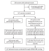
Methods: In this prospective study, 200 patients with 237 adnexal masses underwent MRI between February 2014 and February 2016 and were followed until February 2017. Two radiologists calculated ADNEX MR scores using an MRI protocol with a simplified dynamic study, not a high temporal resolution study, as originally proposed. Sensitivity, specificity, positive and negative predictive values, likelihood ratios, and the area under the receiver operating characteristic curve were calculated (cutoff for malignancy, score ≥ 4). The reference standard was histopathologic diagnosis or imaging findings during >12 months of follow-up.
Results: Of 237 lesions, 79 (33.3%) were malignant. The ADNEX MR scoring system, using a simplified MRI protocol, showed 94.9% (95% confidence interval [CI], 87.5%-98.6%) sensitivity and 97.5% (95% CI, 93.6%-99.3%) specificity in malignancy prediction; it was thus highly accurate, like the original system. The level of interobserver agreement on simplified scoring was high (κ = 0.91).
Conclusion: In a tertiary cancer center, the ADNEX MR scoring system, even based on a simplified MRI protocol, performed well in the prediction of malignant adnexal masses. This scoring system may enable the standardization of MRI reporting on adnexal masses, thereby improving communication between radiologists and gynecologists.
Conflict of interest statement
The authors declared no conflicts of interest.
Figures


References
-
- Pickhardt PJ, Hanson ME. Incidental adnexal masses detected at low-dose unenhanced CT in asymptomatic women age 50 and older: implications for clinical management and ovarian cancer screening. Radiology. 2010;257:144–150. https://doi.org/10.1148/radiol.10100511. - DOI - PubMed
-
- Menon U, Griffin M, Gentry-Maharaj A. Ovarian cancer screening--current status, future directions. Gynecol Oncol. 2014;132:490–495. https://doi.org/10.1016/j.ygyno.2013.11.030. - DOI - PMC - PubMed
-
- Forstner R, Sala E, Kinkel K, Spencer JA. ESUR guidelines: ovarian cancer staging and follow-up. Eur Radiol. 2010;20:2773–2780. https://doi.org/10.1007/s00330-010-1886-4. - DOI - PubMed
-
- Webb PM, Jordan SJ. Epidemiology of epithelial ovarian cancer. Best Pract Res Clin Obstet Gynaecol. 2017;41:3–14. https://doi.org/10.1016/j.bpobgyn.2016.08.006. - DOI - PubMed
-
- Coccia ME, Rizzello F, Romanelli C, Capezzuoli T. Adnexal masses: what is the role of ultrasonographic imaging? Arch Gynecol Obstet. 2014;290:843–854. https://doi.org/10.1007/s00404-014-3327-0. - DOI - PubMed
MeSH terms
LinkOut - more resources
Full Text Sources
Other Literature Sources
Medical

