. 2018 Mar 6;115(10):2461-2466.
doi: 10.1073/pnas.1721117115. Epub 2018 Feb 21.

Viral insulin-like peptides activate human insulin and IGF-1 receptor signaling: A paradigm shift for host-microbe interactions

Affiliations

Viral insulin-like peptides activate human insulin and IGF-1 receptor signaling: A paradigm shift for host-microbe interactions

Emrah Altindis et al. Proc Natl Acad Sci U S A. .

Abstract

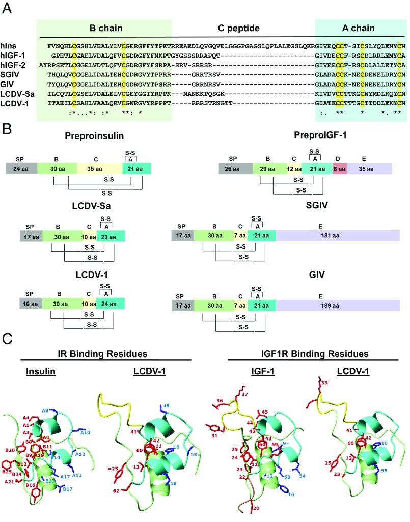
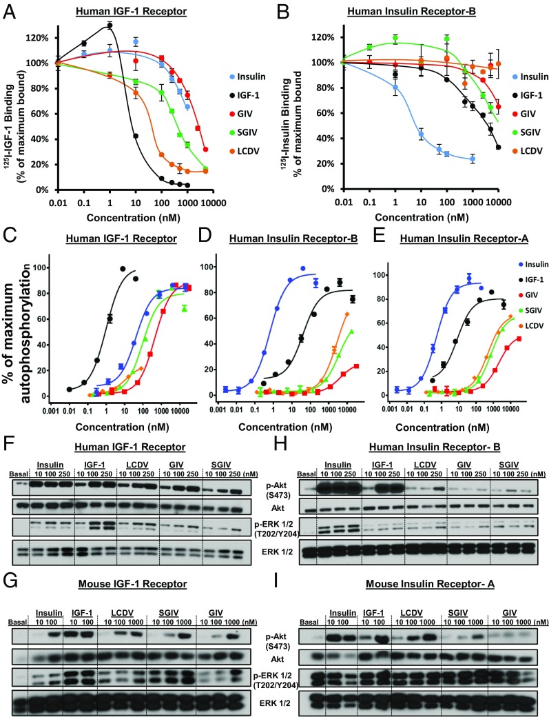
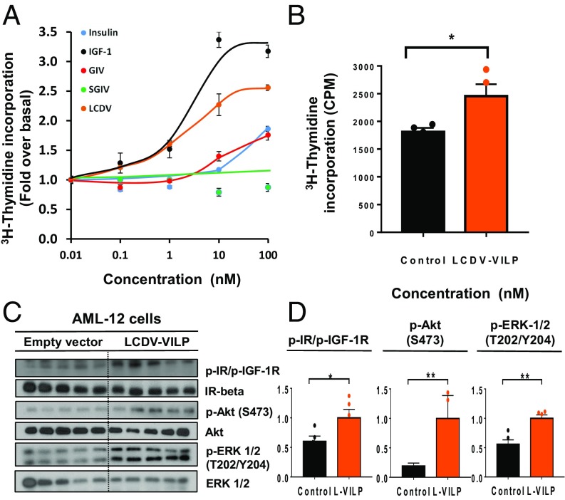
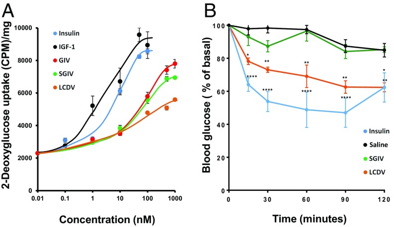
Viruses are the most abundant biological entities and carry a wide variety of genetic material, including the ability to encode host-like proteins. Here we show that viruses carry sequences with significant homology to several human peptide hormones including insulin, insulin-like growth factors (IGF)-1 and -2, FGF-19 and -21, endothelin-1, inhibin, adiponectin, and resistin. Among the strongest homologies were those for four viral insulin/IGF-1-like peptides (VILPs), each encoded by a different member of the family Iridoviridae VILPs show up to 50% homology to human insulin/IGF-1, contain all critical cysteine residues, and are predicted to form similar 3D structures. Chemically synthesized VILPs can bind to human and murine IGF-1/insulin receptors and stimulate receptor autophosphorylation and downstream signaling. VILPs can also increase glucose uptake in adipocytes and stimulate the proliferation of fibroblasts, and injection of VILPs into mice significantly lowers blood glucose. Transfection of mouse hepatocytes with DNA encoding a VILP also stimulates insulin/IGF-1 signaling and DNA synthesis. Human microbiome studies reveal the presence of these Iridoviridae in blood and fecal samples. Thus, VILPs are members of the insulin/IGF superfamily with the ability to be active on human and rodent cells, raising the possibility for a potential role of VILPs in human disease. Furthermore, since only 2% of viruses have been sequenced, this study raises the potential for discovery of other viral hormones which, along with known virally encoded growth factors, may modify human health and disease.

Keywords: diabetes; insulin; insulin-like growth factor; viral hormones; viral pathogenesis.


Conflict of interest statement

Conflict of interest statement: R.D. is currently an employee of Novo-Nordisk, but the work described in this paper was done in collaboration with his academic laboratory at Indiana University and has no commercial support or connection.

Figures

Fig. 1.
Fig. 1.
Viral insulin/IGF-like peptides are structurally a part of the insulin superfamily. (A) Sequence alignment of the B-, C-, and A-chains of insulin, IGF1, IGF2, and four VILPs. Cysteines are highlighted in yellow. Identical residues are denoted by asterisks; low and high degrees of similarity are represented by a period and a colon, respectively. (B) Domain structure of human insulin, IGF1, and four VILPs. The domains are indicated as follows: A, A-chain; B, B-chain; C, C-peptide; S-S, disulfide bonds; and SP, signal peptide. (C) Predicted 3D structure of LCDV-1VILP and comparison with insulin and IGF-I. The A-chain is cyan; the B-chain is light green; the C-peptide is yellow; and the D-domain is pale brown. The conserved or conservatively substituted side chains of residues of the ligands involved in binding to site 1 of the IR/IGF1R are shown in red, and binding site 2 residues are shown in blue. Conservative substitutions are indicated by an equal sign. One substitution that increases affinity in IGF-I (B10 His to Glu) is indicated by a plus sign.
Fig. 2.
Fig. 2.
VILPs bind to hIR-B and hIGF1R and stimulate downstream insulin/IGF1-signaling pathways. (A and B) Binding competition dose–response curves showing the competing effect of VILPs for the hIGF1R (A) and hIR-B (B). The data are plotted as the percentage of the maximal binding of 125I-IGF1 alone or 125I-Insulin alone and are expressed as mean ± SEM (n = 3). (CE) Stimulation of IR and IGF1R autophosphorylation using HEK293 cells overexpressing hIGF1R (C), hIR-B (D), or hIR-A (E) and a phosphotyrosine-specific ELISA. (FI) Western blot analyses of the phosphorylation of Akt and ERK1/2 in lysates of murine DKO brown preadipocytes overexpressing hIGF1R cells (F), mIGF1R (G), hIR-B (H), and mIR-A (I) stimulated with the indicated concentrations of insulin, IGF1, or VILPs for 15 min.
Fig. 3.
Fig. 3.
Endogenous and exogenous mitogenic potency of VILPs illustrated by [3H]-thymidine incorporation and endogenous stimulation of postreceptor signaling. (A) Human fibroblasts (GM00409) were treated with increasing concentrations of the ligands, and [3H]-thymidine incorporation into DNA was assessed. Results are illustrated as the fold-increase over the basal level and are plotted as mean ± SEM (n = 4). (B) AML-12 cells were transiently transfected with either mock vector or plasmid encoding the LCDV-1 VILP gene. Results are shown as raw cpm values (*P < 0.05, t test, n = 4). (C) Immunoblotting of phosphorylation of IR/IGF1R beta subunits, Akt, and ERK1/2 in lysates from AML-12 cells transfected with mock vector or LCDV-1 VILP cDNA (n = 5). (D) Densitometric analysis of phosphorylated IR/IGF1R, Akt, and ERK1/2. Data are shown as mean ± SEM, normalized to each total protein level (*P < 0.05; **P < 0.01; t test; n = 5).
Fig. 4.
Fig. 4.
In vitro and in vivo effects of VILPs on glucose metabolism. (A) Differentiated 3T3-L1 cells were stimulated for 30 min with various concentrations of ligands, and uptake of 2-deoxyglucose was determined. Data are expressed as cpm/mg of protein ± SEM (n = 4). (B) Mice were injected i.p. with LCDV-1 VILP (1 µmol/kg; n = 4), SGIV VILP (1 µmol/kg; n = 4), insulin Humulin R (Eli Lilly), 6 nmol/kg; n = 6], or saline (n = 6). Blood glucose was measured at 0–120 min. Data are shown as mean ± SEM [*P < 0.05; **P < 0.01, ****P < 0.0001; two-way repeated-measures ANOVA (grouped by time) followed by Tukey 6 correction; n = 4 or 6].

Comment in

References

    1. Paez-Espino D, et al. Uncovering earth’s virome. Nature. 2016;536:425–430. - PubMed
    1. Dimitrov DS. Virus entry: Molecular mechanisms and biomedical applications. Nat Rev Microbiol. 2004;2:109–122. - PMC - PubMed
    1. Enard D, Cai L, Gwennap C, Petrov DA. Viruses are a dominant driver of protein adaptation in mammals. eLife. 2016;5:e12469. - PMC - PubMed
    1. Schulz F, et al. Giant viruses with an expanded complement of translation system components. Science. 2017;356:82–85. - PubMed
    1. Gilbert C, et al. Continuous influx of genetic material from host to virus populations. PLoS Genet. 2016;12:e1005838. - PMC - PubMed

Publication types