. 2018 Apr 3;115(14):E3135-E3144.
doi: 10.1073/pnas.1714131115. Epub 2018 Feb 21.

Protonation state of inhibitors determines interaction sites within voltage-gated sodium channels

Affiliations

Protonation state of inhibitors determines interaction sites within voltage-gated sodium channels

Amanda Buyan et al. Proc Natl Acad Sci U S A. .

Abstract

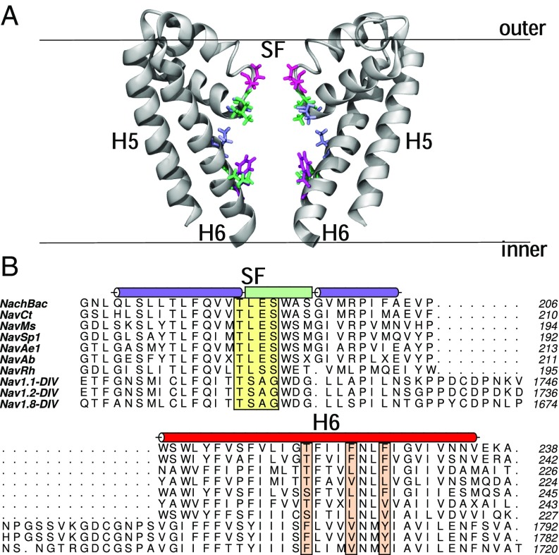
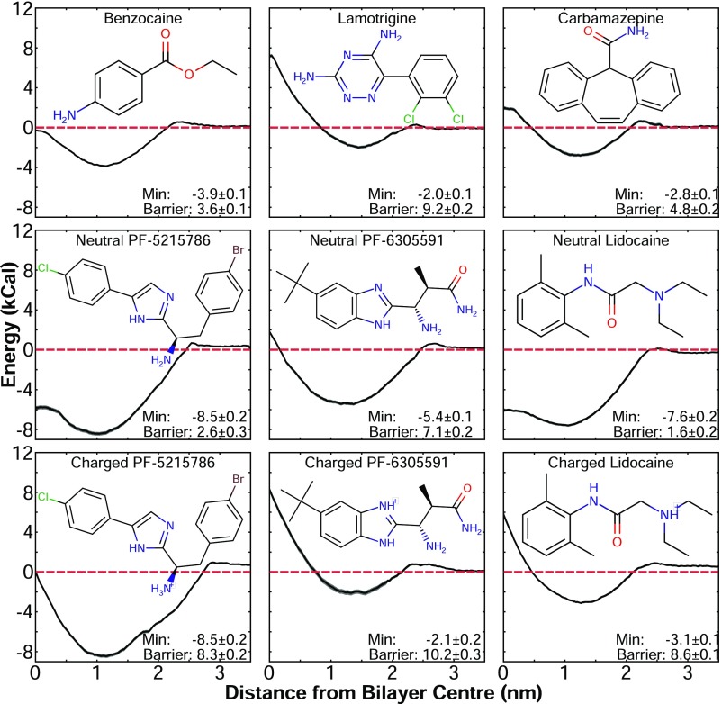
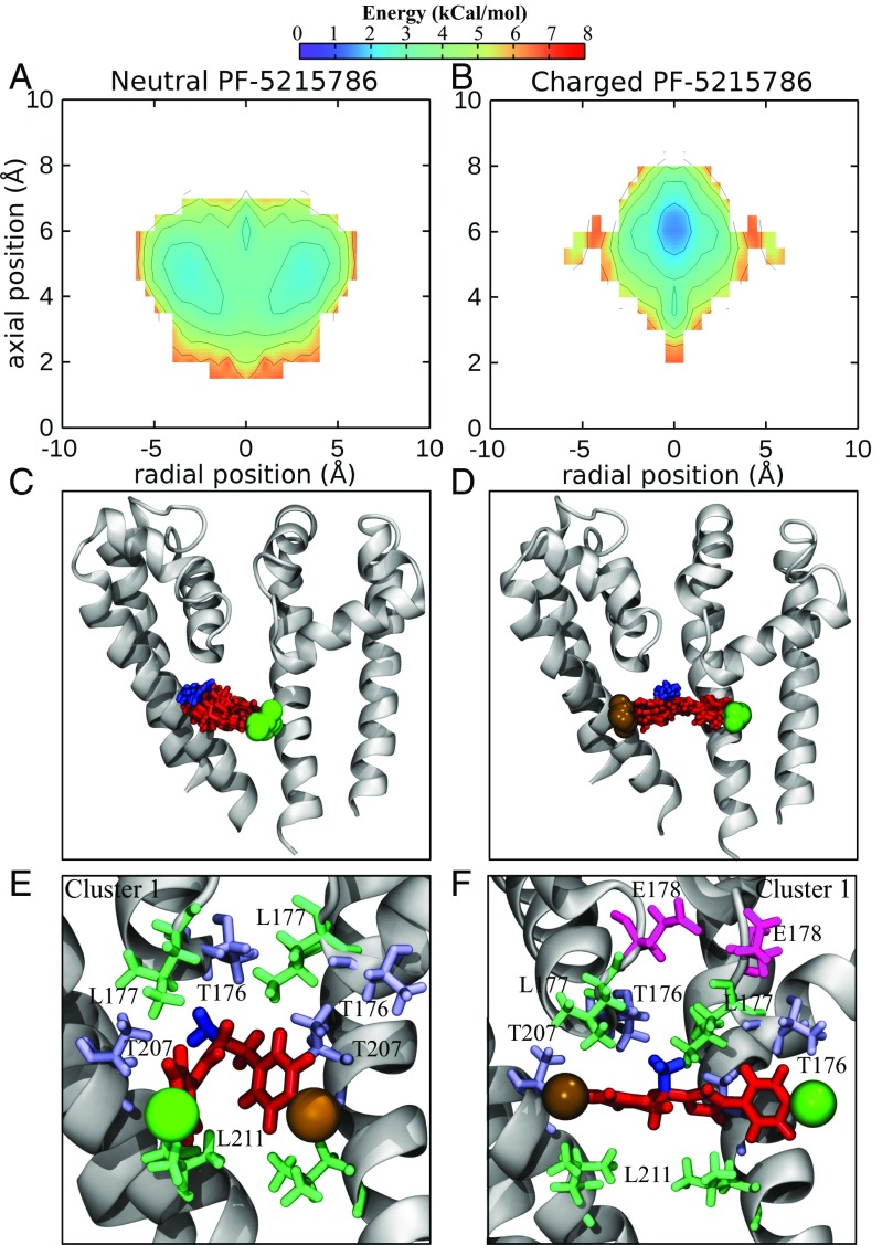
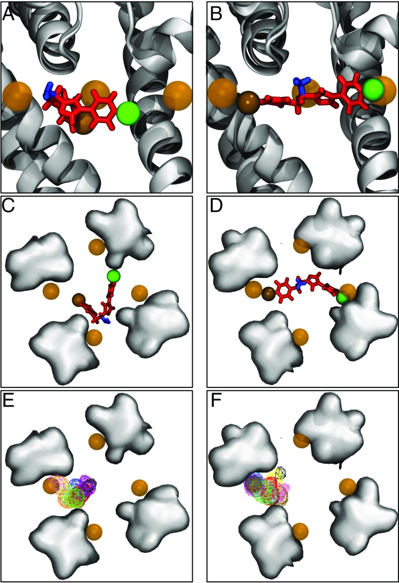
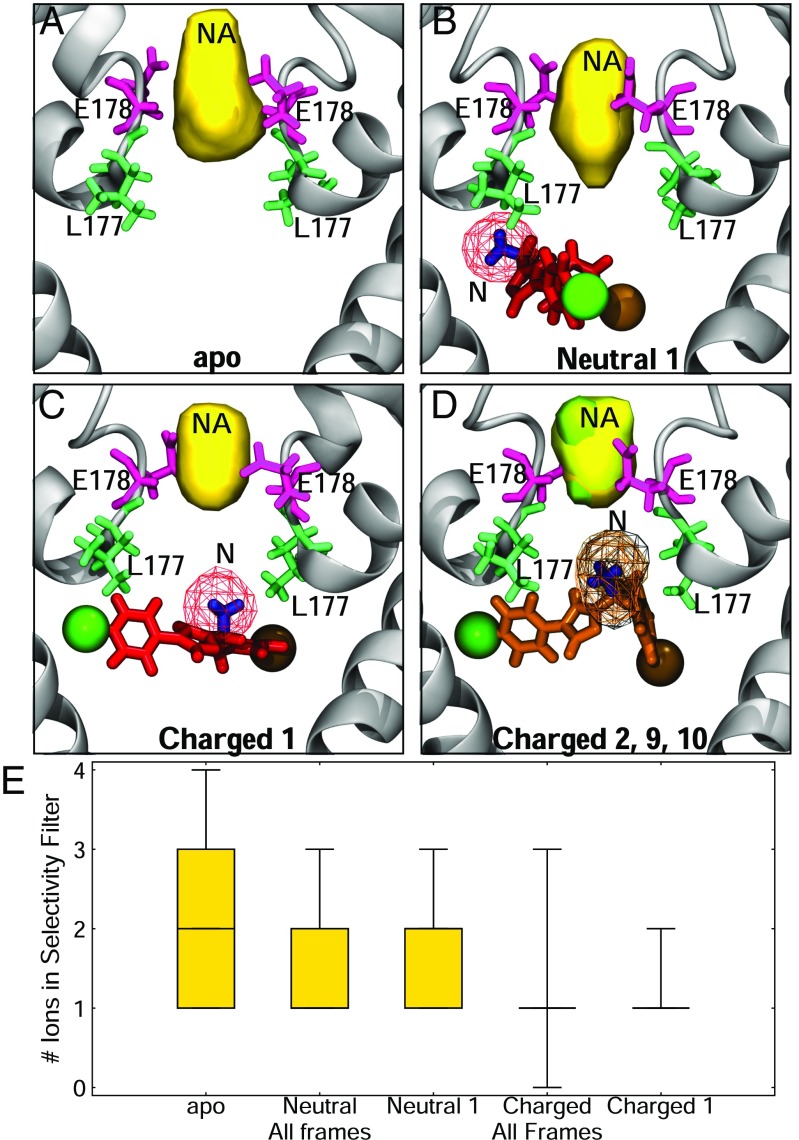
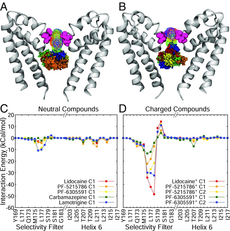
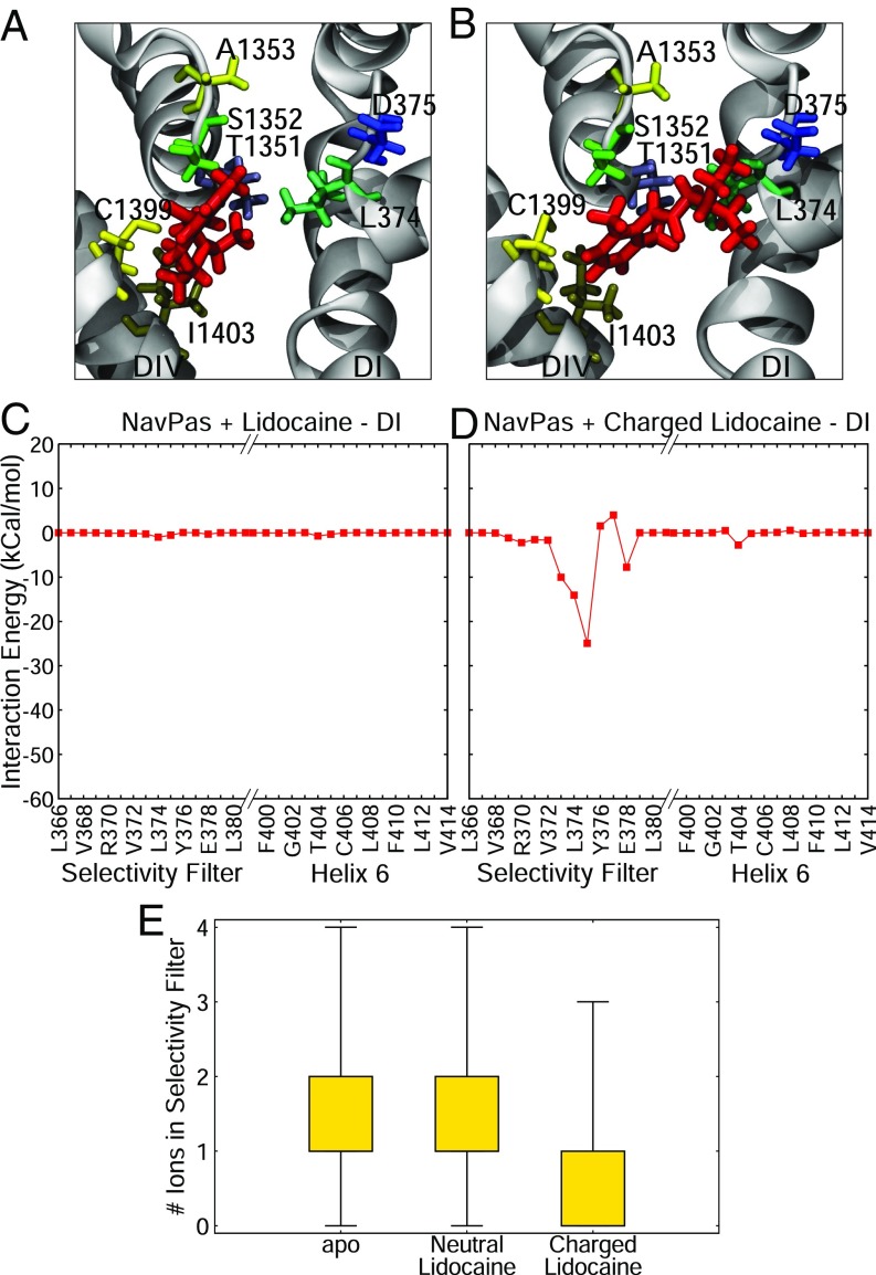
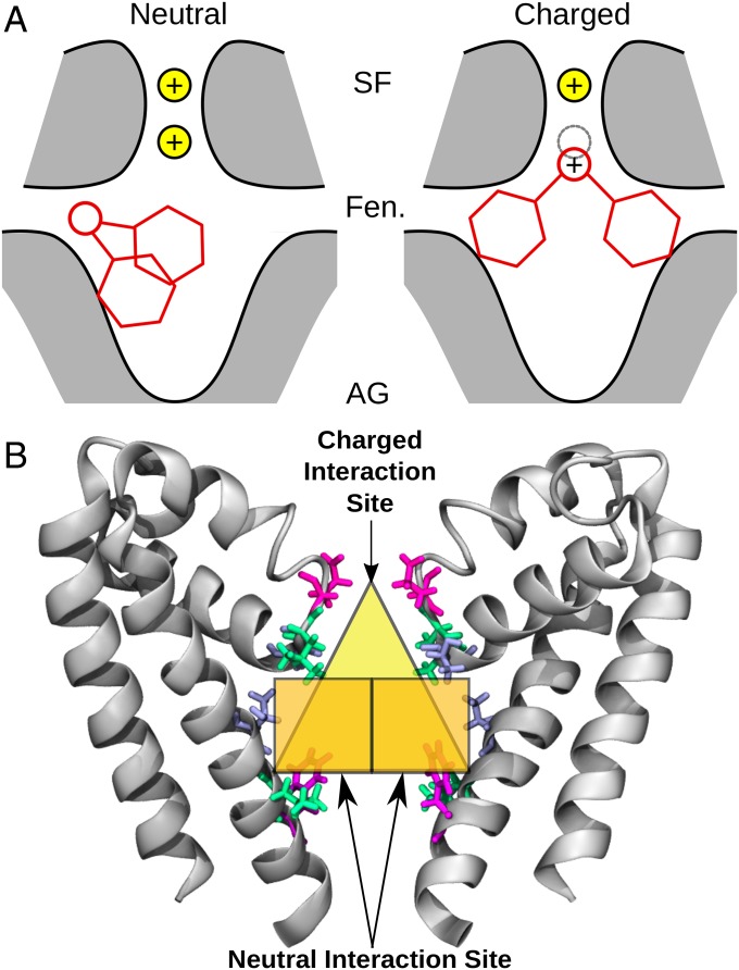
Voltage-gated sodium channels are essential for carrying electrical signals throughout the body, and mutations in these proteins are responsible for a variety of disorders, including epilepsy and pain syndromes. As such, they are the target of a number of drugs used for reducing pain or combatting arrhythmias and seizures. However, these drugs affect all sodium channel subtypes found in the body. Designing compounds to target select sodium channel subtypes will provide a new therapeutic pathway and would maximize treatment efficacy while minimizing side effects. Here, we examine the binding preferences of nine compounds known to be sodium channel pore blockers in molecular dynamics simulations. We use the approach of replica exchange solute tempering (REST) to gain a more complete understanding of the inhibitors' behavior inside the pore of NavMs, a bacterial sodium channel, and NavPas, a eukaryotic sodium channel. Using these simulations, we are able to show that both charged and neutral compounds partition into the bilayer, but neutral forms more readily cross it. We show that there are two possible binding sites for the compounds: (i) a site on helix 6, which has been previously determined by many experimental and computational studies, and (ii) an additional site, occupied by protonated compounds in which the positively charged part of the drug is attracted into the selectivity filter. Distinguishing distinct binding poses for neutral and charged compounds is essential for understanding the nature of pore block and will aid the design of subtype-selective sodium channel inhibitors.

Keywords: local anesthetic; molecular dynamics; pain; sodium channel; structural biology.


Conflict of interest statement

The authors declare no conflict of interest.

Figures

Fig. 1.
Fig. 1.
(A) Structure of the pore of the bacterial sodium channel NavMs. The backbone of the protein is shown in gray cartoon, and residues of interest that are highlighted in B are shown in licorice. The bilayer is represented by a black line. (B) Alignments of sequences of bacterial sodium channels and select eukaryotic channels, consisting of residues in the selectivity filter (SF) to the S6 helix (only domain IV of the eukaryotic channels is shown). The helices on each side of the selectivity filter are shown in a purple cylinder, with the selectivity filter shown as a light green box. The S6 helix is shown in a red cylinder. Residues of interest in the selectivity filter are highlighted in a yellow box, whereas residues on helix S6 are in orange boxes.
Fig. 2.
Fig. 2.
PMF for lipid partitioning of the compounds used in this study, along with their corresponding molecular structure. The dashed red line is at an energy of 0 kcal/mol. The value of the energy minima (Min) and the barrier to cross the bilayer are indicated.
Fig. 3.
Fig. 3.
Binding of PF-5215789 to the NavMs pore. (A and B) Energy landscapes (in kcal/mol) of the centers of mass of neutral PF-5215786 (A) and charged PF-5215786 (B) inside the channel. (C and D) Snapshots from the most populated clusters for neutral PF-5215786 (C) and charged PF-5215786 (D) are shown, with the channel in gray cartoon and the compound in red licorice. (E and F) A representative frame from the cluster shown in C and D of both neutral PF-5215786 (E) and charged PF-5215786 (F) is shown as red licorice, with the bromine atom shown in a brown sphere, the chlorine atom shown as a green sphere, and the protonatable amine highlighted in blue. The channel is shown in gray cartoon, and surrounding residues (T176, L177, E178, T207, and L211) are shown in licorice, with the threonines shown in light blue, the leucines in green, and the glutamatic acid in pink.
Fig. 4.
Fig. 4.
Binding modes of neutral and charged PF-5215786 compared with the crystal structure. (AD) Side views of a representative snapshot from the most populous clusters of neutral PF-5215786 (A) and charged PF-5215786 (B), along with top views of neutral PF-5215786 (C) and charged PF-5215786 (D), when aligned with PDB ID code 4p9o. PF-5215786 is shown in red licorice, with the bromine atom as a solid brown sphere, the chlorine atom as a solid green sphere, and the protonatable amine group as blue licorice. NavMs is shown in cartoon (light gray for the simulation structure, dark gray for the crystal structure). Bromine atoms from the crystal structure are shown in brown transparent spheres. (E and F) Average occupancies of the bromine atoms for each cluster is shown for the neutral (E) and charged PF-5215786 (F), shown in wire mesh and colored according to cluster. For CF, the positions of the S6 helices are shown by the gray surface, with the fenestrations occupying the space between these surfaces.
Fig. 5.
Fig. 5.
Drug binding influences Na+ in the channel. (AD) Densities of Na+ (yellow) and the protonatable amine (mesh) are shown in both the absence (A) and presence (B) of neutral PF-5215786 and charged PF-5215786 cluster 1 (C) and 2, 9, and 10 (D). The protein is shown in gray cartoon, L177 is shown in green licorice, and E178 is shown in pink licorice. (E) The average number of sodium ions present in the selectivity filter are shown for simulations containing no drug (apo) as well as neutral and charged compounds using box-and-whiskers plots. Results are shown for all frames of each compound as well as for just those in the most populous cluster.
Fig. 6.
Fig. 6.
General behavior of neutral and charged anesthetics. (A) Most populous clusters of neutral lidocaine (red licorice), neutral PF-5215786 (orange licorice), neutral PF-6305591 (yellow licorice), carbamazepine (green licorice), and lamotrigine (blue licorice). (B) Clusters of charged lidocaine (red licorice), charged PF-5215786 (orange licorice), and charged PF-6305591 (yellow licorice). For both structures, the channel is shown in silver cartoon, and residue E178 is shown in pink spheres for reference. (C and D) Drug–protein interaction energies of neutral (C) and charged (D) compounds. For both C and D, energies are in kcal/mol, and each cluster is shown as a separate line, and only residues from the selectivity filter (left half of each graph) and the S6 helix (right half of each graph) are shown.
Fig. 7.
Fig. 7.
Binding of lidocaine to NavPas. (A and B) Representative snapshots from the most populous cluster with neutral (A) and charged (B) lidocaine. Lidocaine is shown in red licorice, the backbone of the protein is in gray cartoon, and relevant residues are shown in licorice of varying colors. (C and D) Associated drug–protein interaction energies for neutral (C) and charged (D) lidocaine for DI are shown. (E) The number of ions present in the selectivity filter for apo, NavPas + neutral lidocaine, and NavPas + charged lidocaine REST simulations are shown as a boxplot.
Fig. 8.
Fig. 8.
(A) Schematic of the binding modes of neutral (Left) and charged (Right) compounds. The protein is shown in gray shading with black outline, the drug is shown in red lines, and the sodium ions are shown as yellow spheres, with a gray dotted line outlining a sodium ion site. (B) Neutral and charged interaction sites of local anesthetics. The sodium channel pore is shown in gray cartoon. Select residues involved in binding are shown in blue, green, and pink licorice, and each binding site is highlighted by orange (neutral site) or yellow (charged site) regions. AG, activation gate; Fen, fenestration; SF, selectivity filter.

Comment in

  • Protonation underlies tonic vs. use-dependent block.
    Carnevale V. Carnevale V. Proc Natl Acad Sci U S A. 2018 Apr 3;115(14):3512-3514. doi: 10.1073/pnas.1802178115. Epub 2018 Mar 21. Proc Natl Acad Sci U S A. 2018. PMID: 29563227 Free PMC article. No abstract available.

References

    1. Meisler MH, Kearney JA. Sodium channel mutations in epilepsy and other neurological disorders. J Clin Invest. 2005;115:2010–2017. - PMC - PubMed
    1. Roden DM, George AL. The cardiac ion channels: Relevance to management of arrhythmias. Annu Rev Med. 1996;47:135–148. - PubMed
    1. Ruan Y, Liu N, Priori SG. Sodium channel mutations and arrhythmias. Nat Rev Cardiol. 2009;6:337–348. - PubMed
    1. Waxman SG, Dib-Hajj S, Cummins TR, Black JA. Sodium channels and pain. Proc Natl Acad Sci USA. 1999;96:7635–7639. - PMC - PubMed
    1. Waxman SG, Hains BC. Fire and phantoms after spinal cord injury: Na+ channels and central pain. Trends Neurosci. 2006;29:207–215. - PubMed

Publication types

LinkOut - more resources