

Review
doi: 10.1038/s41556-018-0038-y.
Epub 2018 Feb 21.
Emerging views of the nucleus as a cellular mechanosensor
Affiliations
- PMID: 29467443
- PMCID: PMC6440800
- DOI: 10.1038/s41556-018-0038-y
Item in Clipboard
Review
Emerging views of the nucleus as a cellular mechanosensor
Nat Cell Biol.
2018 Apr.
Abstract
The ability of cells to respond to mechanical forces is critical for numerous biological processes. Emerging evidence indicates that external mechanical forces trigger changes in nuclear envelope structure and composition, chromatin organization and gene expression. However, it remains unclear if these processes originate in the nucleus or are downstream of cytoplasmic signals. Here we discuss recent findings that support a direct role of the nucleus in cellular mechanosensing and highlight novel tools to study nuclear mechanotransduction.
Conflict of interest statement
Competing Financial Interests
The authors have no competing financial interests to declare.
Figures
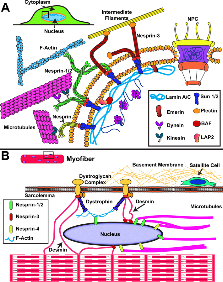
Schematic overview of nuclear envelope proteins involved in force transmission to the nucleus. (A) Force transmission to the nucleus involves interaction of cytoskeletal elements (actin filaments, intermediate filaments, microtubules) with nesprin proteins on the ONM, which transmit force through SUN domain proteins on the INM to the nuclear lamina and interior. (B) Organization of the cytoskeletal network within muscle cells, including the highly ordered actin-myosin structures to form contractile sarcomeres and myofibrils. Nuclei are positioned at the periphery of the cell, where they interact with the muscle-specific proteins dystrophin (through actin filaments) and desmin. Additional proteins such as LINC complex proteins and lamins may be involved in anchoring the myonuclei and place and transmitting forces between the nucleus and cytoskeleton.
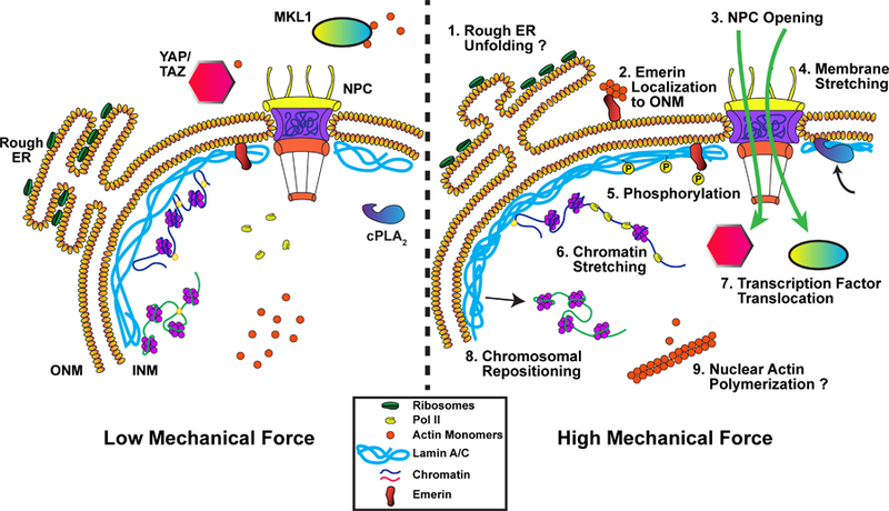
Proposed mechanisms for how the cell nucleus could respond directly to mechanical forces. (1) Stretching of the nuclear membrane could alter the conformation of the rough ER, exposing more ribosomes to the cytoplasm. (2) Force application promotes translocation of emerin from the INM to the ONM, modulating chromatin organization and facilitating actin polymerization at the ONM. (3) Increased membrane tension could open nuclear pore complexes (NPC) and modulate NPC permeability. (4) Stretching of the nuclear membrane recruits cPLA2 to the INM. (5) Force transmission to the nucleus results in post-translational modification and altered dynamics of lamin A/C and INM proteins such as emerin (see also (2)), which can modulate the mechanical properties of the nucleus and induce downstream signaling. (6) External forces can induce chromatin stretching, altering polymerase and transcription factor accessibility and activity. (7) Nuclear pore opening and sequestration at the nuclear envelope can modulate localization and activity of transcription factors. (8) Forces acting on the nucleus may reposition chromatin domains, altering their transcriptional activity. (9) Mechanically induced polymerization of nuclear actin can modulate export and activity of the transcriptional regulator MKL1, and affect other nuclear processes that require monomeric actin.
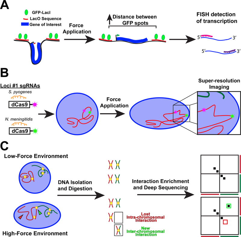
Technologies to study the effect of force transmission to the nucleus on genome organization and gene regulation. (A) Schematic of a reporter transgene to measure chromatin stretching. The transgene is flanked by two fluorescently labeled regions of DNA. An increase in the distance between the fluorescent spots indicates effective chromatin stretching. Changes to the level of transcript of the transgene can be assessed by RNA fluorescence in situ hybridization, allowing to correlate force-induced chromatin stretch with changes in transgene expression. (B) Specific endogenous DNA loci can be fluorescently labeled using CRISPR-dCas9 from different species. Changes to the positioning and spacing between adjacent loci following force application can be determined with high resolution by fluorescence microscopy. (C) Hi-C maps genome-wide chromatin interactions using deep sequencing, with changes to the interaction profile being displayed using heatmaps. Interactions appear as hot spots off the diagonal.
References
-
- Dewey CF Jr., Bussolari SR, Gimbrone MA Jr. & Davies PF The dynamic response of vascular endothelial cells to fluid shear stress. J Biomech Eng 103, 177–85 (1981). - PubMed
-
- Freeman PM, Natarajan RN, Kimura JH & Andriacchi TP Chondrocyte cells respond mechanically to compressive loads. J Orthop Res 12, 311–20 (1994). - PubMed
-
- Magid A & Law DJ Myofibrils bear most of the resting tension in frog skeletal muscle. Science 230, 1280–2 (1985). - PubMed
-
- Le HQ et al. Mechanical regulation of transcription controls Polycomb-mediated gene silencing during lineage commitment. Nat Cell Biol 18, 864–75 (2016). - PubMed
-
- Lecuit T & Lenne PF Cell surface mechanics and the control of cell shape, tissue patterns and morphogenesis. Nat Rev Mol Cell Biol 8, 633–44 (2007). - PubMed
Publication types
MeSH terms
Substances
Grants and funding
LinkOut - more resources
Full Text Sources
Other Literature Sources
