. 2018 Oct;32(10):2152-2166.
doi: 10.1038/s41375-018-0033-0. Epub 2018 Feb 22.

MYC-containing amplicons in acute myeloid leukemia: genomic structures, evolution, and transcriptional consequences

Affiliations

MYC-containing amplicons in acute myeloid leukemia: genomic structures, evolution, and transcriptional consequences

Alberto L Abbate et al. Leukemia. 2018 Oct.

Erratum in

Abstract

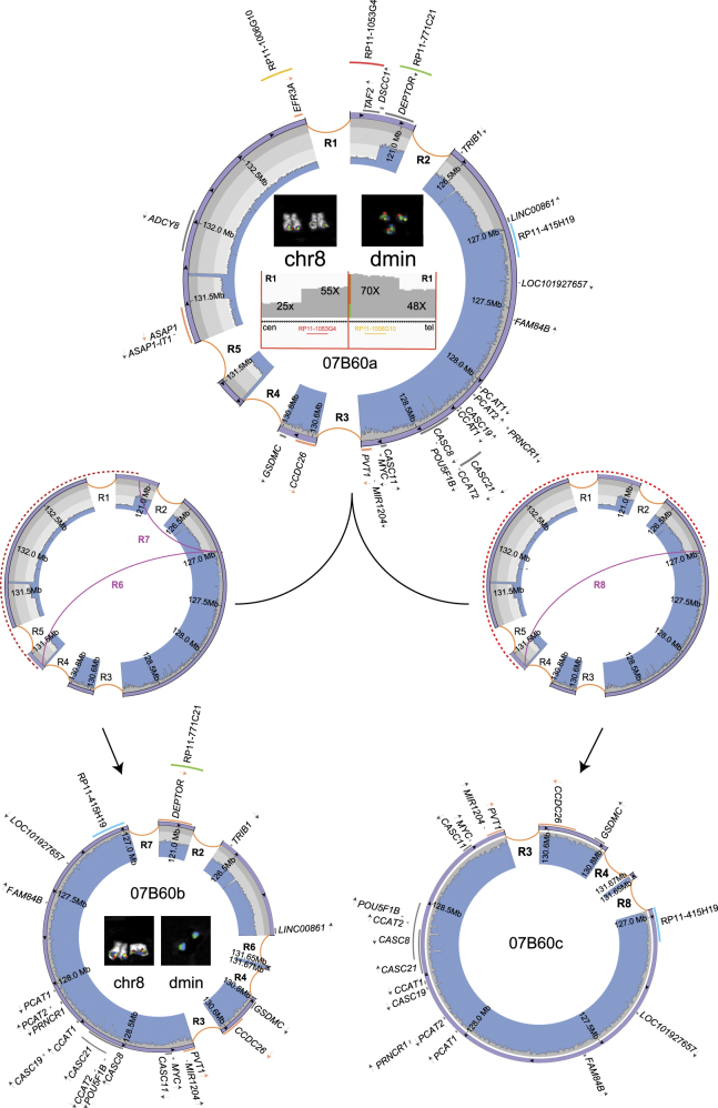
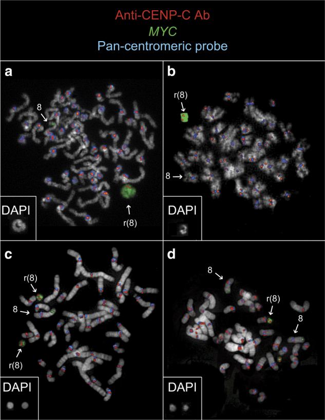
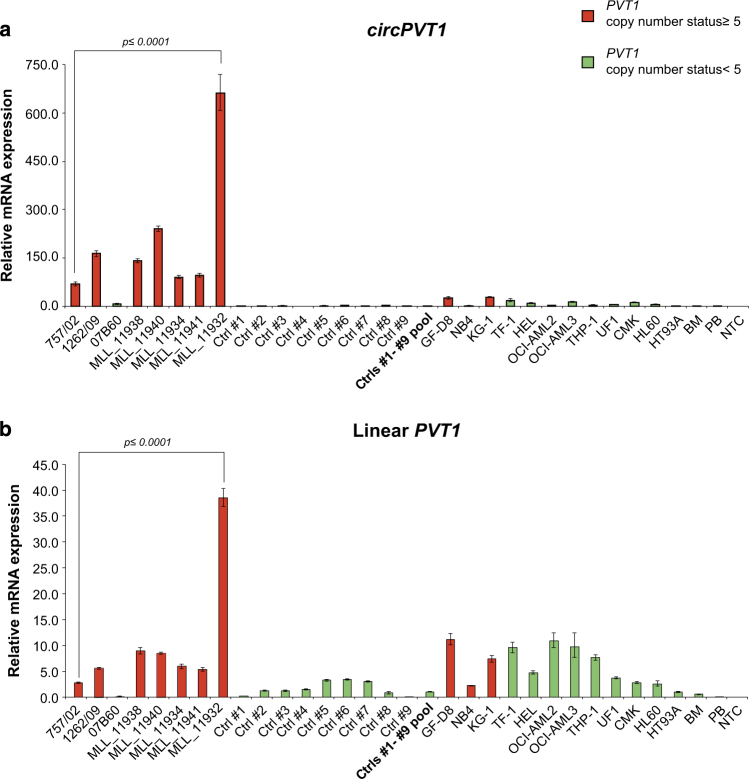
Double minutes (dmin), homogeneously staining regions, and ring chromosomes are vehicles of gene amplification in cancer. The underlying mechanism leading to their formation as well as their structure and function in acute myeloid leukemia (AML) remain mysterious. We combined a range of high-resolution genomic methods to investigate the architecture and expression pattern of amplicons involving chromosome band 8q24 in 23 cases of AML (AML-amp). This revealed that different MYC-dmin architectures can coexist within the same leukemic cell population, indicating a step-wise evolution rather than a single event origin, such as through chromothripsis. This was supported also by the analysis of the chromothripsis criteria, that poorly matched the model in our samples. Furthermore, we found that dmin could evolve toward ring chromosomes stabilized by neocentromeres. Surprisingly, amplified genes (mainly PVT1) frequently participated in fusion transcripts lacking a corresponding DNA template. We also detected a significant overexpression of the circular RNA of PVT1 (circPVT1) in AML-amp cases versus AML with a normal karyotype. Our results show that 8q24 amplicons in AML are surprisingly plastic DNA structures with an unexpected association to novel fusion transcripts and circular RNAs.


Conflict of interest statement

The authors declare that they have no conflict of interest.

Figures

Fig. 1
Fig. 1
Circular amplicons with no or low heterogeneity of structure. Images show the internal structure of amplified segments in cases MLL_11940 (as an example of a patient with no heterogeneous amplicons joined by a duplication) (a) and MLL_12445 (as an example of patient with two coexisting amplicons, one of which derived from the other by a deletion) (b). For each patient: IGV plot of WGS read depth at proximal and distal breakpoints of ancestral amplicons; Circos plots (outer violet circular panel) and WGS read depth (light blue inner panel) of recurrent amplicons, with internal arrows indicating amplicon orientation and genes in dark gray, or in orange if interrupted by a breakpoint (arrows represent the transcriptional orientation). Inside the Circos plots, FISH pseudocolor images showing co-localizing amplified probes, consistently colored as in the Circos plot. Probes mapping respectively upstream and downstream of the amplicons proximal and distal breakpoints are not amplified on dmin and display signal only on normal chromosome 8. The red-dashed semicircles in b represent sequence deletion on the ancestral amplicon to obtain the secondary one
Fig. 2
Fig. 2
Circular amplicons with high heterogeneity of structure. Images show the internal organization and inferred evolutionary path of amplicons in case 07B60, as an example of a patient with clonal amplicon heterogeneity. The ancestral amplicon (07B60a, occurring in the 26.4% (14/53) of cells), at the top of the figure, underwent two independent rearrangement types, indicated as R6/R7 (middle left) or R8 (middle right), originating the secondary amplicons 07B60b (bottom left), observed in 73.6% (39/53) of cells, and 07B60c (bottom right), without cell specificity at the FISH level. Circos plots (in violet) represent all reconstructed amplified structures, with WGS read depth (in light blue) and genes in dark gray, or in orange if interrupted by a breakpoint (arrows represent the transcriptional orientation). Red-dashed semicircles and purple curved lines respectively represent sequence deletions and newly originated SVs labeled by appropriated codes. Inside the 07B60a Circos plot, IGV plots of WGS read depth at amplicon borders (bottom) and FISH pseudocolor images show co-amplification of delimiting amplicons proximal and distal breakpoints (top). The red-dashed semicircles represent sequence deletions on the ancestral amplicon to obtain both secondary ones. It is worth noting that dmin-harboring 07B60a amplicons showed the co-localization of all four indicated probes in the FISH experiments, contrary to those containing 07B60b, which displayed signals only for the green and blue probes, indicating a cell specificity of sequence amplifications
Fig. 3
Fig. 3
Neocentromere at ring chromosomes harboring 8q24 amplicons. Immuno-FISH co-hybridization results in cases MLL_11929 (a), MLL_11933 (b), #18 (c), and #30 (d) display ring chromosomes with MYC amplifications (green) positive for CENP-A (red) but negative for alpha-satellite DNA (blue). Square boxes at the bottom left of each panel show the DAPI image of the rings
Fig. 4
Fig. 4
Chimeric transcripts significantly associated with a peculiar gene expression profile at amplified regions and breakpoints. a Heatmap of 8q24 gene expression levels in our AML-amp cohort. b Box and Whisker plots illustrating differences in the number of breakpoints and chimeras in 8q24 between Groups A and B. c Correlation analysis between the number of genomic breakpoints and chimeras in the 8q24 region identified by CS (left) and FM (right)
Fig. 5
Fig. 5
Circular and linear PVT1 expression. qPCR results obtained for circPVT1 (a) and linear PVT1 (b) in AML cases and cell lines. In red and green, respectively, samples with and without PVT1 exon 2 gain. A pool of nine AML-NK cases was used as calibrator. NTC no template control

References

    1. Turner KM, Deshpande V, Beyter D, Koga T, Rusert J, Lee C, et al. Extrachromosomal oncogene amplification drives tumour evolution and genetic heterogeneity. Nature. 2017;543:122–5. doi: 10.1038/nature21356. - DOI - PMC - PubMed
    1. Mitelman F, Johansson B, Mertens FE Mitelman Database of Chromosome Aberrations and Gene Fusions in Cancer. 2017.
    1. Huh YO, Tang G, Talwalkar SS, Khoury JD, Ohanian M, Bueso-Ramos CE, et al. Double minute chromosomes in acute myeloid leukemia, myelodysplastic syndromes, and chronic myelomonocytic leukemia are associated with micronuclei, MYC or MLL amplification, and complex karyotype. Cancer Genet. 2016;209:313–20. doi: 10.1016/j.cancergen.2016.05.072. - DOI - PubMed
    1. Sait SN, Qadir MU, Conroy JM, Matsui S, Nowak NJ, Baer MR. Double minute chromosomes in acute myeloid leukemia and myelodysplastic syndrome: identification of new amplification regions by fluorescence in situ hybridization and spectral karyotyping. Genes Chromosomes Cancer. 2002;34:42–47. doi: 10.1002/gcc.10038. - DOI - PubMed
    1. Stephens PJ, Greenman CD, Fu B, Yang F, Bignell GR, Mudie LJ, et al. Massive genomic rearrangement acquired in a single catastrophic event during cancer development. Cell. 2011;144:27–40. doi: 10.1016/j.cell.2010.11.055. - DOI - PMC - PubMed

Publication types

Substances

LinkOut - more resources