Review
. 2018 Feb 6:12:44.
doi: 10.3389/fnins.2018.00044. eCollection 2018.

Phospho-Tau Bar Code: Analysis of Phosphoisotypes of Tau and Its Application to Tauopathy

Affiliations
Review

Phospho-Tau Bar Code: Analysis of Phosphoisotypes of Tau and Its Application to Tauopathy

Taeko Kimura et al. Front Neurosci. .

Abstract

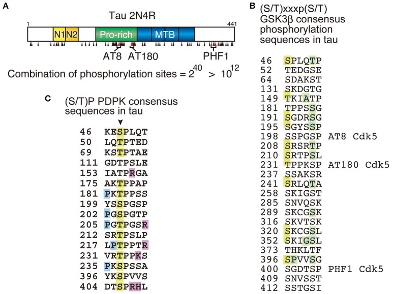
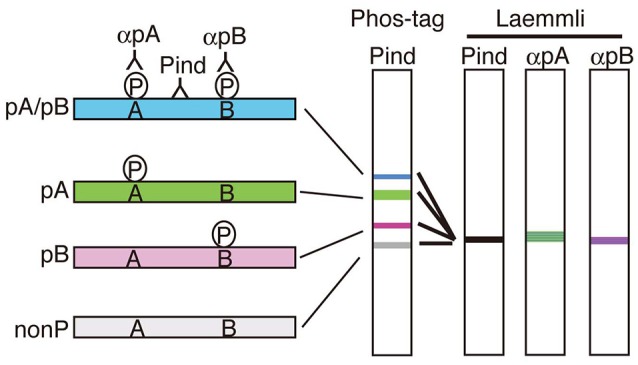
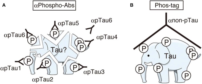
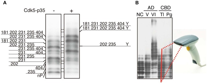
Tau is a microtubule-associated protein which regulates the assembly and stability of microtubules in the axons of neurons. Tau is also a major component of neurofibrillary tangles (NFTs), a pathological hallmark in Alzheimer's disease (AD). A characteristic of AD tau is hyperphosphorylation with more than 40 phosphorylation sites. Aggregates of hyperphosphorylated tau are also found in other neurodegenerative diseases which are collectively called tauopathies. Although a large number of studies have been performed on the phosphorylation of AD tau, it is not known if there is disease-specific phosphorylation among tauopathies. This is due to the lack of a proper method for analyzing tau phosphorylation in vivo. Most previous phosphorylation studies were conducted using a range of phosphorylation site-specific antibodies. These studies describe relative changes of different phosphorylation sites, however, it is hard to estimate total, absolute and collective changes in phosphorylation. To overcome these problems, we have recently applied the Phos-Tag technique to the analysis of tau phosphorylation in vitro and in vivo. This method separates tau into many bands during SDS-PAGE depending on its phosphorylation states, creating a bar code appearance. We propose calling this banding pattern of tau the "phospho-tau bar code." In this review article, we describe what is newly discovered regarding tau phosphorylation through the use of the Phos-Tag. We would like to propose its use for the postmortem diagnosis of tauopathy which is presently done by immunostaining diseased brains with anti-phospho-antibodies. While Phos-tag SDS-PAGE, like other biochemical assays, will lose morphological information, it could provide other types of valuable information such as disease-specific phosphorylation.

Keywords: Alzheimer' disease; Cdk5; GSK3β; phos-tag; phospho-tau bar code; phosphorylation; tau; tauopathy.


Figures

Figure 1
Figure 1
Phosphorylation sites in tau molecule. (A) The longest human tau isoform is composed of 441 amino acids with four microtubule-binding (MTB) repeats in the C-terminal half. Phosphorylation sites are indicated by black arrowheads. AT8 (Ser202 and Thr205), AT180 (Thr231 and Ser235) and PHF1 (Ser396 and Ser404) are phosphospecific antibodies frequently used for the postmortem diagnosis of tauopathy and their epitopes are indicated. The number of phosphorylation combination, if all sites are phosphorylated independently, is indicated below. (B) Amino acid sequences conforming to the GSK3β consensus sequences, (S/T)xx(x)p(S/T), in tau. There are 25 such sequences and 12 sites are reported to be phosphorylated (orange). The site in the C-terminal sides known to be phosphorylated are indicated by green. (C) Ser/Thr-Pro {(S/T)P} sequences in tau targeted by proline-directed protein kinases (PDPK). Arrow indicates Ser or Thr in (S/T)P sequences. Orange is the reported phosphorylation sites, blue is proline (P) conforming to the consensus sequence {Px(S/T)P or P(S/T)P} for MAPK, and magenta is basic amino acids at the C-terminal site which makes Ser or Thr phosphorylation sites favorable for Cdk5.
Figure 2
Figure 2
Separation of phosphoisotypes of a protein on Phos-tag SDS-PAGE. If there are two phosphorylation sites, A and B, in a protein, there would be 4 phosphoisotypes, doubly phosphorylated (blue), two single site-phosphorylated (green and red) and nonphosphorylated (left). These four phosphoisotypes can be separated into four bands on Phos-tag SDS-PAGE by shifting upward differently depending on the number and site of phosphorylation (Phos-tag). Phospho-specific antibodies, αpA and αpB, are required to detect the respective phosphorylation in Laemmli's SDS-PAGE, in contrast, the separation in Phos-tag indicates not only phosphorylation but also the degree of phosphorylation detected with a phosphorylation-independent antibody alone (Pind).
Figure 3
Figure 3
Cartoons representing the images of immunoblotting with many phospho-specific antibodies (A) and a phosphorylation-independent single antibody after Phos-tag SDS-PAGE (B). Phosphospecific antibodies detect only part of phosphorylation at each site and some sites can be masked by another phosphorylated site nearby (here the site for αpTau6 is masked by a phosphorylated site which reacts with αpTau4). Phos-tag shows the whole phosphorylation profile at once. An illustration of an elephant was downloaded from the free illustration site at: http://illpop.com/png_animalhtm/elephant_a04.htm.
Figure 4
Figure 4
Phosphoisotypes of tau on Phos-tag SDS-PAGE. (A) Immunoblotting of tau expressed in COS-7 cells with tau5 in the presence (+) or absence (−) of Cdk5-p35 after Phos-tag-SDS-PAGE, and the phosphorylation sites in each band are indicated by the amino acid number according to the human longest isoform of tau. Y is an unknown phosphorylation site. (B) Immunoblotting of tau in human brains after Phos-tag SDS-PAGE. NC, normal control; AD V and VI, Braak stage V and VI of Alzheimer's disease (AD); CBD Tl and Pg, temporal lobe (Tl) and prefrontal gyrus (Pg) of corticobasal degeneration (CBD). We propose that the banding pattern created using Phos-tag SDS-PAGE could be read by a bar code reader and result in a simple method for diagnosing tauopathy. (A,B) are reproduced and modified from figures reported previously by Kimura et al. (2016a,b), respectively.

References

    1. Ando K., Maruko-Otake A., Ohtake Y., Hayashishita M., Sekiya M., Iijima K. M. (2016). Stabilization of Microtubule-Unbound Tau via Tau Phosphorylation at Ser262/356 by Par-1/MARK Contributes to Augmentation of AD-Related Phosphorylation and Aβ42-Induced Tau Toxicity. PLoS Genet. 2:e1005917 10.1371/journal.pgen.1005917 - DOI - PMC - PubMed
    1. Arendt T., Stieler J. T., Holzer M. (2016). Tau and tauopathies. Brain Res. Bull. 126, 238–292. 10.1016/j.brainresbull.2016.08.018 - DOI - PubMed
    1. Arrasate M., Pérez M., Avila J. (2000). Tau dephosphorylation at tau-1 site correlates with its association to cell membrane. Neurochem. Res. 25, 43–50. 10.1023/A:1007583214722 - DOI - PubMed
    1. Asada A., Yamazaki R., Kino Y., Saito T., Kimura T., Miyake M., et al. . (2014). Cyclin-dependent kinase 5 phosphorylates and induces the degradation of ataxin-2. Neurosci. Lett. 563, 112–117. 10.1016/j.neulet.2014.01.046 - DOI - PubMed
    1. Ballatore C., Lee V. M., Trojanowski J. Q. (2007). Tau-mediated neurodegeneration in Alzheimer's disease and related disorders. Nat. Rev. Neurosci. 8, 663–672. 10.1038/nrn2194 - DOI - PubMed