Review
. 2018 Feb 5:9:31.
doi: 10.3389/fneur.2018.00031. eCollection 2018.

Eye Movement Abnormalities in Multiple Sclerosis: Pathogenesis, Modeling, and Treatment

Affiliations
Review

Eye Movement Abnormalities in Multiple Sclerosis: Pathogenesis, Modeling, and Treatment

Alessandro Serra et al. Front Neurol. .

Abstract

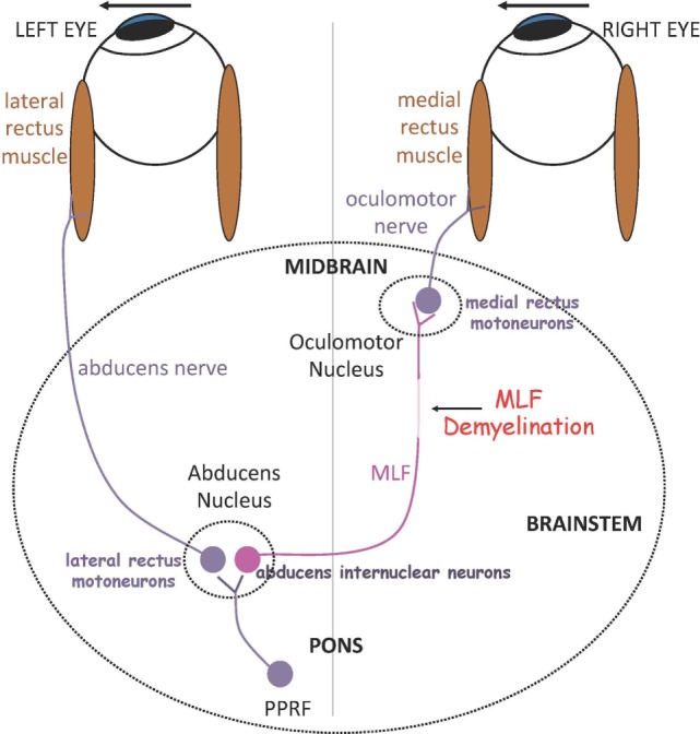
Multiple sclerosis (MS) commonly causes eye movement abnormalities that may have a significant impact on patients' disability. Inflammatory demyelinating lesions, especially occurring in the posterior fossa, result in a wide range of disorders, spanning from acquired pendular nystagmus (APN) to internuclear ophthalmoplegia (INO), among the most common. As the control of eye movements is well understood in terms of anatomical substrate and underlying physiological network, studying ocular motor abnormalities in MS provides a unique opportunity to gain insights into mechanisms of disease. Quantitative measurement and modeling of eye movement disorders, such as INO, may lead to a better understanding of common symptoms encountered in MS, such as Uhthoff's phenomenon and fatigue. In turn, the pathophysiology of a range of eye movement abnormalities, such as APN, has been clarified based on correlation of experimental model with lesion localization by neuroimaging in MS. Eye movement disorders have the potential of being utilized as structural and functional biomarkers of early cognitive deficit, and possibly help in assessing disease status and progression, and to serve as platform and functional outcome to test novel therapeutic agents for MS. Knowledge of neuropharmacology applied to eye movement dysfunction has guided testing and use of a number of pharmacological agents to treat some eye movement disorders found in MS, such as APN and other forms of central nystagmus.

Keywords: eye movements; internuclear ophthalmoplegia; multiple sclerosis; nystagmus; pathologic saccades.


Figures

Figure 1
Figure 1
Summary of a simple model for generating horizontal gaze shifts in INO. Premotor excitatory burst neurons lying in the paramedian pontine reticular formation (PPRF), project a pulse of innervation to the abducens nucleus (CN VI). Abducens motoneurons project the pulse of innervation via the sixth nerve to the right lateral rectus, which contracts rapidly to generate an abducting saccade of the left eye. Abducens internuclear neurons project the pulse of innervation, via the medial longitudinal fasciculus (MLF, internuclear pathway), to medial rectus motoneurons that, in turn, innervate the right medial rectus via the third nerve, to generate an adducting saccade of the right eye. If the MLF is demyelinated, signals are low-pass filtered and delayed, affecting the size and timing of the pulse thus causing adducting saccades of the right eye to be slow. *Adapted with permission from Ref. (6).
Figure 2
Figure 2
Representative horizontal leftward saccade before (A) and after Dalfampridine (B) in one patient. (A) Particularly for the leftward movement the adducting eye requires several small saccades to acquire the target (arrows) and the abducting eye shows dissociated nystagmus. (B) The right eye requires less adducting saccades to acquire the target (arrows) and dissociated nystagmus intensity is decreased. Positive values indicate rightward movements, negative values indicate leftward movements. *Adapted with permission from Ref. (49).

Similar articles

Cited by

References

    1. Meyer-Moock S, Feng Y-S, Maeurer M, Dippel F-W, Kohlmann T. Systematic literature review and validity evaluation of the Expanded Disability Status Scale (EDSS) and the multiple sclerosis functional composite (MSFC) in patients with multiple sclerosis. BMC Neurol (2014) 4:58.10.1186/1471-2377-14-58 - DOI - PMC - PubMed
    1. Derwenskus J, Rucker JC, Serra A, Stahl JS, Downey DL, Adams NL, et al. Abnormal eye movements predict disability in MS: two-year follow-up. Ann N Y Acad Sci (2005) 1039:521–3.10.1196/annals.1325.058 - DOI - PubMed
    1. Moster S, Wilson JA, Galetta SL, Balcer LJ. The King-Devick (K-D) test of rapid eye movements: a bedside correlate of disability and quality of life in MS. J Neurol Sci (2014) 343:105–9.10.1016/j.jns.2014.05.047 - DOI - PubMed
    1. Raphael BA, Galetta KM, Jacobs DA, Markowitz CE, Liu GT, Nano-Schiavi ML, et al. Validation and test characterisitics of a 10-item neuro-ophthalmic supplement to the NEI-VFQ-25. Am J Ophthalmol (2006) 142:1026–35.10.1016/j.ajo.2006.06.060 - DOI - PubMed
    1. Polman CH, Reingold SC, Banwell B, Clanet M, Cohen JA, Filippi M, et al. Diagnostic criteria for multiple sclerosis: 2010 revisions to the McDonald criteria. Ann Neurol (2011) 69(2):292–302.10.1002/ana.22366 - DOI - PMC - PubMed