

Mitochondrial Proteins Coded by Human Tumor Viruses
- PMID: 29467726
- PMCID: PMC5808139
- DOI: 10.3389/fmicb.2018.00081
Mitochondrial Proteins Coded by Human Tumor Viruses
Abstract
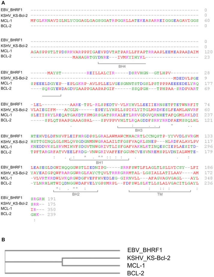
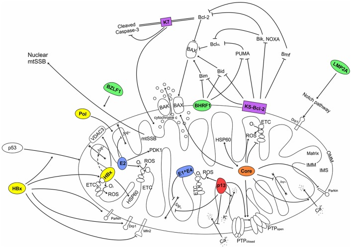
Viruses must exploit the cellular biosynthetic machinery and evade cellular defense systems to complete their life cycles. Due to their crucial roles in cellular bioenergetics, apoptosis, innate immunity and redox balance, mitochondria are important functional targets of many viruses, including tumor viruses. The present review describes the interactions between mitochondria and proteins coded by the human tumor viruses human T-cell leukemia virus type 1, Epstein-Barr virus, Kaposi's sarcoma-associated herpesvirus, human hepatitis viruses B and C, and human papillomavirus, and highlights how these interactions contribute to viral replication, persistence and transformation.
Keywords: EBV; HBV; HCV; HPV; HTLV-1; KSHV; Mitochondria.
Figures
Similar articles
-
Mitochondria as functional targets of proteins coded by human tumor viruses.Adv Cancer Res. 2005;94:87-142. doi: 10.1016/S0065-230X(05)94003-7. Adv Cancer Res. 2005. PMID: 16096000 Review.
-
Interplay Between KSHV and the Host DNA Damage Response.Front Cell Infect Microbiol. 2020 Dec 9;10:604351. doi: 10.3389/fcimb.2020.604351. eCollection 2020. Front Cell Infect Microbiol. 2020. PMID: 33425783 Free PMC article. Review.
-
Oncogenes and RNA splicing of human tumor viruses.Emerg Microbes Infect. 2014 Sep;3(9):e63. doi: 10.1038/emi.2014.62. Epub 2014 Sep 3. Emerg Microbes Infect. 2014. PMID: 26038756 Free PMC article. Review.
-
Oncogenic Virus Infections in the General Population and End-stage Renal Disease Patients With Special Emphasis on Kaposi's Sarcoma Associated Herpes Virus (KSHV) in Northeast of Iran.Jundishapur J Microbiol. 2015 Mar 21;8(3):e14920. doi: 10.5812/jjm.14920. eCollection 2015 Mar. Jundishapur J Microbiol. 2015. PMID: 25834713 Free PMC article.
-
Human Oncogenic Herpesvirus and Post-translational Modifications - Phosphorylation and SUMOylation.Front Microbiol. 2016 Jun 17;7:962. doi: 10.3389/fmicb.2016.00962. eCollection 2016. Front Microbiol. 2016. PMID: 27379086 Free PMC article. Review.
Cited by
-
Metabolic modeling elucidates phenformin and atpenin A5 as broad-spectrum antiviral drugs against RNA viruses.Commun Biol. 2025 May 23;8(1):791. doi: 10.1038/s42003-025-08148-y. Commun Biol. 2025. PMID: 40410544 Free PMC article.
-
Dysregulation of host cell calcium signaling during viral infections: Emerging paradigm with high clinical relevance.Mol Aspects Med. 2021 Oct;81:101004. doi: 10.1016/j.mam.2021.101004. Epub 2021 Jul 23. Mol Aspects Med. 2021. PMID: 34304899 Free PMC article. Review.
-
Functional properties and sequence variation of HTLV-1 p13.Retrovirology. 2020 May 12;17(1):11. doi: 10.1186/s12977-020-00517-1. Retrovirology. 2020. PMID: 32398094 Free PMC article. Review.
-
Virus Control of Cell Metabolism for Replication and Evasion of Host Immune Responses.Front Cell Infect Microbiol. 2019 Apr 18;9:95. doi: 10.3389/fcimb.2019.00095. eCollection 2019. Front Cell Infect Microbiol. 2019. PMID: 31058096 Free PMC article. Review.
-
Warburg Effect, Glutamine, Succinate, Alanine, When Oxygen Matters.Biology (Basel). 2021 Oct 4;10(10):1000. doi: 10.3390/biology10101000. Biology (Basel). 2021. PMID: 34681099 Free PMC article.
References
Publication types
LinkOut - more resources
Full Text Sources
Other Literature Sources