

Advanced imaging techniques for small bowel Crohn's disease: what does the future hold?
- PMID: 29467827
- PMCID: PMC5813850
- DOI: 10.1177/1756283X18757185
Advanced imaging techniques for small bowel Crohn's disease: what does the future hold?
Abstract
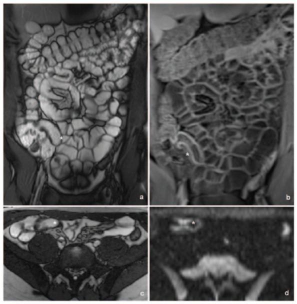
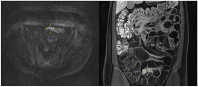
Treatment of Crohn's disease (CD) is intrinsically reliant on imaging techniques, due to the preponderance of small bowel disease and its transmural pattern of inflammation. Ultrasound (US), computed tomography (CT) and magnetic resonance imaging (MRI) are the most widely employed imaging methods and have excellent diagnostic accuracy in most instances. Some limitations persist, perhaps the most clinically relevant being the distinction between inflammatory and fibrotic strictures. In this regard, several methodologies have recently been tested in animal models and human patients, namely US strain elastography, shear wave elastography, contrast-enhanced US, magnetization transfer MRI and contrast dynamics in standard MRI. Technical advances in each of the imaging methods may expand their indications. The addition of oral contrast to abdominal US appears to substantially improve its diagnostic capabilities compared to standard US. Ionizing dose-reduction methods in CT can decrease concern about cumulative radiation exposure in CD patients and diffusion-weighted MRI may reduce the need for gadolinium contrast. Clinical indexes of disease activity and severity are also increasingly relying on imaging scores, such as the recently developed Lémann Index. In this review we summarize some of the recent advances in small bowel CD imaging and how they might affect clinical practice in the near future.
Keywords: Crohn disease; diagnostic imaging; fibrosis; inflammation; magnetic resonance imaging; positron-emission tomography; small intestine; ultrasonography.
Conflict of interest statement
Conflict of interest statement: The authors declare that there is no conflict of interest.
Figures
Similar articles
-
New cross-sectional imaging in IBD.Curr Opin Gastroenterol. 2018 Jul;34(4):194-207. doi: 10.1097/MOG.0000000000000440. Curr Opin Gastroenterol. 2018. PMID: 29846256 Review.
-
Noninvasive Multimodal Methods to Differentiate Inflamed vs Fibrotic Strictures in Patients With Crohn's Disease.Clin Gastroenterol Hepatol. 2019 Nov;17(12):2397-2415. doi: 10.1016/j.cgh.2019.04.025. Epub 2019 Apr 14. Clin Gastroenterol Hepatol. 2019. PMID: 30995529 Review.
-
Assessment of stricturing Crohn's disease: Current clinical practice and future avenues.World J Gastroenterol. 2016 Jan 21;22(3):1008-16. doi: 10.3748/wjg.v22.i3.1008. World J Gastroenterol. 2016. PMID: 26811643 Free PMC article. Review.
-
Real-Time Shear Wave Ultrasound Elastography Differentiates Fibrotic from Inflammatory Strictures in Patients with Crohn's Disease.Inflamm Bowel Dis. 2018 Sep 15;24(10):2183-2190. doi: 10.1093/ibd/izy115. Inflamm Bowel Dis. 2018. PMID: 29718309 Free PMC article.
-
Accuracy of small-intestine contrast ultrasonography, compared with computed tomography enteroclysis, in characterizing lesions in patients with Crohn's disease.Clin Gastroenterol Hepatol. 2013 Aug;11(8):950-5. doi: 10.1016/j.cgh.2013.01.015. Epub 2013 Jan 29. Clin Gastroenterol Hepatol. 2013. PMID: 23375998
Cited by
-
Lemann Index for Assessment of Crohn's Disease: Correlation with the Quality of Life, Endoscopic Disease Activity, Magnetic Resonance Index of Activity and C- Reactive Protein.Open Med (Wars). 2019 Nov 7;14:785-791. doi: 10.1515/med-2019-0092. eCollection 2019. Open Med (Wars). 2019. PMID: 31737782 Free PMC article.
-
Diagnostic Accuracy of Magnetic Resonance Enterography for the Evaluation of Active and Fibrotic Inflammation in Crohn's Disease.Front Surg. 2022 May 13;9:872596. doi: 10.3389/fsurg.2022.872596. eCollection 2022. Front Surg. 2022. PMID: 35647009 Free PMC article.
-
Elastography as a Discriminator Between Fibrotic and Inflammatory Strictures in Crohn's Disease: A Dead End or Bright Future in Clinical Decision-Making? Critical Review.Diagnostics (Basel). 2024 Oct 16;14(20):2299. doi: 10.3390/diagnostics14202299. Diagnostics (Basel). 2024. PMID: 39451622 Free PMC article. Review.
-
Inflammatory bowel disease cross-sectional imaging: What's new?United European Gastroenterol J. 2022 Dec;10(10):1179-1193. doi: 10.1002/ueg2.12343. Epub 2022 Dec 3. United European Gastroenterol J. 2022. PMID: 36461914 Free PMC article. Review.
-
Radiology of fibrosis part II: abdominal organs.J Transl Med. 2024 Jul 2;22(1):610. doi: 10.1186/s12967-024-05346-w. J Transl Med. 2024. PMID: 38956593 Free PMC article. Review.
References
-
- Gomollon F, Dignass A, Annese V, et al. 3rd European Evidence-based Consensus on the Diagnosis and Management of Crohn’s Disease 2016: part 1. Diagnosis and medical management. J Crohns Colitis 2017; 11: 3–25. - PubMed
-
- Horsthuis K, Bipat S, Bennink RJ, et al. Inflammatory bowel disease diagnosed with US, MR, scintigraphy, and CT: meta-analysis of prospective studies. Radiology 2008; 247: 64–79. - PubMed
-
- Panes J, Bouzas R, Chaparro M, et al. Systematic review: the use of ultrasonography, computed tomography and magnetic resonance imaging for the diagnosis, assessment of activity and abdominal complications of Crohn’s disease. Aliment Pharmacol Ther 2011; 34: 125–145. - PubMed
-
- Negaard A, Paulsen V, Sandvik L, et al. A prospective randomized comparison between two MRI studies of the small bowel in Crohn’s disease, the oral contrast method and MR enteroclysis. Eur Radiol 2007; 17: 2294–2301. - PubMed
Publication types
LinkOut - more resources
Full Text Sources
Other Literature Sources
