. 2017 Dec 22;9(6):6771-6779.
doi: 10.18632/oncotarget.23645. eCollection 2018 Jan 23.

Kanglaite inhibits EMT caused by TNF-α via NF-κΒ inhibition in colorectal cancer cells

Affiliations

Kanglaite inhibits EMT caused by TNF-α via NF-κΒ inhibition in colorectal cancer cells

Guiling Shi et al. Oncotarget. .

Abstract

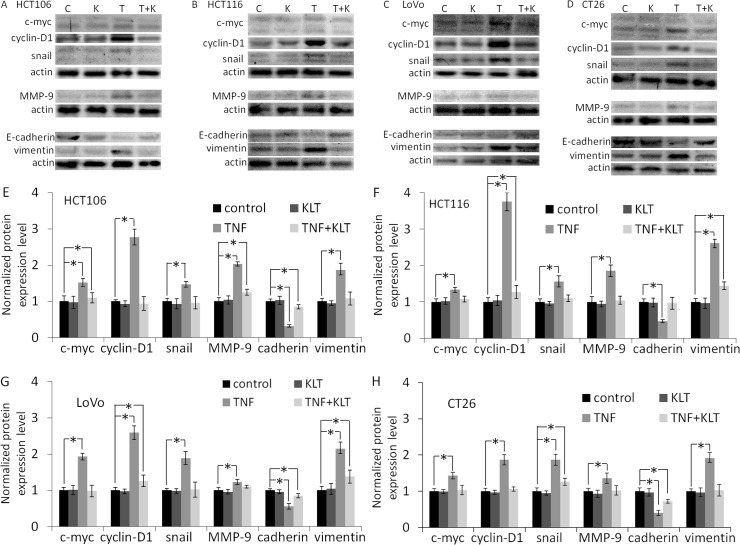
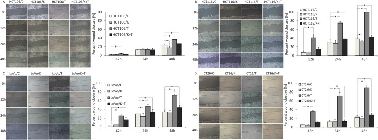
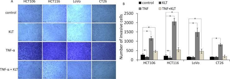
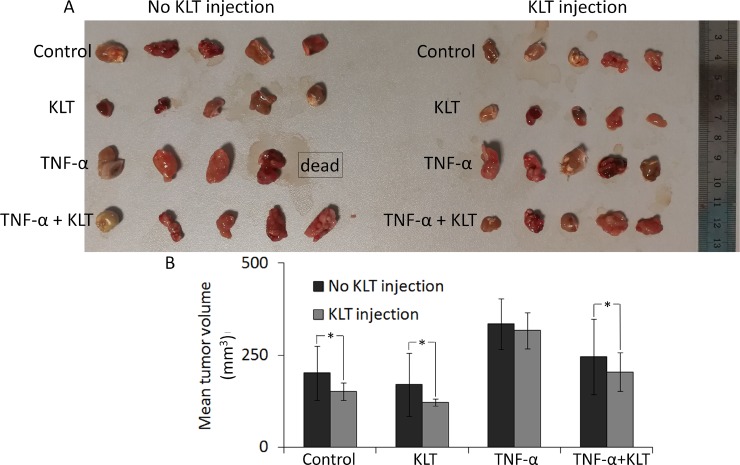
Tumor necrosis factor-alpha is a critical pro-inflammatory cytokine produced by macrophages and was once considered an anti-tumor agent. However, a low dose of tumor necrosis factor-alpha can cause epithelial mesenchymal transition, angiogenesis and metastasis. NF-κΒ contributes to epithelial mesenchymal transition induced by tumor necrosis factor-alpha. Kanglaite, an extract from the Coix lacryma-jobi (adlay) seed, is an NF-κΒ inhibitor. The aim of this study was to investigate whether Kanglaite could inhibit epithelial mesenchymal transition caused by tumor necrosis factor-alpha using four colorectal cancer cell lines, HCT106, HCT116, LoVo and CT26. Our results showed that tumor necrosis factor-alpha -mediated activation of NF-κΒ, caused changes in epithelial mesenchymal transition -related protein expression, and increased migration and invasion in all four cell lines. However, these effects were inhibited by Kanglaite when used in combination with tumor necrosis factor-alpha. In a subcutaneous tumor model of CT26, tumor necrosis factor-alpha enhanced the tumorigenic ability of the cells, and again this was inhibited by Kanglaite. However, treatment with Kanglaite alone caused almost no inhibition of epithelial mesenchymal transition -mediated tumor growth, when cells were pretreated with tumor necrosis factor-alpha prior to injection. These results suggest that Kanglaite inhibits tumor necrosis factor-alpha -mediated epithelial mesenchymal transition in colorectal cancer cell lines via inhibition of NF-κΒ.

Keywords: EMT; NF-κΒ; TNF-α; colorectal cancer cell lines; kanglaite.


Conflict of interest statement

CONFLICTS OF INTEREST None.

Figures

Figure 1
Figure 1. Western blot analysis of NF-κΒ p65, IKKα and IκBα expression
NF-κΒ expression was measured in the cytoplasm, nuclei and whole cell lysate in order to determine whether NF-κΒ is translocated to the nucleus induced by TNF-α. Because IKK and IκB are both cytoplasmic proteins, and also interact with NF-κΒ in the cytoplasm, these proteins could only be detected in the cytoplasmic samples. In blot images, C represents control, K represents KLT only, T represents TNF-α only and T+K represents TNF-α plus KLT. Experiments were performed in triplicate. (A) NF-κΒ p65 expression in the cytoplasm. (B) NF-κΒ p65 expression in the nuclei. (C) NF-κΒ p65 expression in whole cells. (D) IκBα expression in the cytoplasm. (E) IKKα expression in the cytoplasm. (F–I) Bar diagrams of densitometric analysis of NF-κΒ, IKKα and IκΒα expression in the four CRC cell lines. NF-κΒ-c represents NF-κΒ expression in cytoplasm, NF-κΒ-n represents NF-κΒ expression in nuclei, and NF-κΒ-w represents NF-κΒ expression in the whole cell. *P < 0.05 indicates a significant difference compared to the control group. Full length blots of A–E are shown in Supplementary Figure 5.
Figure 2
Figure 2. Western blot analysis of c-myc, cyclin-D1, snail, MMP-9, E-cadherin and vimentin expression
Experiments were performed in triplicate. (AD) Protein expression in the four CRC cell lines after treatment with different reagents, as indicated. C represents control, K represents KLT only, T represents TNF-α only, and T+K represents TNF-α plus KLT. (EH) Bar diagrams of densitometric analysis of data in panels A–D. *P < 0.05 indicates a significant difference compared to the control group. Full length blots of A–D are shown in Supplementary Figure 6.
Figure 3
Figure 3. Migration of the four CRC cell lines after KLT and TNF-α treatment was measured using a wound healing scratch assay (original magnification ×100)
C represents control, K represents KLT only, T represents TNF-α only, and T+K represents TNF-α plus KLT. The scale bars represent 100 μm. Experiments were performed in triplicate. *P < 0.05 indicates a significant difference compared to the control group. Furthermore, for all cell lines, the percent wound closure in the K+T group was significantly lower than for the T group (P < 0.05).
Figure 4
Figure 4. Invasion of the four CRC cell lines after KLT and TNF-α treatment was measured using a transwell assay (original magnification ×100)
The scale bars represent 100 μm. Experiments were performed in triplicate. *P < 0.05 indicates a significant difference compared to the control group. For all cells lines, invasion cell numbers were significantly lower in the TNF+KLT group than in the TNF group (P < 0.05).
Figure 5
Figure 5. Inhibition of tumor growth by KLT
(A) Four different pretreatment strategies of CT26 cells used for subcutaneous inoculation. Each group contained five mice. Tumors were allowed to develop for 15 days, but one mouse from the TNF-α pretreated & no KLT injection group was found dead at 14 days. (B) Columns represent mean volume from 5 tumors, except for the TNF-α pretreated & no KLT injection data, which came from 4 tumors. Average tumor volumes in two adjacent columns were compared using the Student's t-test. *P < 0.05 represents a significant difference.

References

    1. Wajant H, Pfizenmaier K, Scheurich P. Tumor necrosis factor signaling. Cell Death Differ. 2003;10:45–65. - PubMed
    1. Wang X, Lin Y. Tumor necrosis factor and cancer, buddies or foes? Acta Pharmacol Sin. 2008;29:1275–1288. - PMC - PubMed
    1. Balkwill F. Tumor necrosis factor or tumor promoting factor? Cytokine Growth Factor Rev. 2002;13:135–141. - PubMed
    1. Soria G, Ofri-Shahak M, Haas I, Yaal-Hahoshen N, Leider-Trejo L, Leibovich-Rivkin T, Weitzenfeld P, Meshel T, Shabtai E, Gutman M, Ben-Baruch A. Inflammatory mediators in breast cancer: coordinated expression of TNFα & IL-1β with CCL2 & CCL5 and effects on epithelial-to-mesenchymal transition. BMC Cancer. 2011;11:130. https://doi.org/10.1186/1471-2407-11-130. - DOI - PMC - PubMed
    1. Csiszár A, Szentes T, Haraszti B, Zou W, Emilie D, Petrányi G, Pócsik E. Characterisation of cytokine mRNA expression in tumour-infiltrating mononuclear cells and tumour cells freshly isolated from human colorectal carcinomas. Eur Cytokine Netw. 2001;12:87–96. - PubMed

LinkOut - more resources